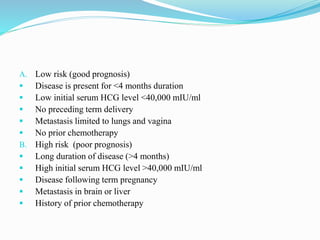
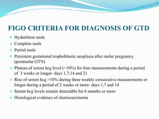
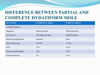
DR.PRIYA SAXENA
DEFINITION
 GTD is a spectrum of abnormal proliferation of trophoblastic tissue
associated with pregnancy
CLASSIFICATION OF GTD BASED ON THE
SEVERITY OF DISEASE
 Hydatiform mole
 Complete
 Partial
 Invasive mole
 Placental site trophoblastic tumor
 Epitheloid trophoblastic tumor
 Choriocarcinoma
 Non metastatic choriocarcinoma (restricted to uterus)
 Metastatic choriocarcinoma (spread beyond uterus)
A. Low risk (good prognosis)
 Disease is present for <4 months duration
 Low initial serum HCG level <40,000 mIU/ml
 No preceding term delivery
 Metastasis limited to lungs and vagina
 No prior chemotherapy
B. High risk (poor prognosis)
 Long duration of disease (>4 months)
 High initial serum HCG level >40,000 mIU/ml
 Disease following term pregnancy
 Metastasis in brain or liver
 History of prior chemotherapy
FIGO CRITERIA FOR DIAGNOSIS OF GTD
 Hydatiform mole
 Complete mole
 Partial mole
 Persistent gestational trophoblastic neoplasia after molar pregnancy
(postmolar GTN)
 Plateau of serum hcg level (>10%) for four measurements during a period
of 3 weeks or longer- days 1,7,14 and 21
 Rise of serum hcg >10% during three weekly consecutive measurements or
longer during a period of 2 weeks or more- days 1,7 and 14
 Serum hcg levels remain detectable for 6 months or more
 Histological evidence of choriocarcinoma
HYDATIFORM MOLE(MOLAR
PREGNANCY,VESICULAR MOLE)
 Definition:
 It is an abnormal condition of the placenta where there are partly
degenerative and partly proliferative changes in the young chorionic villi.
 These result in the formation of clusters of small cysts of varying sizes.
 Because of its superficial resemblance to hydatid cyst, it is named as
hydatidiform mole.
 It is best regarded as a benign neoplasia of the chorion with malignant
potential.
TYPES
 Complete hydatiform mole
 Partial hydatiform mole
DIFFERENCE BETWEEN PARTIALAND
COMPLETE HYDATIFORM MOLE
FEATURES COMPLETE MOLE PARTIAL MOLE
1.Clinical features
Diagnosis Molar gestation Missed abortion
Uterine size 50% large for dates Small for dates
Medical complications Frequent Rare
Theca lutein cysts 25-30% 5-10%
GTN 15% 0.5%
Need of chemotherapy 15% 0.5%
Choriocarcinoma 3% 0.1%
COMPLETE MOLE PARTIAL MOLE
2.Pathology
Fetal tissue Absent Often present
Amnion and fetal RBC Absent Often present
Villus edema Diffuse Variable,focal
Trophoblastic proliferation Variable, slight to severe Variable, focal, slight to moderate
Karyotype 46,XX or 46,XY Usually 69,XXX or 69,XXY
HCG values Markedly increased Moderately increased
ETIOLOGY OF H.MOLE
 Maternal age: Women in the extremes of the reproductive age group are at
an increased risk.
 The incidence seems to change with race and ethnicity being more common
in Asians than Caucasians
 Dietary factors: Decreased consumption of carotene, fats,animal fats,
vitamin and proteins may be associated with progressively increased risk of
molar pregnancies.
 Previous H.Mole: The risk of H.Mole after one previous H. Mole is 1-2%
whereas after two molar pregnancies it rises sharply to 15-28%
 Cytogenetic abnormality: Fertilization of an empty ovum(complete
mole),triploidy (partial mole)
 Miscellaneous factors: like smoking, use of oral contraception,previous
miscarriages, increased gammaglobulin levels and AB blood group are other
high risk factors for molar pregnancy
PATHOLOGY
 It is principally a disease of the chorion.
 Death of the ovum or failure of the embryo to grow is essential to develop
complete (classic) hydatidiform mole.
 The secretion from the hyperplastic cells and transferred substances from
the maternal blood accumulate in the stroma of the villi which are devoid of
blood vessels.
 This results in distension of the villi to form small vesicles.
 The distension may also be due to edema and liquefaction of the stroma.
 Vesicle fluid is interstitial fluid and is almost similar to ascitic or edema
fluid, but rich in hCG.
 Naked eye appearance :
 The mass filling the uterus is made of multiple chains and clusters
 of cysts of varying sizes.
 There is no trace of embryo or the amniotic sac.
 Hemorrhage, if occurs, takes place in the decidual space.
 Microscopic appearance:
The basic findings are—
(1) There is marked proliferation of the syncytial and cytotrophoblastic
epithelium.
(2) Marked thinning of the stromal tissue due to hydropic degeneration.
(3) There is absence of blood vessels in the villi which seems primary rather
than due to pressure atrophy.
(4) The villous pattern is distinctly maintained.
 Ovarian changes:
 Bilateral lutein cysts are present in about 50%.
 These are due to excessive production of chorionic gonadotropin and they
are also observed in multiple pregnancy.
 These regress spontaneously within 2 months after expulsion of mole.
 The contained fluid is rich in chorionic gonadotropin. It also contains
estrogen and progesterone.
SYMPTOMS
 Amenorrhoea: varying duration (usually 1-2 months)
 Hyperemesis: common due to high levels of HCG
 Vaginal bleeding: most common presentation (90% cases) varying from
spotting to profuse bleeding. The blood may be mixed with a gelatinous
fluid from ruptured cysts giving the appearance of discharge “white currant
in red currant juice”.
 Abdominal pain:
Causes of pain in hydatiform mole are:
 Excessive distension of the uterus by the mole
 Onset of uterine contractions in an attempt to expel the mole
 Sepsis
 Concealed uterine hemorrhage with maternal anemia
 Perforating invasive mole
 General symptoms:
 Dyspnea: due to embolization of the trophoblastic cells in the lungs
 Thyrotoxicosis and thyroid storm: due to thyrotropin like effect of HCG
whose levels are excessive in hydatiform mole
 Quickening is absent
 Symptoms of early onset pre-eclampsia
 Passage of grape like vesicles vaginally: diagnostic of a molar pregnancy
SIGNS
 The patient appears sick and may be dyspneic
 Anemia (mostly microcytic hypochromic due to iron deficiency but may be
megaloblastic due to folate and cyanocobalamine deficiency)
 Early onset pre-eclampsia (present in about 25% cases): The pre-eclamptic
process may be due to uterine enlargement or more probably due to
trophoblastic proliferation
 Per abdomen: On abdominal palpation
 The uterus is bigger than the gestational period in 50-70% cases, equivalent
in 20-35% cases, smaller in rest 10-15% cases.
 It feels doughy as uterus is full of mole without much amniotic fluid.
 Due to absence of fetus in a complete mole, fetal parts, fetal movement,
external ballotment are missing
 Fetal heart cannot be heard
 Vaginal examination:
 Internal ballotment is absent
 Theca lutein cysts of the ovary may be felt in many cases(50%)
 Presence of grape like vesicles on vaginal examination makes diagnosis of
hydatiform mole certain
 If finger can be negotiated through cervix, fetal parts and membranes are
not felt byt vesicles and blood clots are felt
INVESTIGATIONS
 Complete hemogram and coagulation profile
 Blood group and Rh type
 Liver, kidney and thyroid function tests are carried out in indicated cases
 Ultrasound:
 The typical ultrasound picture of complete mole is the presence of multiple
small sonolucencies called the snow storm appearance with no fetus or
amniotic sac
 In case of a partial mole, some fetal tissues may be visualized along with
cystic dilation in the placenta
 Ultrasound also detects theca lutein cysts in the ovaries
 Quantitative estimation of chorionic gonadotropin
 X-ray abdomen: rarely performed in centers not equipped with ultrasound
 X-ray chest: Posterior-anterior view is performed in all cases for pulmonary
embolization and to rule out lung metastasis and as a baseline for future
follow-up of these cases.
 CT and MRI: performed in metastatic disease
 Ultrasound is the modality of choice for diagnosis of hydatiform mole as
confirmed diagnosis is made by it and the patient is managed on its basis.
 HCG estimation is more useful for follow-up.
MANAGEMENT
 The principles in the management are:
 Suction evacuation (SE) of the uterus as early as the diagnosis is made.
 Supportive therapy: Correction of anemia and infection, if there is any.
 Counseling for regular follow-up
 The patients are grouped into two:
 Group A: The mole is in process of expulsion—less common.
 Group B: The uterus remains inert (early diagnosis with ultrasonography).
INDICATIONS OF HYSTERECTOMY
(i) Patients with age over 35.
(ii) Patient completed her family irrespective of age.
(iii) Uncontrolled hemorrhage or perforation during surgical evacuation.
INDICATIONS OF HYSTEROTOMY
(i) profuse vaginal bleeding,
(ii) cervix is unfavorable for immediate vaginal evacuation and
(iii) accidental perforation of the uterus during surgical evacuation.
CHEMOTHERAPY IN THE MOLE
 INDICATIONS:
 High levels of HCG for more than 4 weeks post evacuation
 Urine HCG >30,000 IU/24 hours or serum HCG >20,000 IU/24 hours
 Rising HCG levels
 Plateauing of HCG
 Invasive mole
 Histological evidence of choriocarcinoma
 Evidence of metastasis
 When hormonal follow-up is unreliable or not available in a high risk
patient with complete mole
 REGIMENS:
 A total of 3 courses at intervals of 2 weeks are given with intramuscular
Methotrexate (1 mg/kg body weight) on days 1,3,5 and 7 and 0.1 mg/kg
folinic acid on days 2,4,6 and 8
 Beta HCG level should decrease by atleast 15%,4-7 days after
Methotrexate.
 Alternatively intravenous Actinomycin D 12 micro/kg body weight daily for
5 days may be given. It is less toxic than Methotrexate.
POSTMOLAR PROTOCOL
 Prevent pregnancy for atleast 6 months as new pregnancy will interfere with
the monitoring causing increase in beta HCG.
 Baseline serum beta HCG level is obtained within 48 hours after evacuation
and then weekly till 3 negative values are obtained.
 Levels usually become negative in 9 weeks in complete mole and 7 weeks
in partial mole
 Once the HCG level falls to a normal level, beta HCG levels are performed
monthly for 6 months and then follow-up is discontinued and pregnancy
allowed after 1 year.
 Baseline X-ray chest is performed to exclude chest metastasis and as
baseline for comparison in future.
 At each visit, she is asked history of irregular vaginal bleeding, cough,
hemoptysis and dyspnea. Clinical examination is performed for uterine size,
ovarian cysts and any suburethral vaginal metastasis. Ultrasound is
indicated for uterine size and ovarian cysts on both sides.
CONTRACEPTION
 IUD is contraindicated, because of its frequent association of irregular
bleeding—a feature often coexists with choriocarcinoma.
 Combined oral pills can be used after the hCG value has become normal.
Injection DMPA can be used safely.
 Barrier method of contraception can also be used.
 Surgical sterilization is another alternative when she has completed her
family.
 Unfavorable manifestations:
(1) Persistent ill health.
(2) Irregular vaginal bleeding or continuing amenorrhea.
(3) Appearance of respiratory symptoms.
(4) Subinvolution.
(5) Appearance of secondary metastasis in the vagina.
(6) Chest radiograph showing positive finding of “cannon ball” shadow.
(7) hCG titers remain elevated or there is reelevation after a negative report.
hCG levels should be checked 3 weeks after the end of any pregnancy,
subsequent to a molar one.
PROGNOSIS
 Spontaneous regression occurs in 80% cases
 About 20% develop Gestational trophoblastic neoplasia
 The risk of recurrence of hydatiform mole in future pregnancy is 1-4%
COMPLICATIONS OF MOLAR
PREGNANCYAND EVACUATION
 EARLY COMPLICATIONS:
 Excessive bleeding, hemorrhage and shock
 Pre-eclampsia or eclampsia
 Acute respiratory distress
 Uterine perforation
 Infection
 DIC
 Thyroid storm
 LATE COMPLICATIONS:
 Malignant transformation occurs in approximately 15% of complete molar
pregnancies and 0.5% of partial molar pregnancies.
THANK YOU

Gestational trophoblastic disease

  • 1.
  • 2.
    DEFINITION  GTD isa spectrum of abnormal proliferation of trophoblastic tissue associated with pregnancy
  • 3.
    CLASSIFICATION OF GTDBASED ON THE SEVERITY OF DISEASE  Hydatiform mole  Complete  Partial  Invasive mole  Placental site trophoblastic tumor  Epitheloid trophoblastic tumor  Choriocarcinoma  Non metastatic choriocarcinoma (restricted to uterus)  Metastatic choriocarcinoma (spread beyond uterus)
  • 4.
    A. Low risk(good prognosis)  Disease is present for <4 months duration  Low initial serum HCG level <40,000 mIU/ml  No preceding term delivery  Metastasis limited to lungs and vagina  No prior chemotherapy B. High risk (poor prognosis)  Long duration of disease (>4 months)  High initial serum HCG level >40,000 mIU/ml  Disease following term pregnancy  Metastasis in brain or liver  History of prior chemotherapy
  • 5.
    FIGO CRITERIA FORDIAGNOSIS OF GTD  Hydatiform mole  Complete mole  Partial mole  Persistent gestational trophoblastic neoplasia after molar pregnancy (postmolar GTN)  Plateau of serum hcg level (>10%) for four measurements during a period of 3 weeks or longer- days 1,7,14 and 21  Rise of serum hcg >10% during three weekly consecutive measurements or longer during a period of 2 weeks or more- days 1,7 and 14  Serum hcg levels remain detectable for 6 months or more  Histological evidence of choriocarcinoma
  • 6.
    HYDATIFORM MOLE(MOLAR PREGNANCY,VESICULAR MOLE) Definition:  It is an abnormal condition of the placenta where there are partly degenerative and partly proliferative changes in the young chorionic villi.  These result in the formation of clusters of small cysts of varying sizes.  Because of its superficial resemblance to hydatid cyst, it is named as hydatidiform mole.  It is best regarded as a benign neoplasia of the chorion with malignant potential.
  • 7.
    TYPES  Complete hydatiformmole  Partial hydatiform mole
  • 8.
    DIFFERENCE BETWEEN PARTIALAND COMPLETEHYDATIFORM MOLE FEATURES COMPLETE MOLE PARTIAL MOLE 1.Clinical features Diagnosis Molar gestation Missed abortion Uterine size 50% large for dates Small for dates Medical complications Frequent Rare Theca lutein cysts 25-30% 5-10% GTN 15% 0.5% Need of chemotherapy 15% 0.5% Choriocarcinoma 3% 0.1%
  • 9.
    COMPLETE MOLE PARTIALMOLE 2.Pathology Fetal tissue Absent Often present Amnion and fetal RBC Absent Often present Villus edema Diffuse Variable,focal Trophoblastic proliferation Variable, slight to severe Variable, focal, slight to moderate Karyotype 46,XX or 46,XY Usually 69,XXX or 69,XXY HCG values Markedly increased Moderately increased
  • 10.
    ETIOLOGY OF H.MOLE Maternal age: Women in the extremes of the reproductive age group are at an increased risk.  The incidence seems to change with race and ethnicity being more common in Asians than Caucasians  Dietary factors: Decreased consumption of carotene, fats,animal fats, vitamin and proteins may be associated with progressively increased risk of molar pregnancies.  Previous H.Mole: The risk of H.Mole after one previous H. Mole is 1-2% whereas after two molar pregnancies it rises sharply to 15-28%  Cytogenetic abnormality: Fertilization of an empty ovum(complete mole),triploidy (partial mole)
  • 11.
     Miscellaneous factors:like smoking, use of oral contraception,previous miscarriages, increased gammaglobulin levels and AB blood group are other high risk factors for molar pregnancy
  • 12.
    PATHOLOGY  It isprincipally a disease of the chorion.  Death of the ovum or failure of the embryo to grow is essential to develop complete (classic) hydatidiform mole.  The secretion from the hyperplastic cells and transferred substances from the maternal blood accumulate in the stroma of the villi which are devoid of blood vessels.  This results in distension of the villi to form small vesicles.  The distension may also be due to edema and liquefaction of the stroma.  Vesicle fluid is interstitial fluid and is almost similar to ascitic or edema fluid, but rich in hCG.
  • 13.
     Naked eyeappearance :  The mass filling the uterus is made of multiple chains and clusters  of cysts of varying sizes.  There is no trace of embryo or the amniotic sac.  Hemorrhage, if occurs, takes place in the decidual space.
  • 14.
     Microscopic appearance: Thebasic findings are— (1) There is marked proliferation of the syncytial and cytotrophoblastic epithelium. (2) Marked thinning of the stromal tissue due to hydropic degeneration. (3) There is absence of blood vessels in the villi which seems primary rather than due to pressure atrophy. (4) The villous pattern is distinctly maintained.
  • 15.
     Ovarian changes: Bilateral lutein cysts are present in about 50%.  These are due to excessive production of chorionic gonadotropin and they are also observed in multiple pregnancy.  These regress spontaneously within 2 months after expulsion of mole.  The contained fluid is rich in chorionic gonadotropin. It also contains estrogen and progesterone.
  • 16.
    SYMPTOMS  Amenorrhoea: varyingduration (usually 1-2 months)  Hyperemesis: common due to high levels of HCG  Vaginal bleeding: most common presentation (90% cases) varying from spotting to profuse bleeding. The blood may be mixed with a gelatinous fluid from ruptured cysts giving the appearance of discharge “white currant in red currant juice”.  Abdominal pain: Causes of pain in hydatiform mole are:  Excessive distension of the uterus by the mole  Onset of uterine contractions in an attempt to expel the mole  Sepsis  Concealed uterine hemorrhage with maternal anemia  Perforating invasive mole
  • 17.
     General symptoms: Dyspnea: due to embolization of the trophoblastic cells in the lungs  Thyrotoxicosis and thyroid storm: due to thyrotropin like effect of HCG whose levels are excessive in hydatiform mole  Quickening is absent  Symptoms of early onset pre-eclampsia  Passage of grape like vesicles vaginally: diagnostic of a molar pregnancy
  • 18.
    SIGNS  The patientappears sick and may be dyspneic  Anemia (mostly microcytic hypochromic due to iron deficiency but may be megaloblastic due to folate and cyanocobalamine deficiency)  Early onset pre-eclampsia (present in about 25% cases): The pre-eclamptic process may be due to uterine enlargement or more probably due to trophoblastic proliferation  Per abdomen: On abdominal palpation  The uterus is bigger than the gestational period in 50-70% cases, equivalent in 20-35% cases, smaller in rest 10-15% cases.  It feels doughy as uterus is full of mole without much amniotic fluid.  Due to absence of fetus in a complete mole, fetal parts, fetal movement, external ballotment are missing  Fetal heart cannot be heard
  • 19.
     Vaginal examination: Internal ballotment is absent  Theca lutein cysts of the ovary may be felt in many cases(50%)  Presence of grape like vesicles on vaginal examination makes diagnosis of hydatiform mole certain  If finger can be negotiated through cervix, fetal parts and membranes are not felt byt vesicles and blood clots are felt
  • 20.
    INVESTIGATIONS  Complete hemogramand coagulation profile  Blood group and Rh type  Liver, kidney and thyroid function tests are carried out in indicated cases  Ultrasound:  The typical ultrasound picture of complete mole is the presence of multiple small sonolucencies called the snow storm appearance with no fetus or amniotic sac  In case of a partial mole, some fetal tissues may be visualized along with cystic dilation in the placenta  Ultrasound also detects theca lutein cysts in the ovaries
  • 21.
     Quantitative estimationof chorionic gonadotropin  X-ray abdomen: rarely performed in centers not equipped with ultrasound  X-ray chest: Posterior-anterior view is performed in all cases for pulmonary embolization and to rule out lung metastasis and as a baseline for future follow-up of these cases.  CT and MRI: performed in metastatic disease  Ultrasound is the modality of choice for diagnosis of hydatiform mole as confirmed diagnosis is made by it and the patient is managed on its basis.  HCG estimation is more useful for follow-up.
  • 22.
    MANAGEMENT  The principlesin the management are:  Suction evacuation (SE) of the uterus as early as the diagnosis is made.  Supportive therapy: Correction of anemia and infection, if there is any.  Counseling for regular follow-up  The patients are grouped into two:  Group A: The mole is in process of expulsion—less common.  Group B: The uterus remains inert (early diagnosis with ultrasonography).
  • 24.
    INDICATIONS OF HYSTERECTOMY (i)Patients with age over 35. (ii) Patient completed her family irrespective of age. (iii) Uncontrolled hemorrhage or perforation during surgical evacuation.
  • 25.
    INDICATIONS OF HYSTEROTOMY (i)profuse vaginal bleeding, (ii) cervix is unfavorable for immediate vaginal evacuation and (iii) accidental perforation of the uterus during surgical evacuation.
  • 26.
    CHEMOTHERAPY IN THEMOLE  INDICATIONS:  High levels of HCG for more than 4 weeks post evacuation  Urine HCG >30,000 IU/24 hours or serum HCG >20,000 IU/24 hours  Rising HCG levels  Plateauing of HCG  Invasive mole  Histological evidence of choriocarcinoma  Evidence of metastasis  When hormonal follow-up is unreliable or not available in a high risk patient with complete mole
  • 27.
     REGIMENS:  Atotal of 3 courses at intervals of 2 weeks are given with intramuscular Methotrexate (1 mg/kg body weight) on days 1,3,5 and 7 and 0.1 mg/kg folinic acid on days 2,4,6 and 8  Beta HCG level should decrease by atleast 15%,4-7 days after Methotrexate.  Alternatively intravenous Actinomycin D 12 micro/kg body weight daily for 5 days may be given. It is less toxic than Methotrexate.
  • 28.
    POSTMOLAR PROTOCOL  Preventpregnancy for atleast 6 months as new pregnancy will interfere with the monitoring causing increase in beta HCG.  Baseline serum beta HCG level is obtained within 48 hours after evacuation and then weekly till 3 negative values are obtained.  Levels usually become negative in 9 weeks in complete mole and 7 weeks in partial mole  Once the HCG level falls to a normal level, beta HCG levels are performed monthly for 6 months and then follow-up is discontinued and pregnancy allowed after 1 year.  Baseline X-ray chest is performed to exclude chest metastasis and as baseline for comparison in future.
  • 29.
     At eachvisit, she is asked history of irregular vaginal bleeding, cough, hemoptysis and dyspnea. Clinical examination is performed for uterine size, ovarian cysts and any suburethral vaginal metastasis. Ultrasound is indicated for uterine size and ovarian cysts on both sides.
  • 30.
    CONTRACEPTION  IUD iscontraindicated, because of its frequent association of irregular bleeding—a feature often coexists with choriocarcinoma.  Combined oral pills can be used after the hCG value has become normal. Injection DMPA can be used safely.  Barrier method of contraception can also be used.  Surgical sterilization is another alternative when she has completed her family.
  • 31.
     Unfavorable manifestations: (1)Persistent ill health. (2) Irregular vaginal bleeding or continuing amenorrhea. (3) Appearance of respiratory symptoms. (4) Subinvolution. (5) Appearance of secondary metastasis in the vagina. (6) Chest radiograph showing positive finding of “cannon ball” shadow. (7) hCG titers remain elevated or there is reelevation after a negative report. hCG levels should be checked 3 weeks after the end of any pregnancy, subsequent to a molar one.
  • 32.
    PROGNOSIS  Spontaneous regressionoccurs in 80% cases  About 20% develop Gestational trophoblastic neoplasia  The risk of recurrence of hydatiform mole in future pregnancy is 1-4%
  • 33.
    COMPLICATIONS OF MOLAR PREGNANCYANDEVACUATION  EARLY COMPLICATIONS:  Excessive bleeding, hemorrhage and shock  Pre-eclampsia or eclampsia  Acute respiratory distress  Uterine perforation  Infection  DIC  Thyroid storm
  • 34.
     LATE COMPLICATIONS: Malignant transformation occurs in approximately 15% of complete molar pregnancies and 0.5% of partial molar pregnancies.
  • 35.