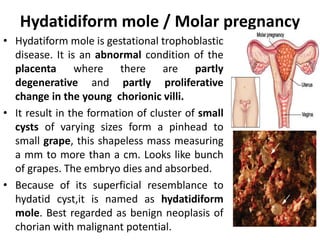
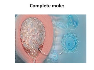
Molar Pregnancy
Prepared by : Shiksha khanal
Hydatidiform mole / Molar pregnancy
• Hydatiform mole is gestational trophoblastic
disease. It is an abnormal condition of the
placenta where there are partly
degenerative and partly proliferative
change in the young chorionic villi.
• It result in the formation of cluster of small
cysts of varying sizes form a pinhead to
small grape, this shapeless mass measuring
a mm to more than a cm. Looks like bunch
of grapes. The embryo dies and absorbed.
• Because of its superficial resemblance to
hydatid cyst,it is named as hydatidiform
mole. Best regarded as benign neoplasis of
chorian with malignant potential.
Etiology
The etiology is unknown although there may be an
ovular defect or a nutritional deficiency. However the
following factors are hypothesized.
– Prevalence is highest in teenage pregnancy in those
women over 35 years of age.
– Faulty nutrition caused by inadequate intake of
protein, animal fat. Low carotein intake in diet is
associated with increased risk. ·
– Woman at higher risk for hydatidiform mole
formation are those who have undergone ovulation
stimulation with Clomiphene.
• Cytogenic abnormality; complete mole have
46 XX, the molar chromosomes are derived
entirely from the father. The ovum nucleus
may be either absent (empty ovum) or
inactivated which has been fertilized by
haploid sperm.
• History of prior hydatidiform mole increases
the chance of recurrence.
Types
• Partial mole • Complete mole
Partial mole
A partial mole often has embryonic or fetal
part and an amniotic sac present congenital
anomalies are usually present. The potential
for malignant transformation. is much less
than that associated with complete hydatiform
mole.
A part of trophoblastic tissue only shows
molar change. There is a fetus or at least an
amniotic sac.
 The uterus is distended by thin walled, translucent,
grape like vesicles of different sizes. These are
degenerated chorionic villi filled with fluid.
 There is no vasculature in the chorionic villi leads to
early death and absorption of the embryo.
 There is trophoblastic proliferation, with mitotic
activity affecting both syncytial and
cytotrophobalstic layers. This causes excessive
secretion of hCG, chronic thyrotrophin and
progesterone. On the other hand, oestrogen
production is low due to absence of the fetal supply
of precursors.
Complete mole:
The whole conceptus is transformed into a mass of
vesicles. No embryo is present. The complete mole
results from fertilization of an egg whose nucleus has
been lost or inactivated. The mole resembles a bunch
of white grapes. The fluid filled vesicles grow rapidly
,causing the uterus to be larger then expected for the
duration of the pregnancy. Usually, the complete mole
contains no fetus, placenta, amniotic membranes, or
fluid. Maternal blood has no placenta to receive it
,hemorrhage into the uterine cavity and vaginal
bleeding ,therefore occurs. In approximately 20% of
cases of complete mole,progressin toward
choriocarcinoma occurs.
• Cytogenic studies have demonstrated that complete
moles usally have a 46xx karyotype,and the molar
chromosomes are entire of paternal origin.
• Complete mole appers to arise from an ovum that has
been fertilized by a haploid sperm, which then duplicates
its chromosomes ,and the ovum nucleus may be either
absent or inactivated.
• Although most complete moles have a 46xx
chromosomal pattern, approximately 10% have a 46xy
karyotype.
• Chromosomes in 46xy complete mole als appear to be
entire of paternal origin but in this circumstance ,an
empty egg is fertilized by two sperm.
Difference between partial mole and complete mole
Features Partial mole Complete mole
karyotype Tripliod (69xxy) Diploid (46xx/xy)
Fetus Present Absent
Trophoblastic proliferation Focal Diffuse
Trophoblastic scalloping Present Absent
Inclusion bodies Present Absent
USG Missed abortion Snow storming
appearance
Uterine size Size<POG Size >POG
Beta HCG <105IU <105IU
Theca lutein cysts Rare Common
Post molar malignant sequelae <5% 20%
Immuno staining Positive Negative
Clinical Features
Symptoms:
• Vaginal bleeding is the commonest presentation. It may
be preceeded by a brownish or watery discharge.
• Varying degree of lower abdominal pain may be due to;
 over distention of the uterus
concealed hemorrhage
 rarely perforation of the uterus by the invasive
mole.
Infection or
Uterine contractions to expel out
• Expulsion of grape like vesicle per vagina is diagnostic
of vesicular mole.
• History of quickening is absent
• Other symptoms are;
the woman become sick without any apparent
reason.
vomiting of pregnancy becomes excessive to
the stage of hyperemesis.
 breathlessness due to pulmonary embolism of
trophoblastic cells.
Signs:
• Features sugestive of early months of pregnancy are
evident.
• Woman looks more ill
• Pallor
• Features of pre-eclampsia (hypertension, edema, or
proteinuria) between 9 and 12 weeks.
Per-abdomen
• The size of the uterus is more than expected period
of amenorrhea (50% of case).
• The feel of the uterus is firm elastic (doughy). This is
due to absence of amniotic fluid sac.
• Fetal parts are not felt, nor any fetal movement.
• Absence of fetal heart sound.
Vaginal Examination
• Internal ballottement can not be elicited.
• Unilateral or bilateral enlargement of ovary.
• Findings of vesicles in the vaginal discharge
• If the cervical os is open, instead of membranes,
blood clot or the vesicles may be felt.
Investigation
• Full blood count, ABO and Rh grouping.
• Hepatic, renal, and thyroid function test
• Sonography , characteristics of molar
pregnancy is snow storm appearance.
• Estimation of serum chorionic gonadotrophin.
Urine pregnancy test is positive in high
dilution, 1/200 is highly suggestive, 1/500 is
surely diagnostic. In normal pregnancy it is
positive in dilutions up to 1/100.
• Straight x-ray abdomen, if the uterus is more
than 16 weeks.
• Straight x-ray chest should also be carried out
for evidence of pulmonary embolization.
• CT and MRI
Complications
Immediate
1. Hemorrhage and shock: The cause of
hemorrhage are;
• Separation of vesicles from its attachment to the
decidua. The hemorrhage may be concealed or
revealed.
• Massive intra peritoneal hemorrhages which may
be first feature of perforation mole.
• During evacuation of the mole due to atonic
uterus or uterine injury.
2. Sepsis: Increased risk of sepsis is due to;
 There is no protective membrane
 Presence of degenerated vesicles, sloughing decidua and
old blood.
 Increased operative interference
3. Perforation of uterus due to;
 perforating mole
 during vaginal evacuation by D and E
4. Pre-eclampsia
5. Acute pulmonary insufficiency due to
pulmonary embolization of the trophoblastic cell.
6. Coagulation failure due to pulmonary
embolization of trophoblastic cells
Late:
• Development of choriocarcinoma following
hydatidiform mole
Management
Nursing assessment during prenatal visits should
include observation for signs of molar pregnancy
during the first 24 weeks. If hydatiform mole is
suspected, ultrasound and serial ß-hCG
immunoassays are used to confirm the diagnosis.
The sonographic pattern of molar pregnancy is
characterized by diffuse "snowstrom" pattern. A -B-
hCG titer will remain high or rise above normal
peak after the time at which it is normal drops (70
to 100 days)
Principles of management
1. Supportive therapy to restore the blood loss
and to prevent infection.
2. Evacuate the uterus as soon as diagnosis is
made
3. Regular follow up for early detection of
persistent trophoblastic disease.
1. Supportive therapy:
Woman usually presents with variable amount of
bleeding. Therefore;
• Start ringer's lacted solution IV infusion
• Arrangement is made for blood transfusion.
• Blood should also be kept ready prior to elective
evacuation of the uterus.
2. Evacuation of uterus
• Although most moles abort spontaneously,
suction/vacuum aspiration is safe, rapid, and effective
method of evacuation of hydatidiform mole if necessary.
The risk of perforation using metal curette is high.
• Digital exploration and removal of the mole by ovum
forceps may be done to evacuate them.
• Infuse oxytocin 20 units in 1 liter IV fluids (NS or RL) at
60 drops per minute to prevent hemorrhage once
evacuation is under way. Induction of labor with oxytocin
or prostaglandins is not recommended because of the
increased risk of embolization of trophoblastic tissue.
• Administration of Rho (D) immune globulin to
women who are Rh negative is necessary to prevent
isoimmunization.
• The nurse provides information to the woman and
her family about the disease process, necessity for
long course of follow up, and the possible
consequences of the disease.
3.Follow up management
• Include frequent physical and pelvic examinations and
biweekly measurement of ß-hCG level until the level
drops to normal and remains normal for 3 weeks.
• Monthly measurements are taken for 6 month and then
every 2 months for a total of one year.
• A rising titer and an enlarging uterus may indicate
choriocarcinoma.
• To nurse help the woman to understand and cope with
pregnancy loss and recognize that the pregnancy was
abnormal.
• Explanation about importance of the need to
postpone a subsequent pregnancy and contraceptive
counselling are provided for consistent and reliable
method of family planning.
• The avoid confusion with sign of pregnancy,
pregnancy should be avoided for one year. Any
contraceptive method except intrauterine device is
acceptable, oral contraceptives are highly effective.

molar pregnancy.pptx

  • 1.
  • 2.
    Hydatidiform mole /Molar pregnancy • Hydatiform mole is gestational trophoblastic disease. It is an abnormal condition of the placenta where there are partly degenerative and partly proliferative change in the young chorionic villi. • It result in the formation of cluster of small cysts of varying sizes form a pinhead to small grape, this shapeless mass measuring a mm to more than a cm. Looks like bunch of grapes. The embryo dies and absorbed. • Because of its superficial resemblance to hydatid cyst,it is named as hydatidiform mole. Best regarded as benign neoplasis of chorian with malignant potential.
  • 3.
    Etiology The etiology isunknown although there may be an ovular defect or a nutritional deficiency. However the following factors are hypothesized. – Prevalence is highest in teenage pregnancy in those women over 35 years of age. – Faulty nutrition caused by inadequate intake of protein, animal fat. Low carotein intake in diet is associated with increased risk. · – Woman at higher risk for hydatidiform mole formation are those who have undergone ovulation stimulation with Clomiphene.
  • 4.
    • Cytogenic abnormality;complete mole have 46 XX, the molar chromosomes are derived entirely from the father. The ovum nucleus may be either absent (empty ovum) or inactivated which has been fertilized by haploid sperm. • History of prior hydatidiform mole increases the chance of recurrence.
  • 5.
    Types • Partial mole• Complete mole
  • 6.
    Partial mole A partialmole often has embryonic or fetal part and an amniotic sac present congenital anomalies are usually present. The potential for malignant transformation. is much less than that associated with complete hydatiform mole. A part of trophoblastic tissue only shows molar change. There is a fetus or at least an amniotic sac.
  • 7.
     The uterusis distended by thin walled, translucent, grape like vesicles of different sizes. These are degenerated chorionic villi filled with fluid.  There is no vasculature in the chorionic villi leads to early death and absorption of the embryo.  There is trophoblastic proliferation, with mitotic activity affecting both syncytial and cytotrophobalstic layers. This causes excessive secretion of hCG, chronic thyrotrophin and progesterone. On the other hand, oestrogen production is low due to absence of the fetal supply of precursors.
  • 10.
  • 11.
    The whole conceptusis transformed into a mass of vesicles. No embryo is present. The complete mole results from fertilization of an egg whose nucleus has been lost or inactivated. The mole resembles a bunch of white grapes. The fluid filled vesicles grow rapidly ,causing the uterus to be larger then expected for the duration of the pregnancy. Usually, the complete mole contains no fetus, placenta, amniotic membranes, or fluid. Maternal blood has no placenta to receive it ,hemorrhage into the uterine cavity and vaginal bleeding ,therefore occurs. In approximately 20% of cases of complete mole,progressin toward choriocarcinoma occurs.
  • 12.
    • Cytogenic studieshave demonstrated that complete moles usally have a 46xx karyotype,and the molar chromosomes are entire of paternal origin. • Complete mole appers to arise from an ovum that has been fertilized by a haploid sperm, which then duplicates its chromosomes ,and the ovum nucleus may be either absent or inactivated. • Although most complete moles have a 46xx chromosomal pattern, approximately 10% have a 46xy karyotype. • Chromosomes in 46xy complete mole als appear to be entire of paternal origin but in this circumstance ,an empty egg is fertilized by two sperm.
  • 14.
    Difference between partialmole and complete mole Features Partial mole Complete mole karyotype Tripliod (69xxy) Diploid (46xx/xy) Fetus Present Absent Trophoblastic proliferation Focal Diffuse Trophoblastic scalloping Present Absent Inclusion bodies Present Absent USG Missed abortion Snow storming appearance Uterine size Size<POG Size >POG Beta HCG <105IU <105IU Theca lutein cysts Rare Common Post molar malignant sequelae <5% 20% Immuno staining Positive Negative
  • 16.
    Clinical Features Symptoms: • Vaginalbleeding is the commonest presentation. It may be preceeded by a brownish or watery discharge. • Varying degree of lower abdominal pain may be due to;  over distention of the uterus concealed hemorrhage  rarely perforation of the uterus by the invasive mole. Infection or Uterine contractions to expel out
  • 17.
    • Expulsion ofgrape like vesicle per vagina is diagnostic of vesicular mole. • History of quickening is absent • Other symptoms are; the woman become sick without any apparent reason. vomiting of pregnancy becomes excessive to the stage of hyperemesis.  breathlessness due to pulmonary embolism of trophoblastic cells.
  • 18.
    Signs: • Features sugestiveof early months of pregnancy are evident. • Woman looks more ill • Pallor • Features of pre-eclampsia (hypertension, edema, or proteinuria) between 9 and 12 weeks.
  • 19.
    Per-abdomen • The sizeof the uterus is more than expected period of amenorrhea (50% of case). • The feel of the uterus is firm elastic (doughy). This is due to absence of amniotic fluid sac. • Fetal parts are not felt, nor any fetal movement. • Absence of fetal heart sound.
  • 20.
    Vaginal Examination • Internalballottement can not be elicited. • Unilateral or bilateral enlargement of ovary. • Findings of vesicles in the vaginal discharge • If the cervical os is open, instead of membranes, blood clot or the vesicles may be felt.
  • 21.
    Investigation • Full bloodcount, ABO and Rh grouping. • Hepatic, renal, and thyroid function test • Sonography , characteristics of molar pregnancy is snow storm appearance. • Estimation of serum chorionic gonadotrophin. Urine pregnancy test is positive in high dilution, 1/200 is highly suggestive, 1/500 is surely diagnostic. In normal pregnancy it is positive in dilutions up to 1/100.
  • 22.
    • Straight x-rayabdomen, if the uterus is more than 16 weeks. • Straight x-ray chest should also be carried out for evidence of pulmonary embolization. • CT and MRI
  • 23.
    Complications Immediate 1. Hemorrhage andshock: The cause of hemorrhage are; • Separation of vesicles from its attachment to the decidua. The hemorrhage may be concealed or revealed. • Massive intra peritoneal hemorrhages which may be first feature of perforation mole. • During evacuation of the mole due to atonic uterus or uterine injury.
  • 24.
    2. Sepsis: Increasedrisk of sepsis is due to;  There is no protective membrane  Presence of degenerated vesicles, sloughing decidua and old blood.  Increased operative interference 3. Perforation of uterus due to;  perforating mole  during vaginal evacuation by D and E 4. Pre-eclampsia 5. Acute pulmonary insufficiency due to pulmonary embolization of the trophoblastic cell. 6. Coagulation failure due to pulmonary embolization of trophoblastic cells
  • 25.
    Late: • Development ofchoriocarcinoma following hydatidiform mole
  • 26.
    Management Nursing assessment duringprenatal visits should include observation for signs of molar pregnancy during the first 24 weeks. If hydatiform mole is suspected, ultrasound and serial ß-hCG immunoassays are used to confirm the diagnosis. The sonographic pattern of molar pregnancy is characterized by diffuse "snowstrom" pattern. A -B- hCG titer will remain high or rise above normal peak after the time at which it is normal drops (70 to 100 days)
  • 27.
    Principles of management 1.Supportive therapy to restore the blood loss and to prevent infection. 2. Evacuate the uterus as soon as diagnosis is made 3. Regular follow up for early detection of persistent trophoblastic disease.
  • 28.
    1. Supportive therapy: Womanusually presents with variable amount of bleeding. Therefore; • Start ringer's lacted solution IV infusion • Arrangement is made for blood transfusion. • Blood should also be kept ready prior to elective evacuation of the uterus.
  • 29.
    2. Evacuation ofuterus • Although most moles abort spontaneously, suction/vacuum aspiration is safe, rapid, and effective method of evacuation of hydatidiform mole if necessary. The risk of perforation using metal curette is high. • Digital exploration and removal of the mole by ovum forceps may be done to evacuate them. • Infuse oxytocin 20 units in 1 liter IV fluids (NS or RL) at 60 drops per minute to prevent hemorrhage once evacuation is under way. Induction of labor with oxytocin or prostaglandins is not recommended because of the increased risk of embolization of trophoblastic tissue.
  • 30.
    • Administration ofRho (D) immune globulin to women who are Rh negative is necessary to prevent isoimmunization. • The nurse provides information to the woman and her family about the disease process, necessity for long course of follow up, and the possible consequences of the disease.
  • 31.
    3.Follow up management •Include frequent physical and pelvic examinations and biweekly measurement of ß-hCG level until the level drops to normal and remains normal for 3 weeks. • Monthly measurements are taken for 6 month and then every 2 months for a total of one year. • A rising titer and an enlarging uterus may indicate choriocarcinoma. • To nurse help the woman to understand and cope with pregnancy loss and recognize that the pregnancy was abnormal.
  • 32.
    • Explanation aboutimportance of the need to postpone a subsequent pregnancy and contraceptive counselling are provided for consistent and reliable method of family planning. • The avoid confusion with sign of pregnancy, pregnancy should be avoided for one year. Any contraceptive method except intrauterine device is acceptable, oral contraceptives are highly effective.