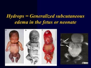
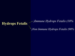
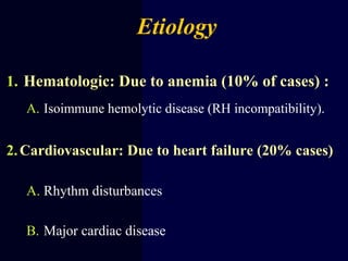
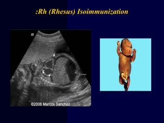
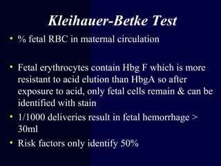
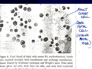
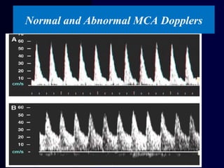
Hydrops Fetalis
Dr Manal Behery
2014
Hydrops = Generalized subcutaneous
edema in the fetus or neonate
Definition
1. Excess serous fluid in at least
one space (ascites, pleural effusion, or
pericardial effusion) + skin edema (> 5 mm thick)
1. Excess fluid in two spaces without edema
Hydrops Fetalis
Non Immune Hydrops Fetalis (90%(
Immune Hydrops Fetalis (10%(
Etiology
1. Hematologic: Due to anemia (10% of cases) :
A. Isoimmune hemolytic disease (RH incompatibility).
2.Cardiovascular: Due to heart failure (20% cases)
A. Rhythm disturbances
B. Major cardiac disease
3. Infection (10% of cases) TORCH-Syphilis-
Congenital Hepatitis, Parvovirus….
4. Chromosomal (5% of cases) : Turner
syndrome, trisomy 13,18,21
5. Pulmonary (5% of cases) Chylothorax,
diaphragmatic hernia
6. Gastrointestinal (5% of cases) : Meconium
peritonitis
7. Renal (5% of cases) Nephrosis, RVT, urinary
obstruction
…..
7. Maternal conditions (5% of cases) :
Toxemia, diabetes, thyrotoxicosis
8. Miscellaneous (10% of cases) : Cystic
hygroma, wilms’ tumor – teratoma
9. -Unknown (20% of cases) :
Rh (Rhesus) Isoimmunization:
Four blood types ( A, B, AB, and O)
Each blood type is additionally classified according to the presence or
absence of the Rh factor
CDE (Rhesus) System
• Clinically Important
• Includes c, C, D, e, E
• Rh negative status indicates the absence of D
antigen
• 87% of Caucasians carry the D antigen
•Rh Disease
•Alloimmunization
•Isoimmunization
•HDFN
•Erythroblastosis Fetalis
Confusing Terminology
When the mother produces Abs
directed against fetus RBC surface Ag.
Isoimmunization
Rh Incompatibility
Exposure to fetal antigens
causes the mother to
produce antibodies
The placenta usually acts as a barrier to fetal
blood entering the maternal circulation.
Fetal cells can enter the maternal circulation
through a “break” in the “placental barrier”.
Maternal production of Rhesus antibodies
following introduction of Rhesus positive blood
Maternal production of Rhesus antibodies
following introduction of Rhesus positive blood
Causes of RBC Transfer:
“A break in the barrier”
• Abortion/Ectopic
Pregnancy
• Partial molar pregnancy
• Blighted ovum
• Antepartum bleeding
• Procedures
(amniocentesis,
cordocentesis, CVS)
• External version
• Platelet transfusion
• Abdominal trauma
• Inadvertent transfusion
Rh+ blood
• Postpartum (Rh+baby)
Sensitized pregnancy Non- Sensitized pregnancy
Rh Incompatibility
Sensitization = Rh neg person exponsed to the Rh (D) antigen and makes
antibodies against that protein (antigen).
Rh Negative Women Man Rh positive
⇓
Fetus
Rh positive Fetus
→
Rh+ve R.B.C.s enter Maternal
circulation⇐
previously sensitized 2nd
immune response
IgM…IgG antibodies
⇓
Non sensitized Mother Primary
immune response
1st
Baby usually escapes. Mother
gets sensitized? ±
Fetus
Haemolysis
⇓
Pathogenesis Of Rh Iso - immunisation
Rh –VE Fetus
no harm
Presentation
Mild jaundice
Erythroblastosis Fetalis
Generalized Edema
Hepatomegaly
Ascites
Natural History
•50%of affected infants have mild
anemia, requiring either phototherapy
or no treatment.
•25%have hepatosplenomegaly ,
moderate anemia and progressive
jundice ending in kernicterus, neonatal
death
•25%are hydropic and die in utero or in
the neonatal period
RhoGAM has decreased HDFN caused by anti-D
Give 300 mcg dose within 72 hrs of delivery to
unsensitized Rh (-) women (Rh positive infant)
• ACOG: 300 mcg at 28 weeks UNLESS father known
to be Rh (-)
Mechanism of action
• Administered antibodies will
bind the fetal Rh- positive cells
• Spleen captured these cells by
Fc-receptors
• Suppressor T cell response is
stimulated
• Spleen remove anti-D coated
red cells prior to contact with
antigen presenting cells
“antigen deviation”
Kleihauer-Betke Test
• % fetal RBC in maternal circulation
• Fetal erythrocytes contain Hbg F which is more
resistant to acid elution than HbgA so after
exposure to acid, only fetal cells remain & can be
identified with stain
• 1/1000 deliveries result in fetal hemorrhage >
30ml
• Risk factors only identify 50%
1.1-Direct Coomb’s Test (DAT(
Detects RBCs that have
already been sensitized with
IgG
Demonstrates that in vivo
coating of RBC by Ab has
occurred but does NOT
identify the antibody
Deepa Babin @TMC Kollam 28
 Detects antibodies to RBC antigens
present in the patient’s serum
 Detects in vitro red cell sensitization
if red cells contain antigen
corresponding to serum antibody
 Procedure:
 STEP 1:
patient’s serum (with unknown Ab) +
RBC (with known Ag)
 STEP 2: product of step 1 + Coomb’s
reagent
IAT((2. Indirect Coomb’s Test
Deepa Babin @TMC Kollam 29
Recognition of pregnancy at risk
•First ante-natal visit check blood group,
antibody screening.
• If indirect coombs test is positive, the
father’s Rh should be tested.
• Serial maternal Anti D titers should be
done every 2- 4 weeks.
• If titer is less than 1/16 the fetus is not
at risk.
• If titer is more than 1/16 then
severity of condition should be
evaluated.
Prevention
• Test for excessive fetal-maternal hemorrhage
after blunt trauma, abruption, cordocentesis,
and bleeding with previa
• Give RhoGAM for partial molar pregnancy,
ectopic, chorionic villus sampling,
amniocentesis, external version
MCA Doppler and Fetal Anemia
• Fetuses with anemia show an
• increased peak velocity of systolic
blood flow in MCA.
• MCA Doppler is also used to follow
fetal response to intrauterine transfusion
and to assist in timing subsequent transfusions.
• Non-invasive,
• no risk for worsening isoimmunization
Normal and Abnormal MCA Dopplers
Amniocentesis
- There is an excellent correlation
between the amount of bilirubin in
amniotic fluid and fetal hematocrit.
-
• - Perform serial amniocenteses
• to the optical density deviation at 450
nm measures the amniotic fluid
unconjugated bilirubin.
• Plot values on Liley Curve
at 16 weeks of gestation
Liley Curve
•Measures the level of bilirubin and predicts
severity of hemolytic disease after 27 weeks
Suggested management after amniocentesis for ΔOD 450
Serial Amniocentesis
Lily zone I
Lower Zone II
Upper Zone II Zone III
Hydramnios & Hydrops
Repeat
Amniocentesis every
2-4 weeks
Delivery at or near term
Repeat Amniocentesis in 7
days or FBS
Hct < 25%
Hct > 25%
Intrauterine
Transfusion
Repeat Sampling
7to 14 days
<35to 36 weeks
And Fetal lung
immaturity
>35to 36 weeks
Lung maturity
present
Intrauterine
Transfusion
Delivery
Cordocentesis
• Gold standard for detection of fetal anemia
• Complications
• 2.7% total risk of fetal loss
• Reserved for patients with
increased MCA-PSV or delta OD450
Intrauterine transfusion
Exchange Transfusion
•.
 Considered if the total serum bilirubin level is approaching 20
mg/dL and continues to rise despite intense in-hospital
phototherapy.
 Mortality rate 1%
Diagnosis and Management contd.
Review of Management for Rh
Isoimmunization
• Monthly indirect coombs titer (in first sensitized
pregnancy)
• If critical titer reached, determine paternal and fetal
antigen status
• Amniocentesis and delta OD450 OR MCA-PSV
** For 2nd
or greater sensitized pregnancy,
initiate amnio or MCA at 18-20 weeks**
Hydrops fetails for  undergranuate

Hydrops fetails for undergranuate

  • 1.
  • 2.
    Hydrops = Generalizedsubcutaneous edema in the fetus or neonate
  • 3.
    Definition 1. Excess serousfluid in at least one space (ascites, pleural effusion, or pericardial effusion) + skin edema (> 5 mm thick) 1. Excess fluid in two spaces without edema
  • 4.
    Hydrops Fetalis Non ImmuneHydrops Fetalis (90%( Immune Hydrops Fetalis (10%(
  • 5.
    Etiology 1. Hematologic: Dueto anemia (10% of cases) : A. Isoimmune hemolytic disease (RH incompatibility). 2.Cardiovascular: Due to heart failure (20% cases) A. Rhythm disturbances B. Major cardiac disease
  • 6.
    3. Infection (10%of cases) TORCH-Syphilis- Congenital Hepatitis, Parvovirus…. 4. Chromosomal (5% of cases) : Turner syndrome, trisomy 13,18,21
  • 7.
    5. Pulmonary (5%of cases) Chylothorax, diaphragmatic hernia 6. Gastrointestinal (5% of cases) : Meconium peritonitis 7. Renal (5% of cases) Nephrosis, RVT, urinary obstruction …..
  • 8.
    7. Maternal conditions(5% of cases) : Toxemia, diabetes, thyrotoxicosis 8. Miscellaneous (10% of cases) : Cystic hygroma, wilms’ tumor – teratoma 9. -Unknown (20% of cases) :
  • 9.
  • 10.
    Four blood types( A, B, AB, and O) Each blood type is additionally classified according to the presence or absence of the Rh factor
  • 11.
    CDE (Rhesus) System •Clinically Important • Includes c, C, D, e, E • Rh negative status indicates the absence of D antigen • 87% of Caucasians carry the D antigen
  • 12.
  • 13.
    When the motherproduces Abs directed against fetus RBC surface Ag. Isoimmunization
  • 14.
    Rh Incompatibility Exposure tofetal antigens causes the mother to produce antibodies
  • 15.
    The placenta usuallyacts as a barrier to fetal blood entering the maternal circulation.
  • 16.
    Fetal cells canenter the maternal circulation through a “break” in the “placental barrier”.
  • 17.
    Maternal production ofRhesus antibodies following introduction of Rhesus positive blood
  • 18.
    Maternal production ofRhesus antibodies following introduction of Rhesus positive blood
  • 19.
    Causes of RBCTransfer: “A break in the barrier” • Abortion/Ectopic Pregnancy • Partial molar pregnancy • Blighted ovum • Antepartum bleeding • Procedures (amniocentesis, cordocentesis, CVS) • External version • Platelet transfusion • Abdominal trauma • Inadvertent transfusion Rh+ blood • Postpartum (Rh+baby)
  • 20.
    Sensitized pregnancy Non-Sensitized pregnancy Rh Incompatibility Sensitization = Rh neg person exponsed to the Rh (D) antigen and makes antibodies against that protein (antigen).
  • 21.
    Rh Negative WomenMan Rh positive ⇓ Fetus Rh positive Fetus → Rh+ve R.B.C.s enter Maternal circulation⇐ previously sensitized 2nd immune response IgM…IgG antibodies ⇓ Non sensitized Mother Primary immune response 1st Baby usually escapes. Mother gets sensitized? ± Fetus Haemolysis ⇓ Pathogenesis Of Rh Iso - immunisation Rh –VE Fetus no harm
  • 22.
  • 23.
    Natural History •50%of affectedinfants have mild anemia, requiring either phototherapy or no treatment. •25%have hepatosplenomegaly , moderate anemia and progressive jundice ending in kernicterus, neonatal death •25%are hydropic and die in utero or in the neonatal period
  • 24.
    RhoGAM has decreasedHDFN caused by anti-D Give 300 mcg dose within 72 hrs of delivery to unsensitized Rh (-) women (Rh positive infant) • ACOG: 300 mcg at 28 weeks UNLESS father known to be Rh (-)
  • 25.
    Mechanism of action •Administered antibodies will bind the fetal Rh- positive cells • Spleen captured these cells by Fc-receptors • Suppressor T cell response is stimulated • Spleen remove anti-D coated red cells prior to contact with antigen presenting cells “antigen deviation”
  • 26.
    Kleihauer-Betke Test • %fetal RBC in maternal circulation • Fetal erythrocytes contain Hbg F which is more resistant to acid elution than HbgA so after exposure to acid, only fetal cells remain & can be identified with stain • 1/1000 deliveries result in fetal hemorrhage > 30ml • Risk factors only identify 50%
  • 28.
    1.1-Direct Coomb’s Test(DAT( Detects RBCs that have already been sensitized with IgG Demonstrates that in vivo coating of RBC by Ab has occurred but does NOT identify the antibody Deepa Babin @TMC Kollam 28
  • 29.
     Detects antibodiesto RBC antigens present in the patient’s serum  Detects in vitro red cell sensitization if red cells contain antigen corresponding to serum antibody  Procedure:  STEP 1: patient’s serum (with unknown Ab) + RBC (with known Ag)  STEP 2: product of step 1 + Coomb’s reagent IAT((2. Indirect Coomb’s Test Deepa Babin @TMC Kollam 29
  • 30.
    Recognition of pregnancyat risk •First ante-natal visit check blood group, antibody screening. • If indirect coombs test is positive, the father’s Rh should be tested. • Serial maternal Anti D titers should be done every 2- 4 weeks. • If titer is less than 1/16 the fetus is not at risk. • If titer is more than 1/16 then severity of condition should be evaluated.
  • 31.
    Prevention • Test forexcessive fetal-maternal hemorrhage after blunt trauma, abruption, cordocentesis, and bleeding with previa • Give RhoGAM for partial molar pregnancy, ectopic, chorionic villus sampling, amniocentesis, external version
  • 32.
    MCA Doppler andFetal Anemia • Fetuses with anemia show an • increased peak velocity of systolic blood flow in MCA. • MCA Doppler is also used to follow fetal response to intrauterine transfusion and to assist in timing subsequent transfusions. • Non-invasive, • no risk for worsening isoimmunization
  • 33.
    Normal and AbnormalMCA Dopplers
  • 34.
    Amniocentesis - There isan excellent correlation between the amount of bilirubin in amniotic fluid and fetal hematocrit. - • - Perform serial amniocenteses • to the optical density deviation at 450 nm measures the amniotic fluid unconjugated bilirubin. • Plot values on Liley Curve at 16 weeks of gestation
  • 35.
    Liley Curve •Measures thelevel of bilirubin and predicts severity of hemolytic disease after 27 weeks
  • 36.
    Suggested management afteramniocentesis for ΔOD 450 Serial Amniocentesis Lily zone I Lower Zone II Upper Zone II Zone III Hydramnios & Hydrops Repeat Amniocentesis every 2-4 weeks Delivery at or near term Repeat Amniocentesis in 7 days or FBS Hct < 25% Hct > 25% Intrauterine Transfusion Repeat Sampling 7to 14 days <35to 36 weeks And Fetal lung immaturity >35to 36 weeks Lung maturity present Intrauterine Transfusion Delivery
  • 37.
    Cordocentesis • Gold standardfor detection of fetal anemia • Complications • 2.7% total risk of fetal loss • Reserved for patients with increased MCA-PSV or delta OD450
  • 38.
  • 39.
    Exchange Transfusion •.  Consideredif the total serum bilirubin level is approaching 20 mg/dL and continues to rise despite intense in-hospital phototherapy.  Mortality rate 1%
  • 40.
  • 41.
    Review of Managementfor Rh Isoimmunization • Monthly indirect coombs titer (in first sensitized pregnancy) • If critical titer reached, determine paternal and fetal antigen status • Amniocentesis and delta OD450 OR MCA-PSV ** For 2nd or greater sensitized pregnancy, initiate amnio or MCA at 18-20 weeks**