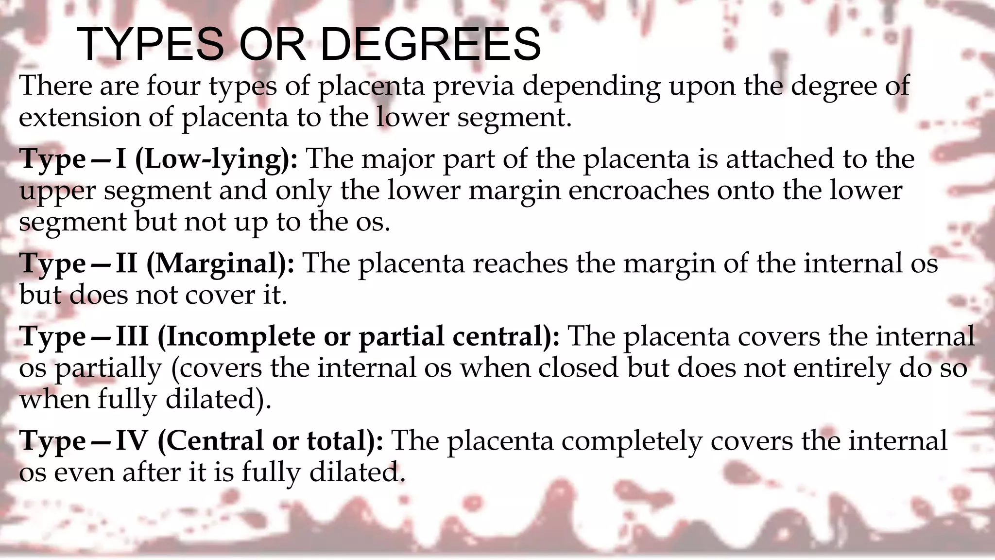
Antepartum Hemorrhage
Mrs. P. Christena
M.Sc-OBG Nursing
RNRM
Assistant Professor
DEFINITION
It is defined as bleeding from or into the genital tract after the
28th week of pregnancy but before the birth of the baby (the first
and second stage of labor are thus included)
PLACENTA PRAEVIA
DEFINITION
• A low implantation of the placenta in the uterus causing it to lie alongside
or Infront of the presenting part
• ANNAMMA JACOB
• Placenta implanted to lower uterine segment ,often causing painless
profuse third trimester bleeding
• UN PANDA
CAUSES
• UNKNOWN
• 3 POSTULATED THEORIES,
1
• DROPPING DOWN THEORY
2
• MUTLIPLE PREGNANCY
3
• DEFECTIVE DECIDUA
PREDISPOSING FACTOR
• Increased placental size
• Previous caesarian section
• Multiparity
• Advanced maternal age
• Previous reproduction surgery
• Placental abnormality
• Leiomyomas distorting the
uterine cavity
• Congenital malformation of
uterus
PATHOLOGICAL ANATOMY
• Placenta—The placenta may be large and thin. There is often a tongue shaped
extension from the main placental mass. Extensive areas of degeneration with
infarction and calcification may be evident. The placenta may be morbidly adherent
due to poor decidua formation in the lower segment.
• Umbilical cord—The cord may be attached to the margin (battledore) or into the
membranes (velamentous). The insertion of the cord may be close to the internal os
or the fetal vessels may run across the internal os in velamentous insertion giving
rise to vasa previa, which may rupture along with rupture of the membranes.
• Lower uterine segment—Due to increased vascularity, the lower uterine segment
and the cervix becomes soft and more friable
TYPES OR DEGREES
There are four types of placenta previa depending upon the degree of
extension of placenta to the lower segment.
Type—I (Low-lying): The major part of the placenta is attached to the
upper segment and only the lower margin encroaches onto the lower
segment but not up to the os.
Type—II (Marginal): The placenta reaches the margin of the internal os
but does not cover it.
Type—III (Incomplete or partial central): The placenta covers the internal
os partially (covers the internal os when closed but does not entirely do so
when fully dilated).
Type—IV (Central or total): The placenta completely covers the internal
os even after it is fully dilated.
Dangerous
placenta previa is
the name given to
the type-II posterior
placenta previa
Reasons for calling out Dangerous
placenta previa
• (1) Because of the curved birth canal major thickness of the placenta (about 2.5 cm)
overlies the sacral promontory, thereby diminishing the anteroposterior diameter of
the inlet and prevents engagement of the presenting part. This hinders effective
compression of the separated placenta to stop bleeding.
• (2) Placenta is more likely to be compressed, if vaginal delivery is allowed.
• (3) More chance of cord compression or cord prolapse. The last two may produce
fetal anoxia or even death.
CLINICAL FEATURES
• The only symptom of placenta previa is vaginal bleeding
• sudden onset, painless, apparently causeless and recurrent
• The bleeding is unassociated with pain unless labor starts simultaneously
• In majority of cases, bleeding occurs before 38 weeks and earlier bleeding is more likely to
occur in major degrees.
• SIGNS: General condition and anemia are proportionate to the visible blood loss
• Abdominal examination: The size of the uterus is proportionate to the period of gestation.
• The uterus feels relaxed, soft and elastic without any localized area of tenderness.
• Persistence of malpresentation
• There is also increased frequency of twin pregnancy.
• Persistent displacement of the fetal head is very suggestive. The head cannot be pushed
down into the pelvis.
• Fetal heart sound is usually present
• (Stallworthy’s sign) Slowing of the fetal heart rate on pressing the head down into the
pelvis which soon recovers promptly as the pressure is released is suggestive of the
presence of low lying placenta especially of posterior type .But this sign is not always
significant because it may be due to fetal head compression even in an otherwise normal
case.
• Vulval inspection: Only inspection is to be done to note whether the
bleeding is still occurring or has ceased
• In placenta praevia, the blood is bright red as the bleeding occurs from
the separated utero-placental sinuses close to the cervical opening and
escapes out immediately.
• Vaginal examination must not be done outside the operation theater in
the hospital, as it can provoke further separation of placenta with
torrential hemorrhage and may be fatal
COMPLICATIONS OF PLACENTA
PREVIA
MATERNAL:
• During pregnancy—
• Antepartum hemorrhage with varying degrees of shock
• Co-existent placental abruption is about 10%.
Malpresentation: There is increased incidence of breech presentation
and transverse lie. The lie often becomes unstable.
Premature labor either spontaneous or induced is common.
During labor
• Early rupture of the membranes
• Cord prolapse due to abnormal attachment of the cord
• Slow dilatation of the cervix due to the attachment of placenta
on the lower segment
• Intrapartum hemorrhage due to further separation of placenta
with dilatation of the cervix.
• Increased incidence of operative interference.
• Postpartum hemorrhage
• Retained placenta
Puerperium:
(1) Sepsis is increased due to :
(a) increased operative interference
(b) placental site near to the vagina and
(c) anemia and devitalized state of the patient.
(2) Subinvolution
(3) Embolism.
FETAL COMPLICATIONS IN PLACENTA PREVIA
• Low birth weight babies are quite common (15%)
• Asphyxia is common and it may be the effect of
(a) early separation of placenta
(b) compression of the placenta or
(c) compression of the cord.
• Intrauterine death
• Birth injuries
• Congenital malformation is three times more common in placenta
previa.
MANAGEMENT
• PREVENTION:
• Adequate antenatal care
• Antenatal diagnosis
• Significance of “warning hemorrhage” should not be ignored.
• Color flow Doppler USG in placenta previa is indicated to detect any placenta
accrete
• AT HOME:
• The patient is immediately put to bed
• To assess the blood loss
• (a) inspection of the clothing soaked with blood
• (b) to note the pulse, blood pressure and degree of anemia
• Quick but gentle abdominal examination
• Vaginal examination must not be done
• TRANSFER TO HOSPITAL:
• Arrangement is made to shift the patient to an equipped hospital having facilities of
blood transfusion, emergency cesarean section and neonatal intensive care unit
(NICU)
• ADMISSION TO HOSPITAL:
• All cases of APH, even if the bleeding is slight or absent by the time the patient
reaches the hospital, should be admitted.
• The reasons are:
(1) All the cases of APH should be regarded as due to placenta previa unless proved
otherwise
(2) The bleeding may recur sooner or later and none can predict when it recurs and
how much she will bleed.
ABRUPTIO
PLACENTAE
(Syn : Accidental
Hemorrhage.
Premature
Separation Of
Placenta)
DEFINITION:
It is one form of antepartum hemorrhage where the
bleeding occurs due to premature separation of normally
situated placenta. Out of the various nomenclatures,
abruptio placentae seems to be appropriate one.
VARIETIES
• (1) Revealed : Following separation of the placenta, the blood insinuates
downwards between the membranes and the decidua. Ultimately, the
blood comes out of the cervical canal to be visible externally. This is the
commonest type.
• (2) Concealed : The blood collects behind the separated placenta or
collected in between the membranes and decidua. The collected blood is
prevented from coming out of the cervix by the presenting part which
presses on the lower segment. At times, the blood may percolate into the
amniotic sac after rupturing the membranes. In any of the circumstances
blood is not visible outside. This type is rare.
• (3) Mixed : In this type, some part of the blood collects inside (concealed)
and a part is expelled out (revealed). Usually one variety predominates
over the other. This is quite common
ETIOLOGY:
• The exact cause of separation of a normally situated placenta
remains obscure in majority of cases.
• The prevalence is more with
(a) high birth order pregnancies with gravida 5 and above —
three times more common than in first birth
(b) advancing age of the mother
(c) poor socio-economic condition
(d) malnutrition
(e) smoking (vaso-spasm).
• Hypertension in pregnancy
• Trauma
• Sudden uterine decompression:
• Placental anomaly
• Uterine factor: Placenta implanted over a septum (Septate Uterus) or a
submucous fibroid. Torsion of the uterus leads to increased venous
pressure and rupture of the veins with separation of the placenta.
• Cocaine abuse is associated with increased risk of transient
hypertension, vasospasm and placental abruption.
• Thrombophilias inherited or acquired have been associated with
increased risk of placental infarcts or abruption.
• Prior abruption: Risk of recurrence for a woman with previous abruption
varies between 5 to 17%.
COMPLICATIONS OF ABRUPTIO
PLACENTAE• MATERNAL:
• In revealed type—maternal risk is proportionate to the visible
blood loss and maternal death is rare
• In concealed variety–
• Hemorrhage
• Shock
• Blood coagulation disorders
• Oliguria and anuria
• Postpartum hemorrhage
• Puerperal sepsis
• increased maternal death varies from 2–8%.
• There is failure of lactation (Sheehan’s syndrome) later on.
FETAL:
• In revealed type, the fetal death is to the extent of 25-30%.
• In concealed type, however, the fetal death is appreciably high,
ranging from 50-100%.
• The deaths are due to prematurity and anoxia due to
placental separation.
• With same degree of placental separation, the fetus is put to
more risk in abruptio placentae than in placenta previa.
• This is due to the presence of pre-existing placental
pathology with poor functional reserve in the former, in
contrast to an almost normal placental functions in the latter.
MANAGEMENT OF ABRUPTIO
PLACENTAE
• Prevention:
• The prevention aims at—
(1) elimination of the known factors likely to produce placental
separation
(2) correction of anemia during antenatal period so that the
patient can withstand blood loss and
(3) prompt detection and institution of the therapy to minimise
the grave complications namely shock, blood coagulation
disorders and renal failure.
• Prevention of known factors likely to cause placental separation are
• • Early detection and effective therapy of pre-eclampsia and other hypertensive
disorders of pregnancy.
• • Needle puncture during amniocentesis should be under ultrasound guidance.
• • Avoidance of trauma—specially forceful external cephalic version under
anesthesia.
• • To avoid sudden decompression of the uterus— in acute or chronic
hydramnios, amniocentesis is preferable to artificial rupture of the
membranes.
• • To avoid supine hypotension the patient is advised tolie in the left lateral
position in the later months of pregnancy.
• • Routine administration of folic acid from the early pregnancy — of doubtful
value.
?????? DOUBTS
???????
Aph-Antepartum Hemorrhage

Aph-Antepartum Hemorrhage

  • 1.
    Antepartum Hemorrhage Mrs. P.Christena M.Sc-OBG Nursing RNRM Assistant Professor
  • 2.
    DEFINITION It is definedas bleeding from or into the genital tract after the 28th week of pregnancy but before the birth of the baby (the first and second stage of labor are thus included)
  • 4.
  • 5.
    DEFINITION • A lowimplantation of the placenta in the uterus causing it to lie alongside or Infront of the presenting part • ANNAMMA JACOB • Placenta implanted to lower uterine segment ,often causing painless profuse third trimester bleeding • UN PANDA
  • 6.
    CAUSES • UNKNOWN • 3POSTULATED THEORIES, 1 • DROPPING DOWN THEORY 2 • MUTLIPLE PREGNANCY 3 • DEFECTIVE DECIDUA
  • 7.
    PREDISPOSING FACTOR • Increasedplacental size • Previous caesarian section • Multiparity • Advanced maternal age • Previous reproduction surgery • Placental abnormality • Leiomyomas distorting the uterine cavity • Congenital malformation of uterus
  • 8.
    PATHOLOGICAL ANATOMY • Placenta—Theplacenta may be large and thin. There is often a tongue shaped extension from the main placental mass. Extensive areas of degeneration with infarction and calcification may be evident. The placenta may be morbidly adherent due to poor decidua formation in the lower segment. • Umbilical cord—The cord may be attached to the margin (battledore) or into the membranes (velamentous). The insertion of the cord may be close to the internal os or the fetal vessels may run across the internal os in velamentous insertion giving rise to vasa previa, which may rupture along with rupture of the membranes. • Lower uterine segment—Due to increased vascularity, the lower uterine segment and the cervix becomes soft and more friable
  • 9.
    TYPES OR DEGREES Thereare four types of placenta previa depending upon the degree of extension of placenta to the lower segment. Type—I (Low-lying): The major part of the placenta is attached to the upper segment and only the lower margin encroaches onto the lower segment but not up to the os. Type—II (Marginal): The placenta reaches the margin of the internal os but does not cover it. Type—III (Incomplete or partial central): The placenta covers the internal os partially (covers the internal os when closed but does not entirely do so when fully dilated). Type—IV (Central or total): The placenta completely covers the internal os even after it is fully dilated.
  • 11.
    Dangerous placenta previa is thename given to the type-II posterior placenta previa
  • 12.
    Reasons for callingout Dangerous placenta previa • (1) Because of the curved birth canal major thickness of the placenta (about 2.5 cm) overlies the sacral promontory, thereby diminishing the anteroposterior diameter of the inlet and prevents engagement of the presenting part. This hinders effective compression of the separated placenta to stop bleeding. • (2) Placenta is more likely to be compressed, if vaginal delivery is allowed. • (3) More chance of cord compression or cord prolapse. The last two may produce fetal anoxia or even death.
  • 13.
    CLINICAL FEATURES • Theonly symptom of placenta previa is vaginal bleeding • sudden onset, painless, apparently causeless and recurrent • The bleeding is unassociated with pain unless labor starts simultaneously • In majority of cases, bleeding occurs before 38 weeks and earlier bleeding is more likely to occur in major degrees. • SIGNS: General condition and anemia are proportionate to the visible blood loss • Abdominal examination: The size of the uterus is proportionate to the period of gestation. • The uterus feels relaxed, soft and elastic without any localized area of tenderness.
  • 14.
    • Persistence ofmalpresentation • There is also increased frequency of twin pregnancy. • Persistent displacement of the fetal head is very suggestive. The head cannot be pushed down into the pelvis. • Fetal heart sound is usually present • (Stallworthy’s sign) Slowing of the fetal heart rate on pressing the head down into the pelvis which soon recovers promptly as the pressure is released is suggestive of the presence of low lying placenta especially of posterior type .But this sign is not always significant because it may be due to fetal head compression even in an otherwise normal case.
  • 15.
    • Vulval inspection:Only inspection is to be done to note whether the bleeding is still occurring or has ceased • In placenta praevia, the blood is bright red as the bleeding occurs from the separated utero-placental sinuses close to the cervical opening and escapes out immediately. • Vaginal examination must not be done outside the operation theater in the hospital, as it can provoke further separation of placenta with torrential hemorrhage and may be fatal
  • 18.
    COMPLICATIONS OF PLACENTA PREVIA MATERNAL: •During pregnancy— • Antepartum hemorrhage with varying degrees of shock • Co-existent placental abruption is about 10%. Malpresentation: There is increased incidence of breech presentation and transverse lie. The lie often becomes unstable. Premature labor either spontaneous or induced is common.
  • 19.
    During labor • Earlyrupture of the membranes • Cord prolapse due to abnormal attachment of the cord • Slow dilatation of the cervix due to the attachment of placenta on the lower segment • Intrapartum hemorrhage due to further separation of placenta with dilatation of the cervix. • Increased incidence of operative interference. • Postpartum hemorrhage • Retained placenta
  • 20.
    Puerperium: (1) Sepsis isincreased due to : (a) increased operative interference (b) placental site near to the vagina and (c) anemia and devitalized state of the patient. (2) Subinvolution (3) Embolism.
  • 21.
    FETAL COMPLICATIONS INPLACENTA PREVIA • Low birth weight babies are quite common (15%) • Asphyxia is common and it may be the effect of (a) early separation of placenta (b) compression of the placenta or (c) compression of the cord. • Intrauterine death • Birth injuries • Congenital malformation is three times more common in placenta previa.
  • 22.
    MANAGEMENT • PREVENTION: • Adequateantenatal care • Antenatal diagnosis • Significance of “warning hemorrhage” should not be ignored. • Color flow Doppler USG in placenta previa is indicated to detect any placenta accrete • AT HOME: • The patient is immediately put to bed • To assess the blood loss • (a) inspection of the clothing soaked with blood • (b) to note the pulse, blood pressure and degree of anemia • Quick but gentle abdominal examination • Vaginal examination must not be done
  • 23.
    • TRANSFER TOHOSPITAL: • Arrangement is made to shift the patient to an equipped hospital having facilities of blood transfusion, emergency cesarean section and neonatal intensive care unit (NICU) • ADMISSION TO HOSPITAL: • All cases of APH, even if the bleeding is slight or absent by the time the patient reaches the hospital, should be admitted. • The reasons are: (1) All the cases of APH should be regarded as due to placenta previa unless proved otherwise (2) The bleeding may recur sooner or later and none can predict when it recurs and how much she will bleed.
  • 25.
  • 26.
    DEFINITION: It is oneform of antepartum hemorrhage where the bleeding occurs due to premature separation of normally situated placenta. Out of the various nomenclatures, abruptio placentae seems to be appropriate one.
  • 27.
    VARIETIES • (1) Revealed: Following separation of the placenta, the blood insinuates downwards between the membranes and the decidua. Ultimately, the blood comes out of the cervical canal to be visible externally. This is the commonest type. • (2) Concealed : The blood collects behind the separated placenta or collected in between the membranes and decidua. The collected blood is prevented from coming out of the cervix by the presenting part which presses on the lower segment. At times, the blood may percolate into the amniotic sac after rupturing the membranes. In any of the circumstances blood is not visible outside. This type is rare. • (3) Mixed : In this type, some part of the blood collects inside (concealed) and a part is expelled out (revealed). Usually one variety predominates over the other. This is quite common
  • 29.
    ETIOLOGY: • The exactcause of separation of a normally situated placenta remains obscure in majority of cases. • The prevalence is more with (a) high birth order pregnancies with gravida 5 and above — three times more common than in first birth (b) advancing age of the mother (c) poor socio-economic condition (d) malnutrition (e) smoking (vaso-spasm).
  • 30.
    • Hypertension inpregnancy • Trauma • Sudden uterine decompression: • Placental anomaly • Uterine factor: Placenta implanted over a septum (Septate Uterus) or a submucous fibroid. Torsion of the uterus leads to increased venous pressure and rupture of the veins with separation of the placenta. • Cocaine abuse is associated with increased risk of transient hypertension, vasospasm and placental abruption. • Thrombophilias inherited or acquired have been associated with increased risk of placental infarcts or abruption. • Prior abruption: Risk of recurrence for a woman with previous abruption varies between 5 to 17%.
  • 34.
    COMPLICATIONS OF ABRUPTIO PLACENTAE•MATERNAL: • In revealed type—maternal risk is proportionate to the visible blood loss and maternal death is rare • In concealed variety– • Hemorrhage • Shock • Blood coagulation disorders • Oliguria and anuria • Postpartum hemorrhage • Puerperal sepsis • increased maternal death varies from 2–8%. • There is failure of lactation (Sheehan’s syndrome) later on.
  • 35.
    FETAL: • In revealedtype, the fetal death is to the extent of 25-30%. • In concealed type, however, the fetal death is appreciably high, ranging from 50-100%. • The deaths are due to prematurity and anoxia due to placental separation. • With same degree of placental separation, the fetus is put to more risk in abruptio placentae than in placenta previa. • This is due to the presence of pre-existing placental pathology with poor functional reserve in the former, in contrast to an almost normal placental functions in the latter.
  • 36.
    MANAGEMENT OF ABRUPTIO PLACENTAE •Prevention: • The prevention aims at— (1) elimination of the known factors likely to produce placental separation (2) correction of anemia during antenatal period so that the patient can withstand blood loss and (3) prompt detection and institution of the therapy to minimise the grave complications namely shock, blood coagulation disorders and renal failure.
  • 37.
    • Prevention ofknown factors likely to cause placental separation are • • Early detection and effective therapy of pre-eclampsia and other hypertensive disorders of pregnancy. • • Needle puncture during amniocentesis should be under ultrasound guidance. • • Avoidance of trauma—specially forceful external cephalic version under anesthesia. • • To avoid sudden decompression of the uterus— in acute or chronic hydramnios, amniocentesis is preferable to artificial rupture of the membranes. • • To avoid supine hypotension the patient is advised tolie in the left lateral position in the later months of pregnancy. • • Routine administration of folic acid from the early pregnancy — of doubtful value.
  • 39.

Editor's Notes

  • #7 The exact cause of implantation of the placenta in the lower segment is not known. The following theories are postulated. •Dropping down theory: The fertilized ovum drops down and is implanted in the lower segment. Poor decidual reaction in the upper uterine segment may be the cause. Failure of zona pellucida to disappear in time can be a hypothetical possibility. This explains the formation of central placenta previa. Multiple pregrancy Persistence of chorionic activity in the decidua capsularis and its subsequent development into capsular placenta which comes in contact with decidua vera of the lower segment can explain the formation of lesser degrees of placenta previa. Big surface area of the placenta as in twins may encroach onto the lower segment •Defective decidua, results in spreading of the chorionic villi over a wide area in the uterine wall to get nourishment. During this process, not only the placenta becomes membranous but encroaches onto the lower segment. Such a placenta previa may invade the underlying decidua or myometrium to cause placenta accreta, increta or percreta
  • #15 Abdominal examination: The size of the uterus is proportionate to the period of gestation. • The uterus feels relaxed, soft and elastic without any localized area of tenderness. • Persistence of malpresentation like breech or transverse or unstable lie is more frequent. There is also increased frequency of twin pregnancy. • The head is floating in contrast to the period of gestation. Persistent displacement of the fetal head is very suggestive. The head cannot be pushed down into the pelvis. • Fetal heart sound is usually present, unless there is major separation of the placenta with the patient in exsanguinated condition. Slowing of the fetal heart rate on pressing the head down into the pelvis which soon recovers promptly as the pressure is released is suggestive of the presence of low lying placenta especially of posterior type (Stallworthy’s sign). But this sign is not always significant because it may be due to fetal head compression even in an otherwise normal case.