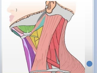
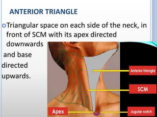
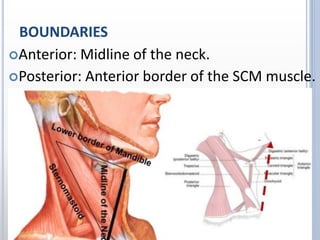
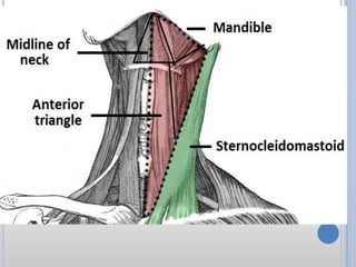
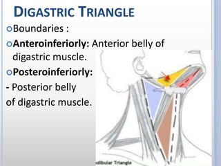
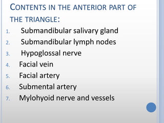
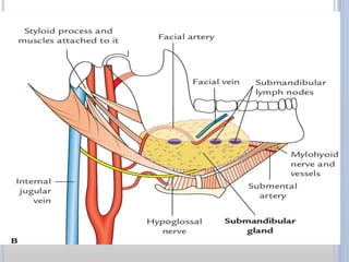
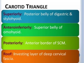
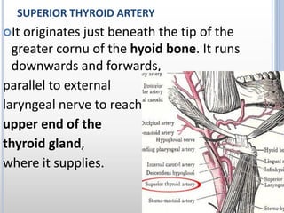
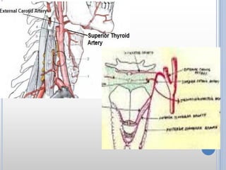
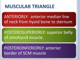
1. The document describes the anatomy of the anterior triangle of the neck, including its boundaries, contents, and structures.
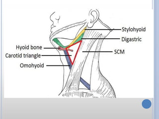
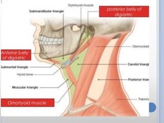
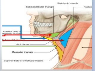
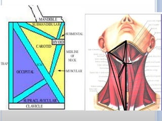
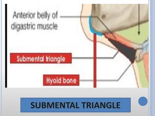
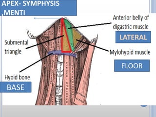
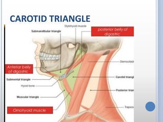
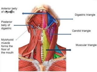
2. It is divided into 4 triangles - submental, digastric, carotid, and muscular. Each triangle contains important muscles, blood vessels, and nerves.
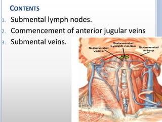
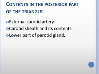
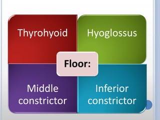
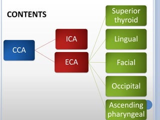
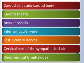
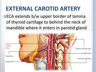
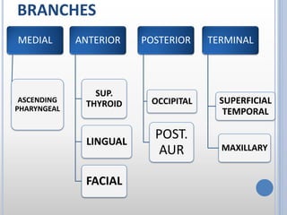
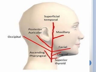
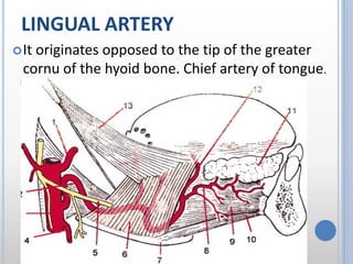
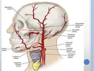
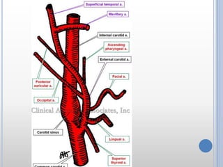
3. The document focuses on the muscles and blood vessels found in each triangle, including the digastric, omohyoid, and infrahyoid muscles as well as branches of the external carotid artery like the lingual and facial arteries.
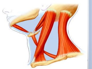
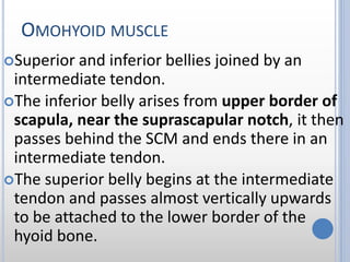
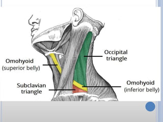
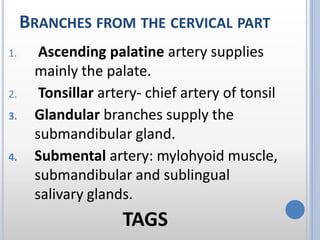
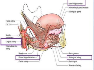


![Scalp[1]](https://cdn.slidesharecdn.com/ss_thumbnails/scalp1-170504174806-thumbnail.jpg?width=640&height=640&fit=bounds)


![Hypothalamus short ppt by Dr. Neha [PT].pptx](https://cdn.slidesharecdn.com/ss_thumbnails/hypothalamusbydr-260124145759-b9f94a93-thumbnail.jpg?width=640&height=640&fit=bounds)


