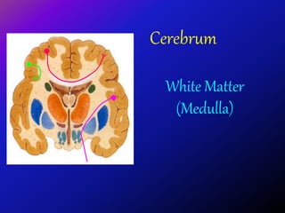
2. White Matter
• Underlies the cortex
• Contains:
• Nerve fibers
(predominantly
myelinated)
• Neuroglia
• Blood vessels
• The nerve fibers
originate, terminate
or sometimes both,
within the cortex
3. • Depending on their origin & termination, these nerve
fibers are classified into three types:
A. Association
B. Projection
C. Commissural
4. Association Fibers
• Unite different parts of
the same hemisphere
• Are of two kinds:
• Short association
fibers: those
connecting adjacent
gyri,
• Long association
fibers: those
connecting more
distant gyri
5. Short Association Fibers
• Lie immediately
beneath the gray
substance of the
cortex
• Connect
together the
adjacent gyri.
6. Long Association Fibers
• Long fibers travel
through white matter
to connect distant
areas of cerebral
cortex
• Link the primary
sensory areas in
parietal, temporal
and occipital lobes to
the association areas
of the cerebral
cortex, and to each
other
7. Superior
longitudinal
fasciculus:
connects the
frontal, parietal,
temporal and
occipital lobes
Uncinate
fasciculus:
connects frontal
to temporal lobe,
contributing to the
regulation of
behavior
Arcuate fasciculus:
connect gyri in
frontal to temporal
lobes, important for
language function
Wernicke’s Area
Broca’s
Area
Arcuate
Fasciculus
8. Cingulum: connects
frontal & parietal
lobes to the para-
hippocampal gyrus
and adjacent
temporal gyri
Inferior longitudinal
fasciculus: connects
occipital to temporal
pole & contributes to
visual recognition
9. Commissural Fibers
• Connect the corresponding
regions of the two
hemispheres
• Include:
• Corpus callosum
• Anterior commissure
• Hippocampal
commissure
(commissure of fornix)
*(Posterior commissure,
not a cerebral
commissure)
Corpus Callosum
F
P
10. Corpus Callosum
• Is a fibrous bridge located
in the depth of the median
longitudinal fissure
• Connects the two cerebral
hemispheres together
• Shorter craniocaudally than
is the hemisphere
• Cranial end is nearer to the
frontal pole of hemisphere
as compared to caudal end
to the occipital pole
11. • The fibers in the c
orpus callosum co
nnect the correspo
nding regions of th
e two hemispheres
with each other (e
xcept the inferior p
art of the temporal
lobes)
C
C
12. • Fibers linking the
two frontal poles
with each other,
curve forward &
form u-shaped
anterior forceps
(forceps minor)
• Fibers linking the
two occipital poles
with each other,
curve backward &
form u-shaped
posterior forceps
(forceps major)
F
P
O
C
C
Anterior
forceps
Posterior
forceps
14. Anterior Commissure
• Bundle of fibers
runs transversely in
front of the anterior
columns of fornix
• Connects the
inferior and middle
temporal gyri & the
olfactory regions of
the two
hemispheres
Anterior
column
of fornix
fornix
IVF
15. Hippocampal Commissure
• Bundle of fibers
runs transversely
between the crura
of the fornix
• Connect the two
hippocampi with
each other
• (note that hippocampo-
mamillary fibers do not
cross)
16. Posterior Commissure
• Rounded band of white
fibers
• Crossing the midline on
the dorsal aspect of
the upper end of the
cerebral aqueduct
(located between superior
colliculus & pineal body)
• Connects the left and
right midbrain. Plays
important role in the
bilateral pupillary reflex
SC
P
IC
Cerebral
aqueduct
17. Projection Fibers
• Fibers running vertically through
the hemispheres
• Consist of:
• Cortical afferent fibers
conveying impulses to the
cerebral cortex: (mainly
thalamo-cortical fibers)
• Cortical efferent fibers carrying
impulses away from the cortex
to the lower centers:
(corticostriate, corticobulbar,
corticopontine, corticospinal, &
descending autonomic fibers)
18. • Deeper to the cortex, these
fibers are arranged radially as
the corona radiata
• Then the fibers converge to
form a sheath, called the
internal capsule, that passes
between the thalamus and the
basal ganglia
• Continue in the:
• Crus of the midbrain
• Basilar part of pons
• Pyramid of medulla
oblongata
• Continue in the spinal cord as
the corticospinal tracts
corona
radiata
19.
20. Internal Capsule
• Bundle of
projection fibers,
passes through
the interval
between the
thalamus and the
basal ganglia
BG
Th
21. Has 5 parts:
1.Anterior limb: between caudate (C
) & lentiform (L) nuclei
2.Genu
3.Posterior limb: between thalamus
(TH) & lentiform nucleus (L)
4.Retrolenticular part: caudal to le
ntiform nucleus
5.Sublenticular part: below lentifor
m nucleus (can not be seen in this s
ection)
C
Th
L
1
2
3
4
22. • Anterior limb contains:
• Thalamocortical
projections that connect
mediodorsal nucleus of
thalamus with the
prefrontal cortex
• Frontopontine fibers
• Genu contains:
• Corticobulbar fibers
which connect the cortex
with cranial nerve motor
nuclei in the brainstem
23. • Posterior limb
contains:
• Corticospinal
• Corticobulbar
• Thalamocortical
projections from:
• VPN to the primary
somatosensory
cortex
• VAN & VLN to
motor regions of
cortex
24. • Retrolenticular part
contains thalamocortical
projections:
• Geniculocalcarine fibers
(visual radiation), from the
lateral geniculate nucleus of
thalamus to the visual
cortex in the occipital lobe
• & few Geniculotemporal
fibers (auditory radiation)
from the medial geniculate
nucleus of thalamus to the
auditory cortex in the
temporal lobe
25. • Sublenticular part
contains thalamocortical
projections: geniculo-
temporal fibers (auditory
radiation) from the
medial geniculate
nucleus of thalamus to
the auditory cortex in
the temporal lobe
26. Clinical Notes
Bilateral lesion of the inferior longitudinal fasciculus (as in
carbon monoxide poisoning), leads to:
• Loss of identification of the:
• Nature of objects (object agnosia)
• Individual faces (prosopagnosia)
• The elementary vision remains intact
Damage to corpus callosum leads to split-brain syndrome.
The two half of the brain behave relatively
autonomously
Damage to splenium of corpus callosum leads to posterior
disconnection syndrome of alexia (cannot understand
written material) without agraphia (can speak and write
without difficulty)