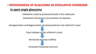
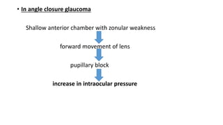
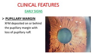
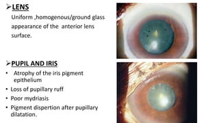
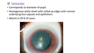
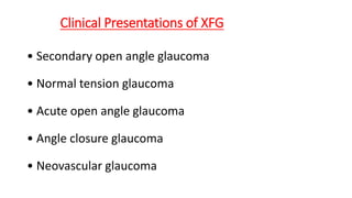
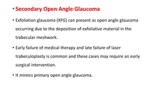
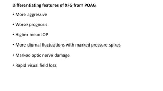
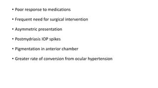
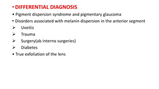
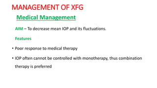
PSEUDOEXFOLIATIVE
GLAUCOMA
DR MITHUN KISHORE
EXFOLIATIVE SYNDROME AND EXFOLIATIVE GLAUCOMA
• Vogt introduced the term ‘exfoliation superficialis capsulae anteriores’
assuming that the flocculent material arose as small flecks peeling off
from the lens capsule.
• Dvorak Theobald called it pseudoexfoliation to distinguish it from
true exfoliation.
• Studies have shown that lens capsule as one of the sources of
exfoliative material and thus termed it as ‘exfoliation syndrome’ or
pseudoexfoliative syndrome and ‘Exfoliative glaucoma’ when
associated with glaucoma.
Exfoliative syndrome (XFS) is an age
related,generalized disorder of the extracellular matrix,
an elastic microfibrillopathy characterized by
progressive production and deposition of abnormal
fibrillar material in ocular and extraocular tissues.
EPIDEMIOLOGY
- AGE
Prevalence of XFS increases with age
- GENDER
• Females have increased predisposition for XFS without glaucoma
• XFG develops earlier ,more frequently and in increased severity
among males
PREVALENCE AGE GROUP
0.6% <65 YRS
2.6% 65-74 YRS
5 % 75-85 YRS
PREVALENCE OF OHT AND GLAUCOMA IN XFS
• 25% of patients with XFS have elevated IOP
• Patients with XFS and OHT are twice likely to convert to glaucoma.
PREVALENCE OF EXFOLIATION GLAUCOMA IN OPEN ANGLE GLAUCOMA
• 20-40% of patients with open angle glaucoma have XFG .
PREVALANCE OF EXFOLIATIVE GLAUCOMA IN ANGLE CLOSURE GLAUCOMA
• Glaucomatous optic neuropathy was found in 28.3% of XFS cases with
primary angle closure glaucoma or occludable angle.
RISK FACTORS FOR CONVERSION FROM XFS TO XFG
• Increased Intraocular pressure
• Presence of XFM in the angle
• Presence of XFM in the other eye
• Decreased pupillary dilation
• Increased trabecular meshwork pigmentation.
• ETIOLOGY AND PATHOGENESIS
• GEOGRAPHICAL FACTORS
• High prevalence rate in Nordic countries suggests that a northern
latitude,cold air contribute to development of XFS
• GENETIC FACTORS
• Thorleifsson et al identified single nucleotide polymorphism (SNP)
associated with XFS.
• Three single nucleotide polymorphism in the coding region of LOX L1
gene located on chromosome 15q24 were strongly associated with XFS
and XFG.
MICROSCOPIC STRUCTURE OF XFM
• It consisits of a tangle of fibrils and filaments embedded in an
amorphous matrix.
• Fibrils are composed of microfibrillar subunits surrounded by an
amorphous matrix.
CHEMICAL STRUCTURE
• It has a protein core surrounded by glycoconjugates
(glycosaminoglycans) which form the amorphous ground substance.
• Protein component includes non collagenous basement membrane
components and epitopes of elastic fibre system.
• Fibrillin -1 – Main component of elastic microfibrils has been
demonstrated in exfoliation fibres and their microfibrillar subunits.
• The latent tumor growth factor beta binding proteins (LTBP-1 andLTBP-2)
 Associated with all exfoliative deposits and co-localized with latent TGF
on exfoliation fibers.
 These LTBPs may have a dual role
a) structural component of exfoliation fibers
b) matrix anchorage of latent TGF β1 to XFM.
• The HNK-1 epitope - involved in the adhesiveness of exfoliation material
deposits on intraocular surfaces acting like a glue and binding the various
components together and to the ocular structures.
•BIOCHEMICAL CHANGES IN AQUEOUS HUMOUR
IN XFS
• -Increased TGF beta1-responsible for overproduction of extracellular
matrix.
• -Increased level of matrix metalloproteinases(MMP)
• -Elevated level of Homocysteine from disturbed methionine metabolism
associated with disruption of elastic fibre component of extracellular
matrix.
• THEORIES OF PATHOGENESIS OF XFS
AMYLOID THEORY
• XFM constituted of crude anti-amyloid serum suggesting amyloid
deposists.
• This theory could not be supported by further tests.
BASEMENT MEMBRANE THEORY
• Exfoliative material have been associated with basement membranes
of cell types.
• Immunohistochemical studies reveal presence of basement
membrane epitopes like laminin, heparin sulphate in XFM aggregates.
• ELASTIC MICRIFIBRIL THEORY
• This theory was based on structural association of XFM associated
with components of elastin system like zonules.
• Immunohistochemical studies demonstrates epitopes of elastin
supports this theory.
• INFECTIOUS ORIGIN THEORY
• XFM after intraocular surgery or ocular trauma suggested the
possibility of this theory
• XFS after penetrating keratoplasty with grafts from elderly supports
this theory.
• No conclusive evidence to support this theory.
STRESSED INDUCED ELASTOSIS THEORY
• XFS is a type of stress induced elastosis
• Associated with excessive production of elastic microfibrills by
elastogenic cells which aggregate to form exfoliative fibrils.
• PROTEIN SINK MODEL THEORY
• Soluble proteins found in biological fluids form insoluble structures
that accumulate to form intra and extracellular aggregates which
form fine dusting on surfaces like lens capsule and angle of anterior
chamber.
• SOURCES OF EXFOLIATIVE MATERIAL
INTRAOCULAR EXTRAOCULAR
Pre-equatorial lens
epithelium
Fibroblasts
Non pigmented ciliary
epithelium
Smooth and striated
muscle cells
Trabecular endothelium Cardiac muscle cells
Vascular endothelium
• EXTRAOCULAR AND SYSTEMIC SITES OF XFM DEPOSITS
EXTRAOCULAR
SITES
SYSTEMIC
SITES
Conjunctiva skin
Orbital tissues Visceral organs
Blood vessels wall
Meninges
• PATHOGENESIS OF GLAUCOMA IN EXFOLIATIVE SYNDROME
In open angle glaucoma
Exfoliation material produced locally in the trabecular
meshwork and passive accumulation via aqueous
disorganization and degeneration of juxtacanalicular and schlemm’s canal
Focal collapse of the schlemm’s canal
Decreased aqueous outflow
Increased intraocular pressure
• In angle closure glaucoma
Shallow anterior chamber with zonular weakness
forward movement of lens
pupillary block
increase in intraocular pressure
CLINICAL FEATURES
EARLY SIGNS
 PUPILLARY MARGIN
XFM deposited on or behind
the pupillary margin with
loss of pupillary ruff.
LENS
Uniform ,homogenous/ground glass
appearance of the anterior lens
surface.
PUPIL AND IRIS
• Atrophy of the iris pigment
epithelium
• Loss of pupillary ruff
• Poor mydriasis
• Pigment dispertion after pupillary
dilatation.
 CILIARY PROCESSES
Deposition of exfoliative material in the ciliary processes is one of
the earliest feature seen on indirect ophthalmoscopy with
indentation.
SIGNS IN ESTABLISHED XFS
 LENS
Deposition of XFM on lens shows a classical pattern which
Consists of 3 zones which is visible after dilatation
 Central disc
• Corresponds to diameter of pupil.
• Homogenous white sheet with rolled up edges with normal
underlying lens capsule and epithelium.
• Absent in 20 % of cases.
 Granular peripheral zone
• Extends equatorially as tongue shaped projections which
merge into normal capsule before reaching the anterior
insertion of zonules.
 Intermediate zone
• 1-2 mm of clear zone devoid of XFM which is formed as a
result of iris scraping the exfoliation material from the
surface of the lens during pupillary movement
 Pre equatorial zone
• This zone is clinically hidden by iris.
• It consists of nodular excrescences covering zonules and
their attachments.
Three zones or Bull’s eye pattern seen on the anterior lens capsule on
retroillumination
 PHACODONESIS
Due to spontaneous subluxation and dislocation of the lens due to
weakness of zonules secondary to deposition of XFM.
 CATARACT
Increased prevalence of cataract (nuclear and subcapsular cataract)
Cause – ocular ischemia and oxidative damage have been proposed.
• IRIS
• Deposition of exfoliative material at the pupillary margin.
• Pigment loss from iris and its deposition on anterior segment structures.
• Transillumination defects at pupillary margin secondary to pigment loss from
iris.
• Diffuse pigment deposition on iris surface.
IRIS TRANSILLUMINATION DEFECT
PUPIL
Eyes with XFS dilate poorly and constrict poorly with pilocarpine due
to fibrotic and degenerative changes in the iris sphincter and dilator
pupillae.
EYES AFTER INSTALLATION OF MYDRIATIC
PEX MATERIAL DEPOSITION ON CORNEAL
ENDOTHELIUM
CORNEAL SIGNS
• Diffuse non specific pigmentation of
central endothelium in the pattern of
krukenberg spindle.
• Altered endothelial cell count and
morphology.
• Keratopathy characterised by
 Diffuse ,irregular thickening of
descemet’s membrane.
 Focal accumulation of exfoliation
material on/ within descemets
membrane.
ANTERIOR CHAMBER ANGLE
• Increased trabecular
pigmentation is a early sign on
gonioscopy.
• Pigment deposited on
Schwalbe’s line and anterior to
it (Sampaolesi’s line).
OPTIC NERVE HEAD AND RNFL LAYER
• The mean optic disc area has been reported to be smaller in eyes with
XFS and exfoliative glaucoma.
• Diffuse cupping compared to POAG.
• The percentage area of optic disc pallor in XFS patients greater than
controls.
• The RNFL has been found to be significantly thinner in XFS eyes
Clinical Presentations of XFG
• Secondary open angle glaucoma
• Normal tension glaucoma
• Acute open angle glaucoma
• Angle closure glaucoma
• Neovascular glaucoma
• Secondary Open Angle Glaucoma
• Exfoliation glaucoma (XFG) can present as open angle glaucoma
occurring due to the deposition of exfoliative material in the
trabecular meshwork.
• Early failure of medical therapy and late failure of laser
trabeculoplasty is common and these cases may require an early
surgical intervention.
• It mimics primary open angle glaucoma.
Differentiating features of XFG from POAG
• More aggressive
• Worse prognosis
• Higher mean IOP
• More diurnal fluctuations with marked pressure spikes
• Marked optic nerve damage
• Rapid visual field loss
• Poor response to medications
• Frequent need for surgical intervention
• Asymmetric presentation
• Postmydriasis IOP spikes
• Pigmentation in anterior chamber
• Greater rate of conversion from ocular hypertension
• ‘Normal Tension’ Exfoliation Glaucoma
• Higher maximum IOP level and a greater IOP fluctuation within
normal range could be significant risk factors for development of
glaucomatous ONH changes in eyes with exfoliation in the presence
of a normal pressure.
• Non-IOP dependant factors include.
 impaired ocular and retrobulbar perfusion
Abnormalities of elastic tissue of the lamina cribrosa.
•Acute Open Angle Glaucoma
• Dispersion of pigment granules and XFM in the anterior
chamber after diagnostic pupillary dilation lead to marked
rise in IOP causing diffuse corneal edema producing a
deceptive clinical picture of acute primary angle closure
glaucoma.
• Presents with IOP rise of more then 50 mm Hg in spite of an
open angle.
• Angle Closure Glaucoma
Mechanisms proposed are
Narrow anterior chamber angle, smaller anterior chamber volume,
along with a minimal subluxation of lens which predisposes to
pupillary block.
Formation of posterior synechiae due to a deranged blood aqueous
barrier leading to pupillary block.
• Neovascular Glaucoma
• CRVO is more common in patients with exfoliation.
• Patients with CRVO can present as neovascular glaucoma.
• DIFFERENTIAL DIAGNOSIS
• Pigment dispersion syndrome and pigmentary glaucoma
• Disorders associated with melanin dispersion in the anterior segment
 Uveitis
 Trauma
 Surgery(ab interno surgeries)
 Diabetes
• True exfoliation of the lens
MANAGEMENT OF XFG
Medical Management
AIM – To decrease mean IOP and its fluctuations.
Features
• Poor response to medical therapy
• IOP often cannot be controlled with monotherapy, thus combination
therapy is preferred
 Aqueous Suppressants
• Beta blockers are effective in controlling initial IOP but significant
diurnal fluctuations have been noted in XFG in contrast to POAG.
• Carbonic anhydrase inhibitors(Dorzolamide) is also effective as timolol
and also has an additive action in combination therapy but should be
avoided in patients with compromised corneal endothelium.
• Alpha adrenergic agonists like brimonidine may also be used in
combination therapy.
 Prostaglandin Analogs
• Prostaglandin analogs increases the aqueous outflow via the
uveoscleral pathway have been found to be effective in lowering IOP
in exfoliation glaucoma.
• Latanoprost provide a better control of diurnal fluctuation as
compared to timolol.
• Both latanoprost and travoprost have been found to reduce IOP at
each time point in a 24 hour study.
 Pilocarpine
• Lower the IOP by increasing the aqueous outflow.
• Pilocarpine limits the pupillary movement, thus prevents iridolenticular
friction and pigment release .
• Hazards of pilocarpine use
- aggravation of the blood aqueous barrier dysfunction
- development of posterior synechiae by restricting pupillary
movement
- aggravation of lens opacities and ciliary block glaucoma in eyes with
marked zonular instability.
• Inter-national Collaborative Exfoliation Syndrome Treatment (ICEST)
study examined treatment with latanoprost and pilocarpine vs timolol
vs fixed-combination timolol and dorzolamide in eyes with exfoliation
syndrome and elevated IOP found that the latanoprost and
pilocarpine lowers IOP and improved aqueous outflow.
Lasers
• XFG patients respond well to laser trabeculoplasty due to the high baseline
IOP as well as the heavily pigmented trabecular meshwork
• Patients with higher baseline IOP, the drop in IOP after ALT is greater in
these eyes than in non-XFG eyes.
• The success rate varies from 70 to 90 % with IOP reduction upto 30%.
Inflammatory reaction and IOP spikes are frequently seen.
• The patients require a IOP lowering and anti-inflammatory medications in
the early post laser period.
• Late failure of laser treatment presenting with high IOP, unexpected disc
damage and visual field progression.
• Selective laser trabeculoplasty
- selectively targets the intracellular melanin granules in the
trabecular meshwork cells.
- IOP reduction is more pronounced if prelaser IOP is high.
- can be done after failed ALT.
• Peripheral laser iridotomy is performed when angle closure is present
along with exfoliation
•SURGICAL MANAGEMENT
• Required earlier and more frequently as compared to
other forms of glaucoma due to the early failure of
medical and laser treatment.
• Surgical options available are
- Traeculectomy
- Non penetrating glaucoma filtering surgery(NPGS)
- cataract surgery
- combined cataract and glaucoma surgery
TRABECULECTOMY
• Trabeculectomy with antimetabolites is the surgery of choice.
• PEXG treated with trabeculectomy associated with increases
perioperative and early postoperative complications like
- Increased risk of choroidal haemorrhage/effusion
. - Increase risk of haemorrhage due to iris vasculopathy at the site of
peripheral iridectomy.
- vitreous obliteration of trabeculectomy opening in eyes with loose
zonules and anterior movement of the lens.
- Increased risk of fibrinous reaction post op period.
NON PENETRATING GLAUCOMA
SURGERY
• Avoids unfavourable bleb related complications and
decreases incidence of post operative inflammation.
• Commonly done Non penetrating glaucoma surgeries are
- Trabeculectomy ab externo
- Deep sclerectomy
COMBINED CATARACT AND GLAUCOMA SURGERY
• Indicated in patients with coexistant cataract and XFG.
• It is associated with
- Greater incidence of spontaneous lens displacement and capsular
rupture.
- Increased incidence of zonular dehiscence and vitreous loss.
- Increased incidence of post capsular opacification.
CATARACT SURGERY
Cataract surgery alone is sufficient in controlling IOP in eyes with little or no
glaucoma damage or in eyes with well controlled glaucoma on minimal
medications.
PREOPERATIVE ASSESSMENT
 Pupillary dilation
 Phacodonesis
 Control of glaucoma
 Zonular status
• INTRAOPERATIVE CONSIDERATIONS
 SMALL PUPIL
• Preoperative NSAIDS given to prevent intraoperative miosis.
• Posterior synechiae if present should be lysed with
capsularhexis forceps.
• Pupil can be expanded using
- Cohesive ophthalmic viscosurgical device.
- Pupil expansion device.
 ZONULAR WEAKNESS
Dependind on severity of zonular dialysis following
can be used
 Capsular tension ring(CTR)
 Cionni modified CTR(m CTR)
 Capsular tension segment(CTS)
 HYDRODISSECTION AND VISCODISSECTION
 Hydrodissection performed with minimal stress on zonules.
 OVD injected between the lens capsule and cortex to prevents
trapping of cortex during CTR insertion and also provides greater
corticocapsular cleavage than hydrodissection alone.
 IOL INSERTION
One piece acrylic IOL of choice- this IOL design allows
surgeon to dial the IOL gently into capsular bag and
place felxible haptic in desired orientation with
minimal capsular and zonular stress.
THANK YOU

Pseudoexfoliative glaucoma seminar

  • 1.
  • 2.
    EXFOLIATIVE SYNDROME ANDEXFOLIATIVE GLAUCOMA • Vogt introduced the term ‘exfoliation superficialis capsulae anteriores’ assuming that the flocculent material arose as small flecks peeling off from the lens capsule. • Dvorak Theobald called it pseudoexfoliation to distinguish it from true exfoliation. • Studies have shown that lens capsule as one of the sources of exfoliative material and thus termed it as ‘exfoliation syndrome’ or pseudoexfoliative syndrome and ‘Exfoliative glaucoma’ when associated with glaucoma.
  • 3.
    Exfoliative syndrome (XFS)is an age related,generalized disorder of the extracellular matrix, an elastic microfibrillopathy characterized by progressive production and deposition of abnormal fibrillar material in ocular and extraocular tissues.
  • 4.
    EPIDEMIOLOGY - AGE Prevalence ofXFS increases with age - GENDER • Females have increased predisposition for XFS without glaucoma • XFG develops earlier ,more frequently and in increased severity among males PREVALENCE AGE GROUP 0.6% <65 YRS 2.6% 65-74 YRS 5 % 75-85 YRS
  • 5.
    PREVALENCE OF OHTAND GLAUCOMA IN XFS • 25% of patients with XFS have elevated IOP • Patients with XFS and OHT are twice likely to convert to glaucoma. PREVALENCE OF EXFOLIATION GLAUCOMA IN OPEN ANGLE GLAUCOMA • 20-40% of patients with open angle glaucoma have XFG . PREVALANCE OF EXFOLIATIVE GLAUCOMA IN ANGLE CLOSURE GLAUCOMA • Glaucomatous optic neuropathy was found in 28.3% of XFS cases with primary angle closure glaucoma or occludable angle.
  • 6.
    RISK FACTORS FORCONVERSION FROM XFS TO XFG • Increased Intraocular pressure • Presence of XFM in the angle • Presence of XFM in the other eye • Decreased pupillary dilation • Increased trabecular meshwork pigmentation.
  • 7.
    • ETIOLOGY ANDPATHOGENESIS • GEOGRAPHICAL FACTORS • High prevalence rate in Nordic countries suggests that a northern latitude,cold air contribute to development of XFS • GENETIC FACTORS • Thorleifsson et al identified single nucleotide polymorphism (SNP) associated with XFS. • Three single nucleotide polymorphism in the coding region of LOX L1 gene located on chromosome 15q24 were strongly associated with XFS and XFG.
  • 8.
    MICROSCOPIC STRUCTURE OFXFM • It consisits of a tangle of fibrils and filaments embedded in an amorphous matrix. • Fibrils are composed of microfibrillar subunits surrounded by an amorphous matrix.
  • 9.
    CHEMICAL STRUCTURE • Ithas a protein core surrounded by glycoconjugates (glycosaminoglycans) which form the amorphous ground substance. • Protein component includes non collagenous basement membrane components and epitopes of elastic fibre system. • Fibrillin -1 – Main component of elastic microfibrils has been demonstrated in exfoliation fibres and their microfibrillar subunits.
  • 10.
    • The latenttumor growth factor beta binding proteins (LTBP-1 andLTBP-2)  Associated with all exfoliative deposits and co-localized with latent TGF on exfoliation fibers.  These LTBPs may have a dual role a) structural component of exfoliation fibers b) matrix anchorage of latent TGF β1 to XFM. • The HNK-1 epitope - involved in the adhesiveness of exfoliation material deposits on intraocular surfaces acting like a glue and binding the various components together and to the ocular structures.
  • 11.
    •BIOCHEMICAL CHANGES INAQUEOUS HUMOUR IN XFS • -Increased TGF beta1-responsible for overproduction of extracellular matrix. • -Increased level of matrix metalloproteinases(MMP) • -Elevated level of Homocysteine from disturbed methionine metabolism associated with disruption of elastic fibre component of extracellular matrix.
  • 12.
    • THEORIES OFPATHOGENESIS OF XFS AMYLOID THEORY • XFM constituted of crude anti-amyloid serum suggesting amyloid deposists. • This theory could not be supported by further tests. BASEMENT MEMBRANE THEORY • Exfoliative material have been associated with basement membranes of cell types. • Immunohistochemical studies reveal presence of basement membrane epitopes like laminin, heparin sulphate in XFM aggregates.
  • 13.
    • ELASTIC MICRIFIBRILTHEORY • This theory was based on structural association of XFM associated with components of elastin system like zonules. • Immunohistochemical studies demonstrates epitopes of elastin supports this theory. • INFECTIOUS ORIGIN THEORY • XFM after intraocular surgery or ocular trauma suggested the possibility of this theory • XFS after penetrating keratoplasty with grafts from elderly supports this theory. • No conclusive evidence to support this theory.
  • 14.
    STRESSED INDUCED ELASTOSISTHEORY • XFS is a type of stress induced elastosis • Associated with excessive production of elastic microfibrills by elastogenic cells which aggregate to form exfoliative fibrils. • PROTEIN SINK MODEL THEORY • Soluble proteins found in biological fluids form insoluble structures that accumulate to form intra and extracellular aggregates which form fine dusting on surfaces like lens capsule and angle of anterior chamber.
  • 15.
    • SOURCES OFEXFOLIATIVE MATERIAL INTRAOCULAR EXTRAOCULAR Pre-equatorial lens epithelium Fibroblasts Non pigmented ciliary epithelium Smooth and striated muscle cells Trabecular endothelium Cardiac muscle cells Vascular endothelium
  • 16.
    • EXTRAOCULAR ANDSYSTEMIC SITES OF XFM DEPOSITS EXTRAOCULAR SITES SYSTEMIC SITES Conjunctiva skin Orbital tissues Visceral organs Blood vessels wall Meninges
  • 17.
    • PATHOGENESIS OFGLAUCOMA IN EXFOLIATIVE SYNDROME In open angle glaucoma Exfoliation material produced locally in the trabecular meshwork and passive accumulation via aqueous disorganization and degeneration of juxtacanalicular and schlemm’s canal Focal collapse of the schlemm’s canal Decreased aqueous outflow Increased intraocular pressure
  • 18.
    • In angleclosure glaucoma Shallow anterior chamber with zonular weakness forward movement of lens pupillary block increase in intraocular pressure
  • 19.
    CLINICAL FEATURES EARLY SIGNS PUPILLARY MARGIN XFM deposited on or behind the pupillary margin with loss of pupillary ruff.
  • 20.
    LENS Uniform ,homogenous/ground glass appearanceof the anterior lens surface. PUPIL AND IRIS • Atrophy of the iris pigment epithelium • Loss of pupillary ruff • Poor mydriasis • Pigment dispertion after pupillary dilatation.
  • 21.
     CILIARY PROCESSES Depositionof exfoliative material in the ciliary processes is one of the earliest feature seen on indirect ophthalmoscopy with indentation. SIGNS IN ESTABLISHED XFS  LENS Deposition of XFM on lens shows a classical pattern which Consists of 3 zones which is visible after dilatation
  • 22.
     Central disc •Corresponds to diameter of pupil. • Homogenous white sheet with rolled up edges with normal underlying lens capsule and epithelium. • Absent in 20 % of cases.
  • 23.
     Granular peripheralzone • Extends equatorially as tongue shaped projections which merge into normal capsule before reaching the anterior insertion of zonules.  Intermediate zone • 1-2 mm of clear zone devoid of XFM which is formed as a result of iris scraping the exfoliation material from the surface of the lens during pupillary movement
  • 24.
     Pre equatorialzone • This zone is clinically hidden by iris. • It consists of nodular excrescences covering zonules and their attachments. Three zones or Bull’s eye pattern seen on the anterior lens capsule on retroillumination
  • 25.
     PHACODONESIS Due tospontaneous subluxation and dislocation of the lens due to weakness of zonules secondary to deposition of XFM.  CATARACT Increased prevalence of cataract (nuclear and subcapsular cataract) Cause – ocular ischemia and oxidative damage have been proposed.
  • 26.
    • IRIS • Depositionof exfoliative material at the pupillary margin. • Pigment loss from iris and its deposition on anterior segment structures. • Transillumination defects at pupillary margin secondary to pigment loss from iris. • Diffuse pigment deposition on iris surface. IRIS TRANSILLUMINATION DEFECT
  • 27.
    PUPIL Eyes with XFSdilate poorly and constrict poorly with pilocarpine due to fibrotic and degenerative changes in the iris sphincter and dilator pupillae. EYES AFTER INSTALLATION OF MYDRIATIC
  • 28.
    PEX MATERIAL DEPOSITIONON CORNEAL ENDOTHELIUM CORNEAL SIGNS • Diffuse non specific pigmentation of central endothelium in the pattern of krukenberg spindle. • Altered endothelial cell count and morphology. • Keratopathy characterised by  Diffuse ,irregular thickening of descemet’s membrane.  Focal accumulation of exfoliation material on/ within descemets membrane.
  • 29.
    ANTERIOR CHAMBER ANGLE •Increased trabecular pigmentation is a early sign on gonioscopy. • Pigment deposited on Schwalbe’s line and anterior to it (Sampaolesi’s line).
  • 30.
    OPTIC NERVE HEADAND RNFL LAYER • The mean optic disc area has been reported to be smaller in eyes with XFS and exfoliative glaucoma. • Diffuse cupping compared to POAG. • The percentage area of optic disc pallor in XFS patients greater than controls. • The RNFL has been found to be significantly thinner in XFS eyes
  • 31.
    Clinical Presentations ofXFG • Secondary open angle glaucoma • Normal tension glaucoma • Acute open angle glaucoma • Angle closure glaucoma • Neovascular glaucoma
  • 32.
    • Secondary OpenAngle Glaucoma • Exfoliation glaucoma (XFG) can present as open angle glaucoma occurring due to the deposition of exfoliative material in the trabecular meshwork. • Early failure of medical therapy and late failure of laser trabeculoplasty is common and these cases may require an early surgical intervention. • It mimics primary open angle glaucoma.
  • 33.
    Differentiating features ofXFG from POAG • More aggressive • Worse prognosis • Higher mean IOP • More diurnal fluctuations with marked pressure spikes • Marked optic nerve damage • Rapid visual field loss
  • 34.
    • Poor responseto medications • Frequent need for surgical intervention • Asymmetric presentation • Postmydriasis IOP spikes • Pigmentation in anterior chamber • Greater rate of conversion from ocular hypertension
  • 35.
    • ‘Normal Tension’Exfoliation Glaucoma • Higher maximum IOP level and a greater IOP fluctuation within normal range could be significant risk factors for development of glaucomatous ONH changes in eyes with exfoliation in the presence of a normal pressure. • Non-IOP dependant factors include.  impaired ocular and retrobulbar perfusion Abnormalities of elastic tissue of the lamina cribrosa.
  • 36.
    •Acute Open AngleGlaucoma • Dispersion of pigment granules and XFM in the anterior chamber after diagnostic pupillary dilation lead to marked rise in IOP causing diffuse corneal edema producing a deceptive clinical picture of acute primary angle closure glaucoma. • Presents with IOP rise of more then 50 mm Hg in spite of an open angle.
  • 37.
    • Angle ClosureGlaucoma Mechanisms proposed are Narrow anterior chamber angle, smaller anterior chamber volume, along with a minimal subluxation of lens which predisposes to pupillary block. Formation of posterior synechiae due to a deranged blood aqueous barrier leading to pupillary block.
  • 38.
    • Neovascular Glaucoma •CRVO is more common in patients with exfoliation. • Patients with CRVO can present as neovascular glaucoma.
  • 39.
    • DIFFERENTIAL DIAGNOSIS •Pigment dispersion syndrome and pigmentary glaucoma • Disorders associated with melanin dispersion in the anterior segment  Uveitis  Trauma  Surgery(ab interno surgeries)  Diabetes • True exfoliation of the lens
  • 40.
    MANAGEMENT OF XFG MedicalManagement AIM – To decrease mean IOP and its fluctuations. Features • Poor response to medical therapy • IOP often cannot be controlled with monotherapy, thus combination therapy is preferred
  • 41.
     Aqueous Suppressants •Beta blockers are effective in controlling initial IOP but significant diurnal fluctuations have been noted in XFG in contrast to POAG. • Carbonic anhydrase inhibitors(Dorzolamide) is also effective as timolol and also has an additive action in combination therapy but should be avoided in patients with compromised corneal endothelium. • Alpha adrenergic agonists like brimonidine may also be used in combination therapy.
  • 42.
     Prostaglandin Analogs •Prostaglandin analogs increases the aqueous outflow via the uveoscleral pathway have been found to be effective in lowering IOP in exfoliation glaucoma. • Latanoprost provide a better control of diurnal fluctuation as compared to timolol. • Both latanoprost and travoprost have been found to reduce IOP at each time point in a 24 hour study.
  • 43.
     Pilocarpine • Lowerthe IOP by increasing the aqueous outflow. • Pilocarpine limits the pupillary movement, thus prevents iridolenticular friction and pigment release . • Hazards of pilocarpine use - aggravation of the blood aqueous barrier dysfunction - development of posterior synechiae by restricting pupillary movement - aggravation of lens opacities and ciliary block glaucoma in eyes with marked zonular instability.
  • 44.
    • Inter-national CollaborativeExfoliation Syndrome Treatment (ICEST) study examined treatment with latanoprost and pilocarpine vs timolol vs fixed-combination timolol and dorzolamide in eyes with exfoliation syndrome and elevated IOP found that the latanoprost and pilocarpine lowers IOP and improved aqueous outflow.
  • 45.
    Lasers • XFG patientsrespond well to laser trabeculoplasty due to the high baseline IOP as well as the heavily pigmented trabecular meshwork • Patients with higher baseline IOP, the drop in IOP after ALT is greater in these eyes than in non-XFG eyes. • The success rate varies from 70 to 90 % with IOP reduction upto 30%. Inflammatory reaction and IOP spikes are frequently seen. • The patients require a IOP lowering and anti-inflammatory medications in the early post laser period. • Late failure of laser treatment presenting with high IOP, unexpected disc damage and visual field progression.
  • 46.
    • Selective lasertrabeculoplasty - selectively targets the intracellular melanin granules in the trabecular meshwork cells. - IOP reduction is more pronounced if prelaser IOP is high. - can be done after failed ALT. • Peripheral laser iridotomy is performed when angle closure is present along with exfoliation
  • 47.
    •SURGICAL MANAGEMENT • Requiredearlier and more frequently as compared to other forms of glaucoma due to the early failure of medical and laser treatment. • Surgical options available are - Traeculectomy - Non penetrating glaucoma filtering surgery(NPGS) - cataract surgery - combined cataract and glaucoma surgery
  • 48.
    TRABECULECTOMY • Trabeculectomy withantimetabolites is the surgery of choice. • PEXG treated with trabeculectomy associated with increases perioperative and early postoperative complications like - Increased risk of choroidal haemorrhage/effusion . - Increase risk of haemorrhage due to iris vasculopathy at the site of peripheral iridectomy. - vitreous obliteration of trabeculectomy opening in eyes with loose zonules and anterior movement of the lens. - Increased risk of fibrinous reaction post op period.
  • 49.
    NON PENETRATING GLAUCOMA SURGERY •Avoids unfavourable bleb related complications and decreases incidence of post operative inflammation. • Commonly done Non penetrating glaucoma surgeries are - Trabeculectomy ab externo - Deep sclerectomy
  • 50.
    COMBINED CATARACT ANDGLAUCOMA SURGERY • Indicated in patients with coexistant cataract and XFG. • It is associated with - Greater incidence of spontaneous lens displacement and capsular rupture. - Increased incidence of zonular dehiscence and vitreous loss. - Increased incidence of post capsular opacification.
  • 51.
    CATARACT SURGERY Cataract surgeryalone is sufficient in controlling IOP in eyes with little or no glaucoma damage or in eyes with well controlled glaucoma on minimal medications. PREOPERATIVE ASSESSMENT  Pupillary dilation  Phacodonesis  Control of glaucoma  Zonular status
  • 52.
    • INTRAOPERATIVE CONSIDERATIONS SMALL PUPIL • Preoperative NSAIDS given to prevent intraoperative miosis. • Posterior synechiae if present should be lysed with capsularhexis forceps. • Pupil can be expanded using - Cohesive ophthalmic viscosurgical device. - Pupil expansion device.
  • 53.
     ZONULAR WEAKNESS Dependindon severity of zonular dialysis following can be used  Capsular tension ring(CTR)  Cionni modified CTR(m CTR)  Capsular tension segment(CTS)  HYDRODISSECTION AND VISCODISSECTION  Hydrodissection performed with minimal stress on zonules.  OVD injected between the lens capsule and cortex to prevents trapping of cortex during CTR insertion and also provides greater corticocapsular cleavage than hydrodissection alone.
  • 54.
     IOL INSERTION Onepiece acrylic IOL of choice- this IOL design allows surgeon to dial the IOL gently into capsular bag and place felxible haptic in desired orientation with minimal capsular and zonular stress.
  • 55.