Downloaded 112 times


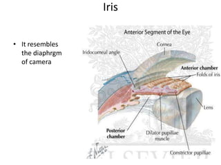


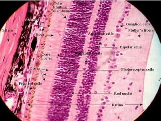


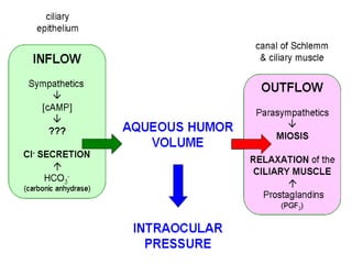


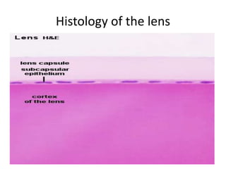

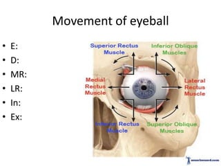

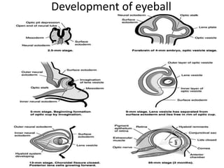


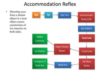


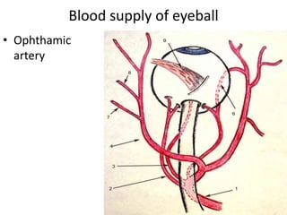


This document provides an overview of the structure and anatomy of the human eyeball. It begins with an introduction to the eyeball's location in the orbital cavity and its resemblance to a camera. The presentation then covers the eyeball's layers (sclera, choroid, retina), internal structures (iris, lens), humors (aqueous, vitreous), blood supply, nerve supply, development, and movements. Examples of applied anatomy are also discussed, such as different types of glaucoma, corneal grafting, and refractive errors like myopia and astigmatism.


