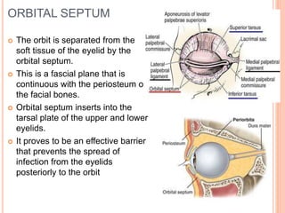
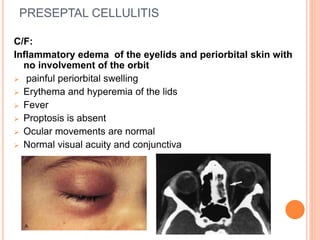
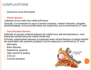
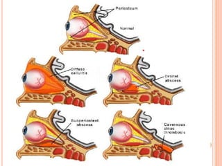
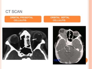
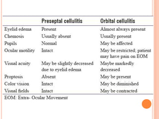
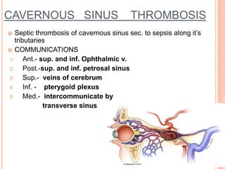
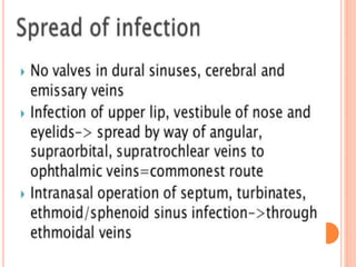
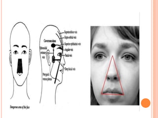
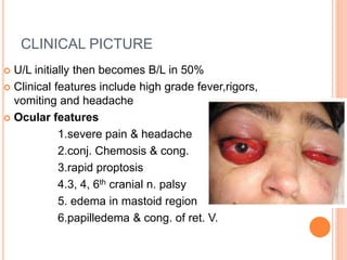
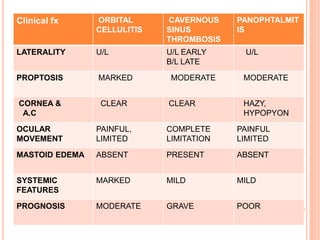
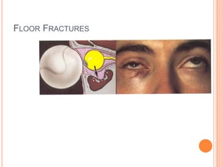
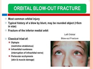
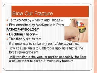
ORBIT
ORBITAL AND PRESEPTAL CELLULITIS
CAVERNOUS SINUS THROMBOSIS
FRACTURE
ORBITAL SEPTUM
 The orbit is separated from the
soft tissue of the eyelid by the
orbital septum.
 This is a fascial plane that is
continuous with the periosteum of
the facial bones.
 Orbital septum inserts into the
tarsal plate of the upper and lower
eyelids.
 It proves to be an effective barrier
that prevents the spread of
infection from the eyelids
posteriorly to the orbit
PRESEPTAL CELLULITUS
 Infection of the subcutaneous tissues anterior to the
orbital septum
 Aetiology : staphylococcus aureus , streptococcus ,
hemophillius influenza
 Mode of infection :
Exogenous : following skin laceration , insect bites
and eyelid operations
Endogenous : hematogenous spread from remote
infection of the middle ear or URT
Extension from local infection : from an acute
hordeolum or acute dacryocystitis
PRESEPTAL CELLULITIS
C/F:
Inflammatory edema of the eyelids and periorbital skin with
no involvement of the orbit
 painful periorbital swelling
 Erythema and hyperemia of the lids
 Fever
 Proptosis is absent
 Ocular movements are normal
 Normal visual acuity and conjunctiva
TREATMENT
 Systemic antibiotics
Mild to moderate cases : amoxiclav 500/125vmg tds
Sever cases : hospitalization , iv ceftriaxone (1-2
g/day in divided doses for 4-5 days )
 Systemic analgesic and anti inflammatory to reduce
pain and swelling
 Warm compression 2-3 times a day
 Surgical exploration and debridement required in
presence of an abscess
ORBITAL CELLULITIS
ORBITAL CELLULITUS
 Refers to an acute infection of the soft tissues of
the orbit behind the orbital septum
 Orbital cellulitis may or may not progress to a
subperiosteal abscess or orbital abscess
 Causative organisms : Those commonly involved
are: Streptococcus pneumoniae, Staphylococcus
aureus, Streptococcus pyogenes and Haemophilus
influenzae.
ETIOLOGY
 EXOGENOUS: penetrating injury associated with
retention of intraorbital foreign body , post operatively
evisceration
 EXTENSION OF INFECTION FROM NEIGHBOURING
structures : paranasal sinuses, teeth, face, lids,
intracranial cavity and intraorbital structures.
It is the commonest mode of orbital infections.
 ENDOGENOUS INFECTION. : metastatic infection
from breast abscess, puerperal sepsis, thrombophlebitis
of legs and septicaemia.
PATHOLOGY
:■Infection establishes early due to absence of
lymphatics in the orbit.
■Rapid spread with extensive necrosis is common
since in most cases infection spreads as
thrombophebitis from the surrounding structures.
■Damage produced is rapid and extensive as orbital
infection is associated with raised intraorbital
pressure due to the tight compartment.
SYMPTOMS :
 Swelling and severe pain (which is increased by
movements of the eyeball or pressure)
 Associated with fever, nausea, vomiting and
prostrations.
 Vision loss and/or diplopia with moderate to
advanced disease
SIGNS
 Swelling of lids, characterised by WOODY
HARDNESS AND REDNESS.
 Chemosis of conjunctiva.
 Axial proptosis
 Restriction of ocular movements
 RAPD may occur due to complications in the form
of optic neuropathy or central retinal artery
occlusion.
 Fundus examination may show congestion of
retinal veins and signs of papillitis or papilloedema.
INVESTIGATIONS
COMPLICATIONS
 Cavernous sinus thrombosis
 Orbital abscess:
collection of pus within the orbital soft tissue.
Clinically, it is suspected by signs of severe proptosis, marked chemosis, complete
ophthalmoplegia, and pus points below the conjunctiva, but is confirmed by CT scan
 Sub-periosteal abscess:
collection of purulent material between the orbital bony wall and periosteum, most
frequently located along the medial orbital wall.
Clinically, subperiosteal abscess is suspected when clinical features of orbital cellulitis
are associated with eccentric proptosis; but the diagnosis is confirmed by CT scan.
 Meningitis
 Brain abscess
 Septicemia, pyaemia
 Optic neuritis & atrophy
 C.R.A.O
 Corneal ulceration
CT SCAN
ORBITAL PRESEPTAL
CELLULITIS
ORBITAL SEPTAL
CELLULITIS
TREATMENT
 I/V ANTIBIOTIC-gram positive coverage by
penicillinase resistant antibiotic,clauvalinic
acid,vancomycin
 Analgesic & anti-inflammatory
 Surgical intervention :
1. Unresponsiveness to antibiotic
2. Decreased vision
3. Orbital or subperiosteal abscess
VENOUS SINUSES
 The dural venous sinuses (also
called dural sinuses, cerebral
sinuses, or cranial sinuses)
are venous channels found between
the endosteal and meningeal layers
of dura mater in the brain.
 They receive blood from internal and
external veins of the brain,
receive cerebrospinal fluid (CSF)
from the subarachnoid
space via arachnoid granulations,
and mainly empty into the internal
jugular vein.
CAVERNOUS SINUS THROMBOSIS
 Septic thrombosis of cavernous sinus sec. to sepsis along it’s
tributaries
 COMMUNICATIONS
1. Ant.- sup. and inf. Ophthalmic v.
2. Post.-sup. and inf. petrosal sinus
3. Sup.- veins of cerebrum
4. Inf. - pterygoid plexus
5. Med.- intercommunicate by
transverse sinus
CLINICAL PICTURE
 U/L initially then becomes B/L in 50%
 Clinical features include high grade fever,rigors,
vomiting and headache
 Ocular features
1.severe pain & headache
2.conj. Chemosis & cong.
3.rapid proptosis
4.3, 4, 6th cranial n. palsy
5. edema in mastoid region
6.papilledema & cong. of ret. V.
INVESTIGATIONS
 Haemogram,sugar,urine routine,c&s
 X-ray paranasal sinus
 USG B scan Orbit
 CT scan brain, orbit,sinus
TREATMENT
 I/V antibiotic in massive doses
 Analgesics & antiinflammatory
 I/V Amphotericin B in fungal inf.
 Corneal protection
 Surgical drainage of abscess
 Treat underlying cause
Clinical fx ORBITAL
CELLULITIS
CAVERNOUS
SINUS
THROMBOSIS
PANOPHTALMIT
IS
LATERALITY U/L U/L EARLY
B/L LATE
U/L
PROPTOSIS MARKED MODERATE MODERATE
CORNEA &
A.C
CLEAR CLEAR HAZY,
HYPOPYON
OCULAR
MOVEMENT
PAINFUL,
LIMITED
COMPLETE
LIMITATION
PAINFUL
LIMITED
MASTOID EDEMA ABSENT PRESENT ABSENT
SYSTEMIC
FEATURES
MARKED MILD MILD
PROGNOSIS MODERATE GRAVE POOR
FACIAL TRAUMA AND FRACTURES
Midfacial fractures
ZMC fracture
Wall and floor fractures
 medial wall- lamina papyracea
 orbital floor- blow out vs rim involvement
 lateral wall and orbital roof- less common
Optic canal fractures
 traumatic optic neuropathy
FLOOR FRACTURES
THANK YOU

Orbit part 2

  • 1.
    ORBIT ORBITAL AND PRESEPTALCELLULITIS CAVERNOUS SINUS THROMBOSIS FRACTURE
  • 2.
    ORBITAL SEPTUM  Theorbit is separated from the soft tissue of the eyelid by the orbital septum.  This is a fascial plane that is continuous with the periosteum of the facial bones.  Orbital septum inserts into the tarsal plate of the upper and lower eyelids.  It proves to be an effective barrier that prevents the spread of infection from the eyelids posteriorly to the orbit
  • 3.
    PRESEPTAL CELLULITUS  Infectionof the subcutaneous tissues anterior to the orbital septum  Aetiology : staphylococcus aureus , streptococcus , hemophillius influenza  Mode of infection : Exogenous : following skin laceration , insect bites and eyelid operations Endogenous : hematogenous spread from remote infection of the middle ear or URT Extension from local infection : from an acute hordeolum or acute dacryocystitis
  • 4.
    PRESEPTAL CELLULITIS C/F: Inflammatory edemaof the eyelids and periorbital skin with no involvement of the orbit  painful periorbital swelling  Erythema and hyperemia of the lids  Fever  Proptosis is absent  Ocular movements are normal  Normal visual acuity and conjunctiva
  • 5.
    TREATMENT  Systemic antibiotics Mildto moderate cases : amoxiclav 500/125vmg tds Sever cases : hospitalization , iv ceftriaxone (1-2 g/day in divided doses for 4-5 days )  Systemic analgesic and anti inflammatory to reduce pain and swelling  Warm compression 2-3 times a day  Surgical exploration and debridement required in presence of an abscess
  • 6.
  • 7.
    ORBITAL CELLULITUS  Refersto an acute infection of the soft tissues of the orbit behind the orbital septum  Orbital cellulitis may or may not progress to a subperiosteal abscess or orbital abscess  Causative organisms : Those commonly involved are: Streptococcus pneumoniae, Staphylococcus aureus, Streptococcus pyogenes and Haemophilus influenzae.
  • 8.
    ETIOLOGY  EXOGENOUS: penetratinginjury associated with retention of intraorbital foreign body , post operatively evisceration  EXTENSION OF INFECTION FROM NEIGHBOURING structures : paranasal sinuses, teeth, face, lids, intracranial cavity and intraorbital structures. It is the commonest mode of orbital infections.  ENDOGENOUS INFECTION. : metastatic infection from breast abscess, puerperal sepsis, thrombophlebitis of legs and septicaemia.
  • 9.
    PATHOLOGY :■Infection establishes earlydue to absence of lymphatics in the orbit. ■Rapid spread with extensive necrosis is common since in most cases infection spreads as thrombophebitis from the surrounding structures. ■Damage produced is rapid and extensive as orbital infection is associated with raised intraorbital pressure due to the tight compartment.
  • 10.
    SYMPTOMS :  Swellingand severe pain (which is increased by movements of the eyeball or pressure)  Associated with fever, nausea, vomiting and prostrations.  Vision loss and/or diplopia with moderate to advanced disease
  • 11.
    SIGNS  Swelling oflids, characterised by WOODY HARDNESS AND REDNESS.  Chemosis of conjunctiva.  Axial proptosis  Restriction of ocular movements  RAPD may occur due to complications in the form of optic neuropathy or central retinal artery occlusion.  Fundus examination may show congestion of retinal veins and signs of papillitis or papilloedema.
  • 12.
  • 13.
    COMPLICATIONS  Cavernous sinusthrombosis  Orbital abscess: collection of pus within the orbital soft tissue. Clinically, it is suspected by signs of severe proptosis, marked chemosis, complete ophthalmoplegia, and pus points below the conjunctiva, but is confirmed by CT scan  Sub-periosteal abscess: collection of purulent material between the orbital bony wall and periosteum, most frequently located along the medial orbital wall. Clinically, subperiosteal abscess is suspected when clinical features of orbital cellulitis are associated with eccentric proptosis; but the diagnosis is confirmed by CT scan.  Meningitis  Brain abscess  Septicemia, pyaemia  Optic neuritis & atrophy  C.R.A.O  Corneal ulceration
  • 15.
  • 16.
    TREATMENT  I/V ANTIBIOTIC-grampositive coverage by penicillinase resistant antibiotic,clauvalinic acid,vancomycin  Analgesic & anti-inflammatory  Surgical intervention : 1. Unresponsiveness to antibiotic 2. Decreased vision 3. Orbital or subperiosteal abscess
  • 18.
    VENOUS SINUSES  Thedural venous sinuses (also called dural sinuses, cerebral sinuses, or cranial sinuses) are venous channels found between the endosteal and meningeal layers of dura mater in the brain.  They receive blood from internal and external veins of the brain, receive cerebrospinal fluid (CSF) from the subarachnoid space via arachnoid granulations, and mainly empty into the internal jugular vein.
  • 19.
    CAVERNOUS SINUS THROMBOSIS Septic thrombosis of cavernous sinus sec. to sepsis along it’s tributaries  COMMUNICATIONS 1. Ant.- sup. and inf. Ophthalmic v. 2. Post.-sup. and inf. petrosal sinus 3. Sup.- veins of cerebrum 4. Inf. - pterygoid plexus 5. Med.- intercommunicate by transverse sinus
  • 24.
    CLINICAL PICTURE  U/Linitially then becomes B/L in 50%  Clinical features include high grade fever,rigors, vomiting and headache  Ocular features 1.severe pain & headache 2.conj. Chemosis & cong. 3.rapid proptosis 4.3, 4, 6th cranial n. palsy 5. edema in mastoid region 6.papilledema & cong. of ret. V.
  • 25.
    INVESTIGATIONS  Haemogram,sugar,urine routine,c&s X-ray paranasal sinus  USG B scan Orbit  CT scan brain, orbit,sinus
  • 26.
    TREATMENT  I/V antibioticin massive doses  Analgesics & antiinflammatory  I/V Amphotericin B in fungal inf.  Corneal protection  Surgical drainage of abscess  Treat underlying cause
  • 27.
    Clinical fx ORBITAL CELLULITIS CAVERNOUS SINUS THROMBOSIS PANOPHTALMIT IS LATERALITYU/L U/L EARLY B/L LATE U/L PROPTOSIS MARKED MODERATE MODERATE CORNEA & A.C CLEAR CLEAR HAZY, HYPOPYON OCULAR MOVEMENT PAINFUL, LIMITED COMPLETE LIMITATION PAINFUL LIMITED MASTOID EDEMA ABSENT PRESENT ABSENT SYSTEMIC FEATURES MARKED MILD MILD PROGNOSIS MODERATE GRAVE POOR
  • 28.
    FACIAL TRAUMA ANDFRACTURES Midfacial fractures ZMC fracture Wall and floor fractures  medial wall- lamina papyracea  orbital floor- blow out vs rim involvement  lateral wall and orbital roof- less common Optic canal fractures  traumatic optic neuropathy
  • 29.
  • 35.