Unlock the power of Unit-I: Fundamentals of UV–Visible Spectroscopy—a specially curated module designed for B.Pharm (7th Semester) students in the Instrumental Methods of Analysis (IMA) course.
In this expertly structured presentation, you will explore:
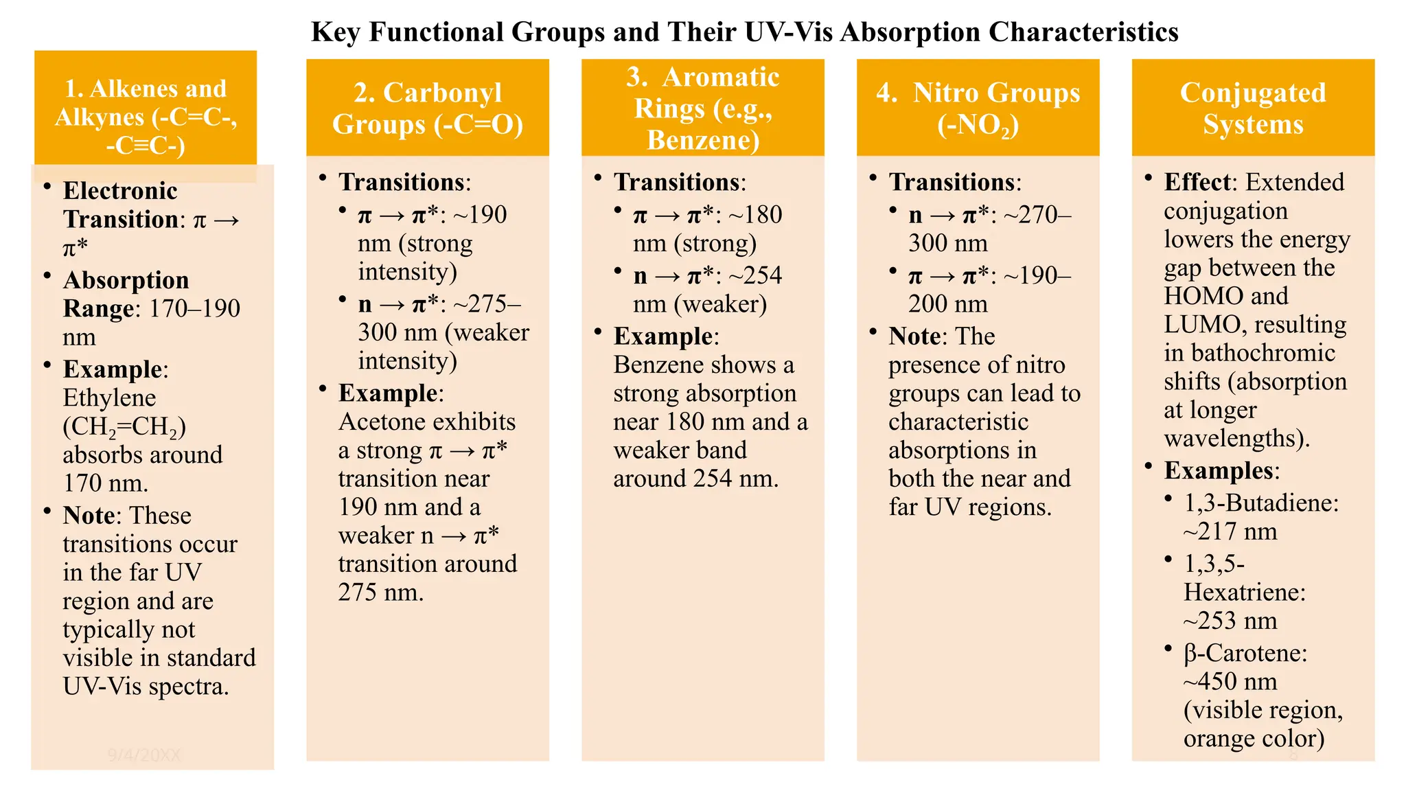
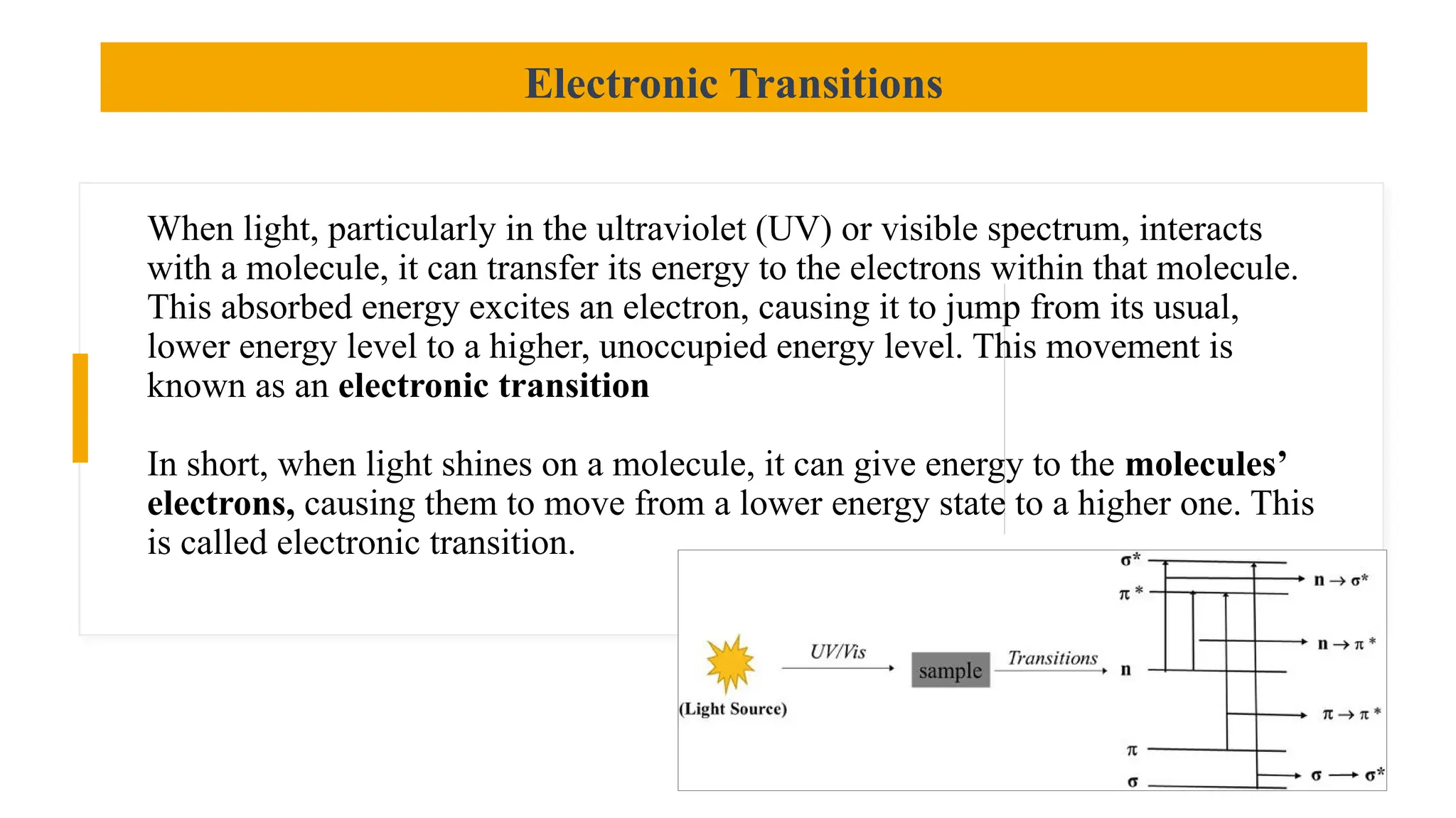
Electronic Transitions: Discover the mechanisms behind σ→σ*, π→π*, n→σ*, and n→π* transitions that form the foundation of UV-Vis spectroscopy.
Wikipedia
Chromophores & Auxochromes: Learn how functional groups absorb light and how auxochromic modifications alter intensity and wavelength for clearer spectral insights.
www.slideshare.net
Wikipedia
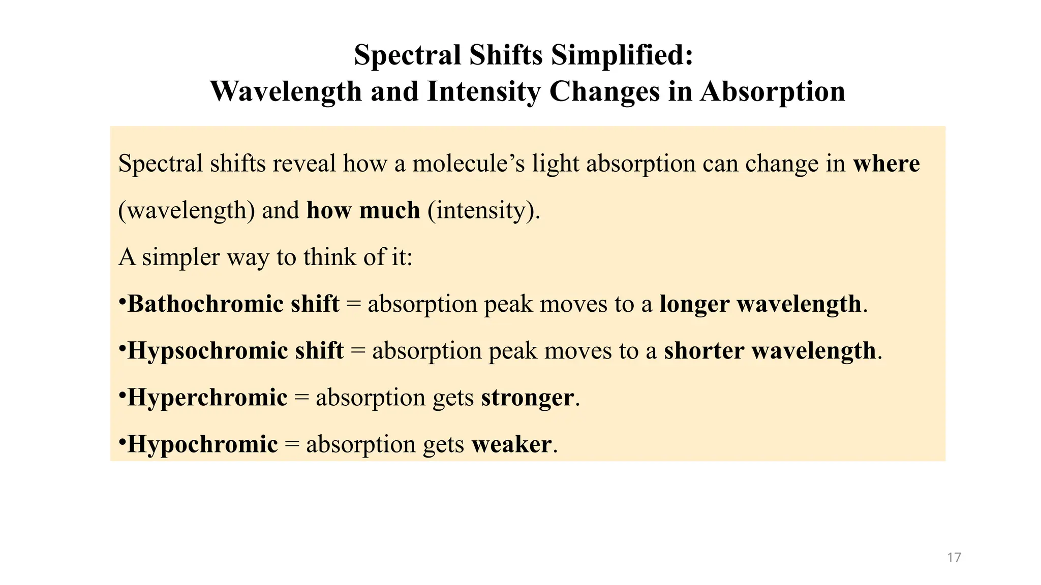
Spectral Shifts: Get to grips with bathochromic (red) and hypsochromic (blue) shifts, understanding how molecular structure and interactions influence absorption behavior.
www.slideshare.net
Solvent Effects: Examine how solvent polarity and hydrogen bonding can shift absorption peaks—crucial for accurate analysis and pharmaceutical applications. Beer–Lambert’s Law: Master this essential quantitative tool—A = ε l c—with clear derivations and discussions of real-world deviations and limitations.
Wikipedia
Why this presentation is ideal:
Clear & Concise: Each subtopic is broken down into easy-to-follow visuals and explanations—perfect for quick learning and effective revision.
Pharma-Relevant Examples: Aligned with PCI syllabus expectations and focused on drug analysis, making learning practical and exam-ready.
PCI
Highly Shareable Content: A rich, visually engaging PPT format that’s optimized for SlideShare—boosting reach among peers, educators, and future pharmacists.


![Presentation Title 6
9/4/20XX
σ → σ* Transition:
Description: An electron
moves from a sigma (σ)
bonding orbital to an
antibonding sigma (σ*)
orbital.
• Energy
Requirement: These
transitions require the
highest amount of energy
because σ bonds are
strong, and the energy gap
between σ and σ* orbitals
is large.
• Observation: Typically
observed in the vacuum
UV region (wavelengths
below 200 nm), making
them less common in
standard UV-Vis
spectroscopy. They occur
in molecules with only
single bonds [2].
n → σ* Transition:
Description: An electron
moves from a non-
bonding (n) orbital (a lone
pair) to an antibonding
sigma (σ*) orbital.
• Energy
Requirement: These
transitions require less
energy than σ → σ*
transitions.
• Observation: Found in
molecules containing
atoms with lone pairs,
such as alcohols, ethers,
amines, and alkyl halides.
These transitions can
occur in the UV
region [2].
π → π* Transition:
Description: An electron
moves from a pi (π)
bonding orbital to an
antibonding pi (π*)
orbital.
• Energy
Requirement: These
transitions require less
energy than σ → σ*
transitions but generally
more than n → σ*
transitions.
• Observation: Common
in molecules with double
or triple bonds
(unsaturated systems) like
alkenes, alkynes, and
aromatic compounds.
These transitions are
frequently observed in the
UV-Vis spectrum and are
very important for
chromophore
identification [2].
n → π* Transition:
• Description: An electron
moves from a non-
bonding (n) orbital (a lone
pair) to an antibonding pi
(π*) orbital.
• Energy
Requirement: These
transitions typically
require the least amount
of energy among the
common types.
• Observation: Occur in
molecules that possess
both lone pairs and pi
bonds, such as carbonyl
compounds (aldehydes,
ketones), nitro
compounds, and imines.
These transitions are often
observed at longer
wavelengths (lower
energy) in the UV-Vis
spectrum compared to π
→ π* transitions [2].
Electronic Transitions](https://image.slidesharecdn.com/uv-visiblespectroscopy-250810080451-fc1bbb8d/75/UV-Visible-spectroscopy-pptx-UV-Visible-Spectroscopy-Electronic-Transitions-Chromophores-Auxochromes-Spectral-Shifts-Solvent-Effects-and-Beer-Lambert-s-Law-6-2048.jpg)