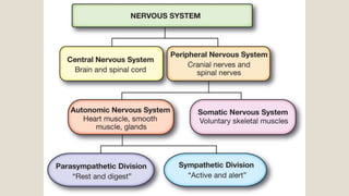
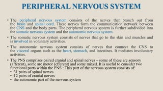
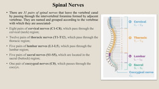
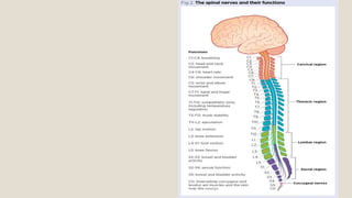
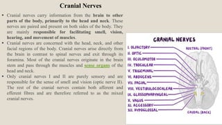
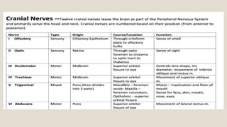
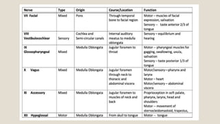
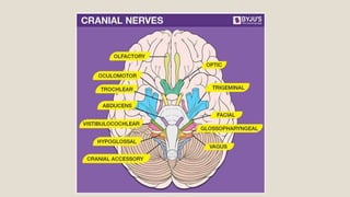
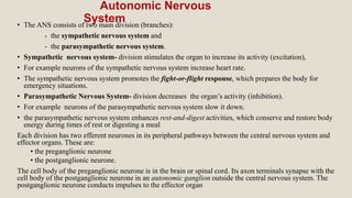
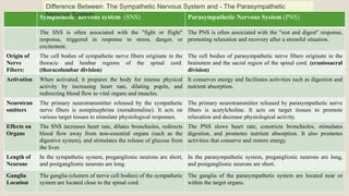
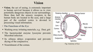
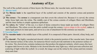
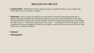
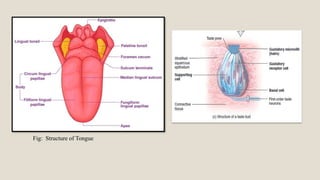
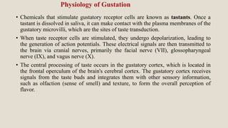
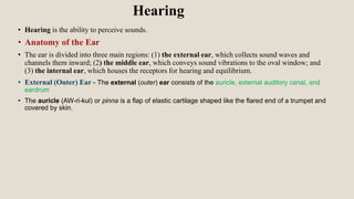
PERIPHERAL NERVOUS
SYSTEM
By- Samruddhi S. Khonde
Asst. Prof
P. R. Patil Institute of Pharmacy
Special Senses
PERIPHERAL NERVOUS SYSTEM
• The peripheral nervous system consists of the nerves that branch out from
the brain and spinal cord. These nerves form the communication network between
the CNS and the body parts. The peripheral nervous system is further subdivided into
the somatic nervous system and the autonomic nervous system.
• The somatic nervous system consists of nerves that go to the skin and muscles and
is involved in voluntary activities.
• The autonomic nervous system consists of nerves that connect the CNS to
the visceral organs such as the heart, stomach, and intestines. It mediates involuntary
activities.
• The PNS comprises paired cranial and spinal nerves – some of these are sensory
(afferent), some are motor (efferent) and some mixed. It is useful to consider two
functional parts within the PNS : This part of the nervous system consists of:
• 31 pairs of spinal nerves
• 12 pairs of cranial nerves
• the autonomic part of the nervous system
Spinal Nerves
• There are 31 pairs of spinal nerves that leave the vertebral canal
by passing through the intervertebral foramina formed by adjacent
vertebrae. They are named and grouped according to the vertebrae
with which they are associated-
• Eight pairs of cervical nerves (C1-C8), which pass through the
cervical (neck) region;
• Twelve pairs of thoracic nerves (T1-T12), which pass through the
thoracic region;
• Five pairs of lumbar nerves (L1-L5), which pass through the
lumbar region;
• Five pairs of sacral nerves (S1-S5), which are located in the
sacral (buttock) region;
• One pair of coccygeal nerves (C0), which passes through the
coccyx.
Cranial Nerves
 Cranial nerves carry information from the brain to other
parts of the body, primarily to the head and neck. These
nerves are paired and present on both sides of the body. They
are mainly responsible for facilitating smell, vision,
hearing, and movement of muscles.
 Cranial nerves are concerned with the head, neck, and other
facial regions of the body. Cranial nerves arise directly from
the brain in contrast to spinal nerves and exit through its
foramina. Most of the cranial nerves originate in the brain
stem and pass through the muscles and sense organs of the
head and neck.
 Only cranial nerves I and II are purely sensory and are
responsible for the sense of smell and vision (optic nerve II).
The rest of the cranial nerves contain both afferent and
efferent fibres and are therefore referred to as the mixed
cranial nerves.
This will help you remember the
following:
1.Only = Olfactory (CN I)
2.One = Optic (CN II)
3.Of = Oculomotor (CN III)
4.The = Trochlear (CN IV)
5.Two = Trigeminal (CN V)
6.Athletes = Abducens (CN VI)
7.Felt = Facial (CN VII)
8.Very = Vestibulocochlear (CN VIII)
9.Good = Glossopharyngeal (CN IX)
10.Victorious = Vagus (CN X)
11.And = Accessory (XI)
12.Healthy = Hypoglossal (XII)
“Only One Of The Two Athletes Felt Very Good, Victorious,
And Healthy”
Autonomic Nervous
System
• The ANS consists of two main division (branches):
- the sympathetic nervous system and
- the parasympathetic nervous system.
• Sympathetic nervous system- division stimulates the organ to increase its activity (excitation),
• For example neurons of the sympathetic nervous system increase heart rate.
• The sympathetic nervous system promotes the fight-or-flight response, which prepares the body for
emergency situations.
• Parasympathetic Nervous System- division decreases the organ’s activity (inhibition).
• For example neurons of the parasympathetic nervous system slow it down.
• the parasympathetic nervous system enhances rest-and-digest activities, which conserve and restore body
energy during times of rest or digesting a meal
Each division has two efferent neurones in its peripheral pathways between the central nervous system and
effector organs. These are:
• the preganglionic neurone
• the postganglionic neurone.
The cell body of the preganglionic neurone is in the brain or spinal cord. Its axon terminals synapse with the
cell body of the postganglionic neurone in an autonomic ganglion outside the central nervous system. The
postganglionic neurone conducts impulses to the effector organ
Sympathetic nervous system (SNS) Parasympathetic Nervous System (PNS)
The SNS is often associated with the "fight or flight"
response, triggered in response to stress, danger, or
excitement.
The PNS is often associated with the "rest and digest" response,
promoting relaxation and recovery after a stressful situation.
Origin of
Nerve
Fibers:
The cell bodies of sympathetic nerve fibers originate in the
thoracic and lumbar regions of the spinal cord.
(thoracolumbar division)
The cell bodies of parasympathetic nerve fibers originate in the
brainstem and the sacral region of the spinal cord. (craniosacral
division)
Activation When activated, it prepares the body for intense physical
activity by increasing heart rate, dilating pupils, and
redirecting blood flow to vital organs and muscles.
It conserves energy and facilitates activities such as digestion and
nutrient absorption.
Neurotran
smitters
The primary neurotransmitter released by the sympathetic
nerve fibers is norepinephrine (noradrenaline). It acts on
various target tissues to stimulate physiological responses.
The primary neurotransmitter released by parasympathetic nerve
fibers is acetylcholine. It acts on target tissues to promote
relaxation and decrease physiological activity.
Effects on
Organs
The SNS increases heart rate, dilates bronchioles, redirects
blood flow away from non-essential organs (such as the
digestive system), and stimulates the release of glucose from
the liver.
The PNS slows heart rate, constricts bronchioles, stimulates
digestion, and promotes nutrient absorption. It also promotes
activities that conserve and restore energy.
Length of
Neurons
In the sympathetic system, preganglionic neurons are short,
and postganglionic neurons are long.
In the parasympathetic system, preganglionic neurons are long,
and postganglionic neurons are short.
Ganglia
Location
The ganglia (clusters of nerve cell bodies) of the sympathetic
system are located close to the spinal cord.
The ganglia of the parasympathetic system are located near or
within the target organs.
Difference Between: The Sympathetic Nervous System and - The Parasympathetic
Nervous System
Special
senses
Vision
• Vision, the act of seeing, is extremely important
to human survival because it allows us to view
potentially dangerous objects in our surroundings.
More than half the sensory receptors in the
human body are located in the eyes, and a large
part of the cerebral cortex is devoted to
processing visual information.
• The Functions of the Eye
 Washing away irritating materials, e.g. Dust, grit
 The bacteriocidal enzyme lysozyme prevents
Microbial infection
 Its oiliness delays evaporation and prevents
drying of the conjunctiva.
 Nourishment of the cornea.
Fig: Anatomy of Eye
Anatomy of Eye
The wall of the eyeball consists of three layers: the fibrous tunic, the vascular tunic, and the retina.
 fibrous tunic
 The fibrous tunic is the superficial layer of the eyeball and consists of the anterior cornea and posterior
sclera
 The cornea - The cornea is a transparent coat that covers the colored iris. Because it is curved, the cornea
helps focus light onto the retina. The middle coat of the cornea consists of collagen fibers and fibroblasts,
and the inner surface is simple squamous epithelium.
 The sclera, the “white” of the eye, is a layer of dense connective tissue made up mostly of collagen fibers
and fibroblasts. The sclera covers the entire eyeball except the cornea; it gives shape to the eyeball, makes it
more rigid, protects its inner parts, and serves as a site of attachment for the extrinsic eye muscles.
 Vascular Tunic
 The vascular tunic is the middle layer of the eyeball. It is composed of three parts: choroid, ciliary body, and
iris.
 The choroid - The highly vascularized choroid, which is the posterior portion of the vascular tunic, lines most
of the internal surface of the sclera. Its numerous blood vessels provide nutrients to the posterior surface of
the retina. The choroid also contains melanocytes that produce the pigment melanin, which causes this layer
to appear dark brown in color. Melanin in the choroid absorbs stray light rays, which prevents reflection and
scattering of light within the eyeball. As a result, the image cast on the retina by the cornea and lens remains
sharp and clear.
 The ciliary body- The ciliary body appears dark brown in color because it contains melanin-producing
melanocytes. the ciliary body consists of ciliary processes and ciliary muscle. The ciliary processes are
protrusions or folds on the internal surface of the ciliary body. They contain blood capillaries that secrete
aqueous humor. The ciliary muscle is a circular band of smooth muscle. Contraction or relaxation of the
ciliary muscle alters the shape of the lens, adapting it for near or far vision.
 The iris, the colored portion of the eyeball, is shaped like a flattened donut. It is suspended between the
cornea and the lens and is attached at its outer margin to the ciliary processes. It consists of melanocytes
and circular and radial smooth muscle fibers. The amount of melanin in the iris determines the eye color.
 The pupil - A principal function of the iris is to regulate the amount of light entering the eyeball through
the pupil, the hole in the center of the iris. The pupil appears black because, as you look through the lens,
you see the heavily pigmented back of the eye (choroid and retina).
 The Retina - The third and inner layer of the eyeball, the retina, lines the posterior three-quarters of the
eyeball and is the beginning of the visual pathway. The retina consists of a pigmented layer and a neural
layer. The pigmented layer is a sheet of melanin-containing epithelial cells
 The optic disc is the site where the optic (II) nerve exits the eyeball, is also called the blind spot.
The macula lutea- yellow spot is in the exact center of the posterior portion of the retina, at the visual axis of
the eye
 vitreous chamber - The larger posterior cavity of the eyeball is the vitreous chamber which lies between
the lens and the retina. Within the vitreous chamber is the vitreous body, a transparent jellylike substance
that holds the retina flush against the choroid, giving the retina an even surface for the reception of clear
images. It occupies about four-fifths of the eyeball.
 Light enters the eye: Light first enters the eye through the cornea, the transparent front part of the eye.
 Pupil and Iris: The pupil, an adjustable opening, controls the amount of light entering the eye.
 The iris, the colored part of the eye, adjusts the size of the pupil based on light conditions.
 Lens Accommodation: The lens, located behind the iris, changes shape to focus the light onto the retina ,
is crucial for clear vision at different distances.
 Retina: Light focused by the lens forms an inverted image on the retina, the light-sensitive layer at the
back of the eye. The retina contains photoreceptor cells, including rods (for low-light vision) and cones
(for color and detail).
 Phototransduction: When light hits the photoreceptor cells, it triggers a chemical reaction
(phototransduction) that converts light energy into electrical signals.
 Transmission through Optic Nerve: Electrical signals generated by photoreceptor cells travel through the
optic nerve, composed of ganglion cell axons.
 Optic Tract: The optic tract carries visual information to the lateral geniculate nucleus (LGN) in the
thalamus.
 Lateral Geniculate Nucleus (LGN): The LGN processes visual information and relays it to the visual
cortex in the brain.
 Visual Cortex: The visual cortex, located in the occipital lobe, interprets the electrical signals to create a
visual perception.
 Perception and Recognition: The brain integrates visual information, allowing the perception and
recognition of objects, shapes, colors, and depth.
Physiology of Vision
DISEASES OF THE EYE
• Conjunctivitis - Inflammation of the conjunctiva may be caused by irritants, such as smoke, dust,
wind, cold or dry air, microbes or antigens.
• Glaucoma - This is a group of conditions in which there is increased intraocular pressure due to
impaired drainage of aqueous fluid through the scleral venous sinus (canal of Schlemm) in the angle
between the iris and cornea in the anterior chamber. Persistently raised intraocular pressure may damage
the optic nerve by: • mechanical compression of the axons • compression of the blood supply causing
ischaemia of the axons. Damage to the optic nerve impairs vision and the extent varies from some visual
impairment to complete blindness.
• Cataract –
• Retinopathies
Sense of Smell / Olfaction
Olfaction, or the sense of smell, is a complex physiological process that
allows organisms to detect and identify odors in their environment. The
physiology of olfaction involves the nose, olfactory receptors, and the
neural pathways that transmit olfactory information to the brain.
Anatomy of Olfaction
• Olfactory Membrane - The olfactory membrane, located in
the superior part of each nostril, folds downward along the
superior septum surface and laterally over the superior
turbinate and a small portion of the middle turbinate's upper
surface. In each nostril, the olfactory membrane has a surface
area of about 2.4 square cm.
• Olfactory Cells - The receptor cells for the smell sensation
are the olfactory cells, which are actually bipolar nerve cells
derived originally from the central nervous system itself.
There are about 100 million of these cells in the olfactory
epithelium interspersed among sustentacular cells.
• The mucosal end of the olfactory cell forms a knob from
which 4 to 25 olfactory hairs (also called olfactory cilia),
project into the mucus that coats the inner surface of the nasal
cavity, projecting into mucus, form a dense mat that reacts to
air odors, stimulating olfactory cells. Spaced among the
olfactory cells in the olfactory membrane are many small
Bowman’s glands that secrete mucus onto the surface of the
olfactory membrane.
Figure : Organization of the olfactory membrane and
olfactory bulb, and connections to the olfactory tract.
• Nasal Cavity: Olfaction begins in the nasal cavity, where specialized sensory cells detect odors, and
is facilitated by a mucous membrane that traps and dissolves odor molecules.
• Olfactory Epithelium: The olfactory epithelium, a nasal cavity tissue, houses millions of olfactory
receptor neurons (ORNs) with cilia, responsible for detecting odors.
• Olfactory Receptors: Olfactory receptors are proteins found in the cilia of olfactory receptor
neurons that can bind to specific odor molecules, initiating a signal transduction cascade.
• Signal Transduction: Odor molecules bind to olfactory receptors, triggering biochemical events
within the neuron, generating action potentials, representing the olfactory stimulus.
• Olfactory Bulb: Olfactory receptor neurons send axons to the olfactory bulb, where they synapse
with mitral and tufted cells, forming the olfactory nerve.
• Olfactory Nerve: The olfactory nerve transmits olfactory information from the bulb to the cortex,
influencing emotional responses linked to smells due to its direct connections to limbic structures.
• Olfactory Cortex: The olfactory cortex, comprising piriform and orbitofrontal regions, processes
and interprets olfactory signals, which are integrated with other sensory input and emotional
responses.
• Olfactory Discrimination: The brain can identify and discriminate various odors based on the
neural activity patterns produced in response to different odor molecules.
Physiology of Olfaction (Nose)
Anatomy of Taste
• The receptors for sensations of taste are located in the taste buds. Most of the nearly 10,000 taste buds of
a young adult are on the tongue, but some are found on the soft palate (posterior portion of the roof of
the mouth), pharynx (throat), and epiglottis (cartilage lid over voice box). The number of taste buds
declines with age.
• Each taste bud is an oval body consisting of three kinds of epithelial cells: supporting cells, gustatory
receptor cells, and basal cells.
• Taste buds are found in elevations on the tongue called papillae, which increase the surface area and
provide a rough texture to the upper surface of the tongue.Three types of papillae contain taste buds:
Circular vallate papillae (val-āt = wall-like) or circumvallate papillae form an inverted v-shaped
row at the back of the tongue. Each of these papillae houses 100–300 taste buds.
Fungiform papillae (mushroomlike) are mushroom shaped elevations scattered over the entire
surface of the tongue that contain about five taste buds each.
Foliate papillae (leaflike) are located in small trenches on the lateral margins of the tongue, but
most of their taste buds degenerate in early childhood.
The entire surface of the tongue has filiform papillae (threadlike). These pointed, threadlike
structures contain tactile receptors but no taste buds. They increase friction between the tongue and
food, making it easier for the tongue to move food in the oral cavity.
Fig: Structure of Tongue
Physiology of Gustation
• Chemicals that stimulate gustatory receptor cells are known as tastants. Once a
tastant is dissolved in saliva, it can make contact with the plasma membranes of the
gustatory microvilli, which are the sites of taste transduction.
• When taste receptor cells are stimulated, they undergo depolarization, leading to
the generation of action potentials. These electrical signals are then transmitted to
the brain via cranial nerves, primarily the facial nerve (VII), glossopharyngeal
nerve (IX), and vagus nerve (X).
• The central processing of taste occurs in the gustatory cortex, which is located in
the frontal operculum of the brain's cerebral cortex. The gustatory cortex receives
signals from the taste buds and integrates them with other sensory information,
such as olfaction (sense of smell) and texture, to form the overall perception of
flavor.
Hearing
• Hearing is the ability to perceive sounds.
• Anatomy of the Ear
• The ear is divided into three main regions: (1) the external ear, which collects sound waves and
channels them inward; (2) the middle ear, which conveys sound vibrations to the oval window; and
(3) the internal ear, which houses the receptors for hearing and equilibrium.
• External (Outer) Ear - The external (outer) ear consists of the auricle, external auditory canal, and
eardrum
• The auricle (AW-ri-kul) or pinna is a flap of elastic cartilage shaped like the flared end of a trumpet and
covered by skin.
Thank you

PERIPHERAL NERVOUS SYSTEM.pptx

  • 1.
    PERIPHERAL NERVOUS SYSTEM By- SamruddhiS. Khonde Asst. Prof P. R. Patil Institute of Pharmacy Special Senses
  • 3.
    PERIPHERAL NERVOUS SYSTEM •The peripheral nervous system consists of the nerves that branch out from the brain and spinal cord. These nerves form the communication network between the CNS and the body parts. The peripheral nervous system is further subdivided into the somatic nervous system and the autonomic nervous system. • The somatic nervous system consists of nerves that go to the skin and muscles and is involved in voluntary activities. • The autonomic nervous system consists of nerves that connect the CNS to the visceral organs such as the heart, stomach, and intestines. It mediates involuntary activities. • The PNS comprises paired cranial and spinal nerves – some of these are sensory (afferent), some are motor (efferent) and some mixed. It is useful to consider two functional parts within the PNS : This part of the nervous system consists of: • 31 pairs of spinal nerves • 12 pairs of cranial nerves • the autonomic part of the nervous system
  • 4.
    Spinal Nerves • Thereare 31 pairs of spinal nerves that leave the vertebral canal by passing through the intervertebral foramina formed by adjacent vertebrae. They are named and grouped according to the vertebrae with which they are associated- • Eight pairs of cervical nerves (C1-C8), which pass through the cervical (neck) region; • Twelve pairs of thoracic nerves (T1-T12), which pass through the thoracic region; • Five pairs of lumbar nerves (L1-L5), which pass through the lumbar region; • Five pairs of sacral nerves (S1-S5), which are located in the sacral (buttock) region; • One pair of coccygeal nerves (C0), which passes through the coccyx.
  • 6.
    Cranial Nerves  Cranialnerves carry information from the brain to other parts of the body, primarily to the head and neck. These nerves are paired and present on both sides of the body. They are mainly responsible for facilitating smell, vision, hearing, and movement of muscles.  Cranial nerves are concerned with the head, neck, and other facial regions of the body. Cranial nerves arise directly from the brain in contrast to spinal nerves and exit through its foramina. Most of the cranial nerves originate in the brain stem and pass through the muscles and sense organs of the head and neck.  Only cranial nerves I and II are purely sensory and are responsible for the sense of smell and vision (optic nerve II). The rest of the cranial nerves contain both afferent and efferent fibres and are therefore referred to as the mixed cranial nerves.
  • 10.
    This will helpyou remember the following: 1.Only = Olfactory (CN I) 2.One = Optic (CN II) 3.Of = Oculomotor (CN III) 4.The = Trochlear (CN IV) 5.Two = Trigeminal (CN V) 6.Athletes = Abducens (CN VI) 7.Felt = Facial (CN VII) 8.Very = Vestibulocochlear (CN VIII) 9.Good = Glossopharyngeal (CN IX) 10.Victorious = Vagus (CN X) 11.And = Accessory (XI) 12.Healthy = Hypoglossal (XII) “Only One Of The Two Athletes Felt Very Good, Victorious, And Healthy”
  • 11.
    Autonomic Nervous System • TheANS consists of two main division (branches): - the sympathetic nervous system and - the parasympathetic nervous system. • Sympathetic nervous system- division stimulates the organ to increase its activity (excitation), • For example neurons of the sympathetic nervous system increase heart rate. • The sympathetic nervous system promotes the fight-or-flight response, which prepares the body for emergency situations. • Parasympathetic Nervous System- division decreases the organ’s activity (inhibition). • For example neurons of the parasympathetic nervous system slow it down. • the parasympathetic nervous system enhances rest-and-digest activities, which conserve and restore body energy during times of rest or digesting a meal Each division has two efferent neurones in its peripheral pathways between the central nervous system and effector organs. These are: • the preganglionic neurone • the postganglionic neurone. The cell body of the preganglionic neurone is in the brain or spinal cord. Its axon terminals synapse with the cell body of the postganglionic neurone in an autonomic ganglion outside the central nervous system. The postganglionic neurone conducts impulses to the effector organ
  • 12.
    Sympathetic nervous system(SNS) Parasympathetic Nervous System (PNS) The SNS is often associated with the "fight or flight" response, triggered in response to stress, danger, or excitement. The PNS is often associated with the "rest and digest" response, promoting relaxation and recovery after a stressful situation. Origin of Nerve Fibers: The cell bodies of sympathetic nerve fibers originate in the thoracic and lumbar regions of the spinal cord. (thoracolumbar division) The cell bodies of parasympathetic nerve fibers originate in the brainstem and the sacral region of the spinal cord. (craniosacral division) Activation When activated, it prepares the body for intense physical activity by increasing heart rate, dilating pupils, and redirecting blood flow to vital organs and muscles. It conserves energy and facilitates activities such as digestion and nutrient absorption. Neurotran smitters The primary neurotransmitter released by the sympathetic nerve fibers is norepinephrine (noradrenaline). It acts on various target tissues to stimulate physiological responses. The primary neurotransmitter released by parasympathetic nerve fibers is acetylcholine. It acts on target tissues to promote relaxation and decrease physiological activity. Effects on Organs The SNS increases heart rate, dilates bronchioles, redirects blood flow away from non-essential organs (such as the digestive system), and stimulates the release of glucose from the liver. The PNS slows heart rate, constricts bronchioles, stimulates digestion, and promotes nutrient absorption. It also promotes activities that conserve and restore energy. Length of Neurons In the sympathetic system, preganglionic neurons are short, and postganglionic neurons are long. In the parasympathetic system, preganglionic neurons are long, and postganglionic neurons are short. Ganglia Location The ganglia (clusters of nerve cell bodies) of the sympathetic system are located close to the spinal cord. The ganglia of the parasympathetic system are located near or within the target organs. Difference Between: The Sympathetic Nervous System and - The Parasympathetic Nervous System
  • 13.
  • 14.
    Vision • Vision, theact of seeing, is extremely important to human survival because it allows us to view potentially dangerous objects in our surroundings. More than half the sensory receptors in the human body are located in the eyes, and a large part of the cerebral cortex is devoted to processing visual information. • The Functions of the Eye  Washing away irritating materials, e.g. Dust, grit  The bacteriocidal enzyme lysozyme prevents Microbial infection  Its oiliness delays evaporation and prevents drying of the conjunctiva.  Nourishment of the cornea. Fig: Anatomy of Eye
  • 15.
    Anatomy of Eye Thewall of the eyeball consists of three layers: the fibrous tunic, the vascular tunic, and the retina.  fibrous tunic  The fibrous tunic is the superficial layer of the eyeball and consists of the anterior cornea and posterior sclera  The cornea - The cornea is a transparent coat that covers the colored iris. Because it is curved, the cornea helps focus light onto the retina. The middle coat of the cornea consists of collagen fibers and fibroblasts, and the inner surface is simple squamous epithelium.  The sclera, the “white” of the eye, is a layer of dense connective tissue made up mostly of collagen fibers and fibroblasts. The sclera covers the entire eyeball except the cornea; it gives shape to the eyeball, makes it more rigid, protects its inner parts, and serves as a site of attachment for the extrinsic eye muscles.  Vascular Tunic  The vascular tunic is the middle layer of the eyeball. It is composed of three parts: choroid, ciliary body, and iris.  The choroid - The highly vascularized choroid, which is the posterior portion of the vascular tunic, lines most of the internal surface of the sclera. Its numerous blood vessels provide nutrients to the posterior surface of the retina. The choroid also contains melanocytes that produce the pigment melanin, which causes this layer to appear dark brown in color. Melanin in the choroid absorbs stray light rays, which prevents reflection and scattering of light within the eyeball. As a result, the image cast on the retina by the cornea and lens remains sharp and clear.
  • 16.
     The ciliarybody- The ciliary body appears dark brown in color because it contains melanin-producing melanocytes. the ciliary body consists of ciliary processes and ciliary muscle. The ciliary processes are protrusions or folds on the internal surface of the ciliary body. They contain blood capillaries that secrete aqueous humor. The ciliary muscle is a circular band of smooth muscle. Contraction or relaxation of the ciliary muscle alters the shape of the lens, adapting it for near or far vision.  The iris, the colored portion of the eyeball, is shaped like a flattened donut. It is suspended between the cornea and the lens and is attached at its outer margin to the ciliary processes. It consists of melanocytes and circular and radial smooth muscle fibers. The amount of melanin in the iris determines the eye color.  The pupil - A principal function of the iris is to regulate the amount of light entering the eyeball through the pupil, the hole in the center of the iris. The pupil appears black because, as you look through the lens, you see the heavily pigmented back of the eye (choroid and retina).  The Retina - The third and inner layer of the eyeball, the retina, lines the posterior three-quarters of the eyeball and is the beginning of the visual pathway. The retina consists of a pigmented layer and a neural layer. The pigmented layer is a sheet of melanin-containing epithelial cells  The optic disc is the site where the optic (II) nerve exits the eyeball, is also called the blind spot. The macula lutea- yellow spot is in the exact center of the posterior portion of the retina, at the visual axis of the eye  vitreous chamber - The larger posterior cavity of the eyeball is the vitreous chamber which lies between the lens and the retina. Within the vitreous chamber is the vitreous body, a transparent jellylike substance that holds the retina flush against the choroid, giving the retina an even surface for the reception of clear images. It occupies about four-fifths of the eyeball.
  • 17.
     Light entersthe eye: Light first enters the eye through the cornea, the transparent front part of the eye.  Pupil and Iris: The pupil, an adjustable opening, controls the amount of light entering the eye.  The iris, the colored part of the eye, adjusts the size of the pupil based on light conditions.  Lens Accommodation: The lens, located behind the iris, changes shape to focus the light onto the retina , is crucial for clear vision at different distances.  Retina: Light focused by the lens forms an inverted image on the retina, the light-sensitive layer at the back of the eye. The retina contains photoreceptor cells, including rods (for low-light vision) and cones (for color and detail).  Phototransduction: When light hits the photoreceptor cells, it triggers a chemical reaction (phototransduction) that converts light energy into electrical signals.  Transmission through Optic Nerve: Electrical signals generated by photoreceptor cells travel through the optic nerve, composed of ganglion cell axons.  Optic Tract: The optic tract carries visual information to the lateral geniculate nucleus (LGN) in the thalamus.  Lateral Geniculate Nucleus (LGN): The LGN processes visual information and relays it to the visual cortex in the brain.  Visual Cortex: The visual cortex, located in the occipital lobe, interprets the electrical signals to create a visual perception.  Perception and Recognition: The brain integrates visual information, allowing the perception and recognition of objects, shapes, colors, and depth. Physiology of Vision
  • 18.
    DISEASES OF THEEYE • Conjunctivitis - Inflammation of the conjunctiva may be caused by irritants, such as smoke, dust, wind, cold or dry air, microbes or antigens. • Glaucoma - This is a group of conditions in which there is increased intraocular pressure due to impaired drainage of aqueous fluid through the scleral venous sinus (canal of Schlemm) in the angle between the iris and cornea in the anterior chamber. Persistently raised intraocular pressure may damage the optic nerve by: • mechanical compression of the axons • compression of the blood supply causing ischaemia of the axons. Damage to the optic nerve impairs vision and the extent varies from some visual impairment to complete blindness. • Cataract – • Retinopathies
  • 19.
    Sense of Smell/ Olfaction Olfaction, or the sense of smell, is a complex physiological process that allows organisms to detect and identify odors in their environment. The physiology of olfaction involves the nose, olfactory receptors, and the neural pathways that transmit olfactory information to the brain.
  • 20.
    Anatomy of Olfaction •Olfactory Membrane - The olfactory membrane, located in the superior part of each nostril, folds downward along the superior septum surface and laterally over the superior turbinate and a small portion of the middle turbinate's upper surface. In each nostril, the olfactory membrane has a surface area of about 2.4 square cm. • Olfactory Cells - The receptor cells for the smell sensation are the olfactory cells, which are actually bipolar nerve cells derived originally from the central nervous system itself. There are about 100 million of these cells in the olfactory epithelium interspersed among sustentacular cells. • The mucosal end of the olfactory cell forms a knob from which 4 to 25 olfactory hairs (also called olfactory cilia), project into the mucus that coats the inner surface of the nasal cavity, projecting into mucus, form a dense mat that reacts to air odors, stimulating olfactory cells. Spaced among the olfactory cells in the olfactory membrane are many small Bowman’s glands that secrete mucus onto the surface of the olfactory membrane. Figure : Organization of the olfactory membrane and olfactory bulb, and connections to the olfactory tract.
  • 21.
    • Nasal Cavity:Olfaction begins in the nasal cavity, where specialized sensory cells detect odors, and is facilitated by a mucous membrane that traps and dissolves odor molecules. • Olfactory Epithelium: The olfactory epithelium, a nasal cavity tissue, houses millions of olfactory receptor neurons (ORNs) with cilia, responsible for detecting odors. • Olfactory Receptors: Olfactory receptors are proteins found in the cilia of olfactory receptor neurons that can bind to specific odor molecules, initiating a signal transduction cascade. • Signal Transduction: Odor molecules bind to olfactory receptors, triggering biochemical events within the neuron, generating action potentials, representing the olfactory stimulus. • Olfactory Bulb: Olfactory receptor neurons send axons to the olfactory bulb, where they synapse with mitral and tufted cells, forming the olfactory nerve. • Olfactory Nerve: The olfactory nerve transmits olfactory information from the bulb to the cortex, influencing emotional responses linked to smells due to its direct connections to limbic structures. • Olfactory Cortex: The olfactory cortex, comprising piriform and orbitofrontal regions, processes and interprets olfactory signals, which are integrated with other sensory input and emotional responses. • Olfactory Discrimination: The brain can identify and discriminate various odors based on the neural activity patterns produced in response to different odor molecules. Physiology of Olfaction (Nose)
  • 22.
    Anatomy of Taste •The receptors for sensations of taste are located in the taste buds. Most of the nearly 10,000 taste buds of a young adult are on the tongue, but some are found on the soft palate (posterior portion of the roof of the mouth), pharynx (throat), and epiglottis (cartilage lid over voice box). The number of taste buds declines with age. • Each taste bud is an oval body consisting of three kinds of epithelial cells: supporting cells, gustatory receptor cells, and basal cells. • Taste buds are found in elevations on the tongue called papillae, which increase the surface area and provide a rough texture to the upper surface of the tongue.Three types of papillae contain taste buds: Circular vallate papillae (val-āt = wall-like) or circumvallate papillae form an inverted v-shaped row at the back of the tongue. Each of these papillae houses 100–300 taste buds. Fungiform papillae (mushroomlike) are mushroom shaped elevations scattered over the entire surface of the tongue that contain about five taste buds each. Foliate papillae (leaflike) are located in small trenches on the lateral margins of the tongue, but most of their taste buds degenerate in early childhood. The entire surface of the tongue has filiform papillae (threadlike). These pointed, threadlike structures contain tactile receptors but no taste buds. They increase friction between the tongue and food, making it easier for the tongue to move food in the oral cavity.
  • 23.
  • 24.
    Physiology of Gustation •Chemicals that stimulate gustatory receptor cells are known as tastants. Once a tastant is dissolved in saliva, it can make contact with the plasma membranes of the gustatory microvilli, which are the sites of taste transduction. • When taste receptor cells are stimulated, they undergo depolarization, leading to the generation of action potentials. These electrical signals are then transmitted to the brain via cranial nerves, primarily the facial nerve (VII), glossopharyngeal nerve (IX), and vagus nerve (X). • The central processing of taste occurs in the gustatory cortex, which is located in the frontal operculum of the brain's cerebral cortex. The gustatory cortex receives signals from the taste buds and integrates them with other sensory information, such as olfaction (sense of smell) and texture, to form the overall perception of flavor.
  • 25.
    Hearing • Hearing isthe ability to perceive sounds. • Anatomy of the Ear • The ear is divided into three main regions: (1) the external ear, which collects sound waves and channels them inward; (2) the middle ear, which conveys sound vibrations to the oval window; and (3) the internal ear, which houses the receptors for hearing and equilibrium. • External (Outer) Ear - The external (outer) ear consists of the auricle, external auditory canal, and eardrum • The auricle (AW-ri-kul) or pinna is a flap of elastic cartilage shaped like the flared end of a trumpet and covered by skin.
  • 26.