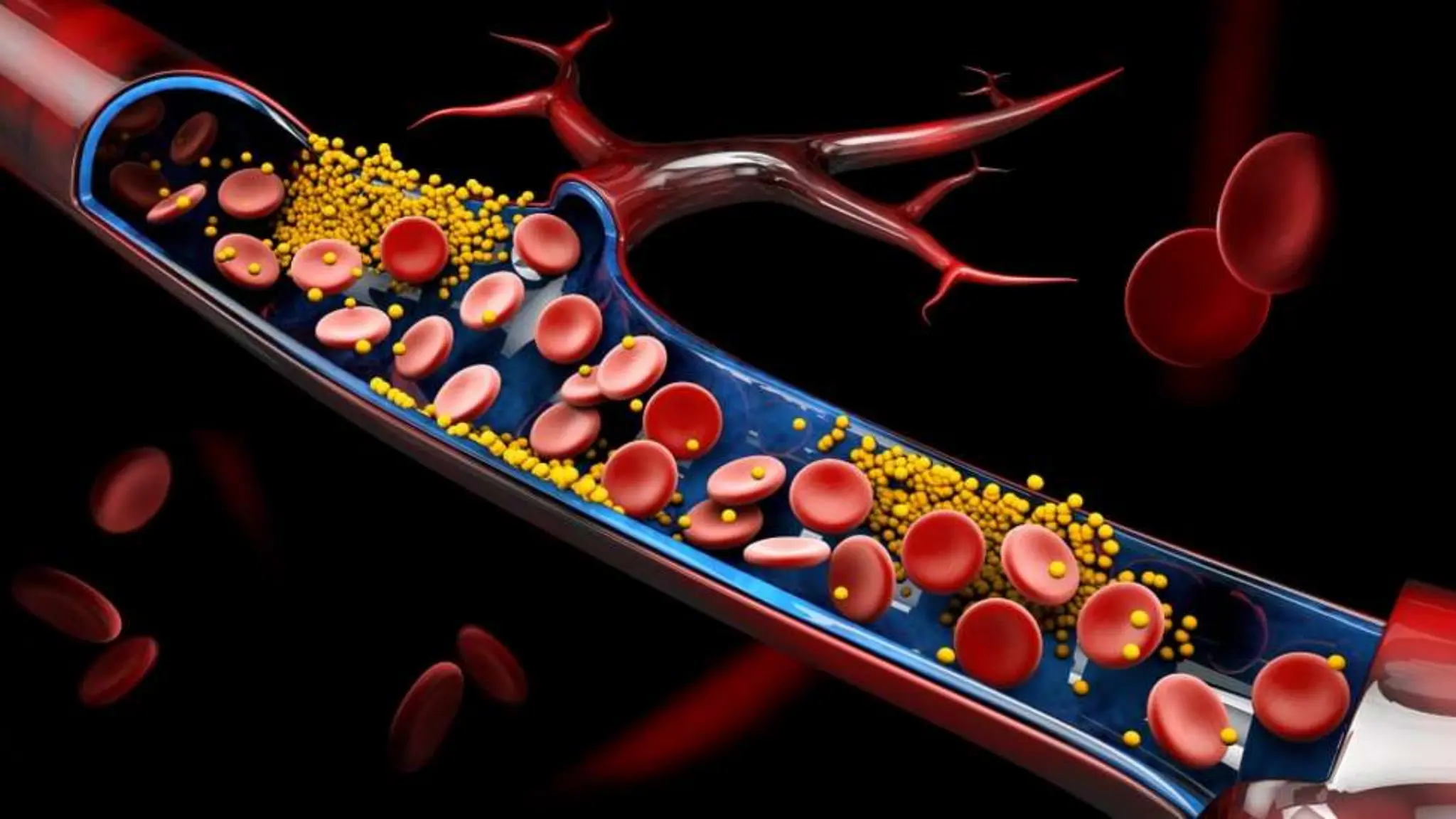
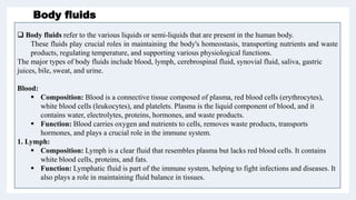
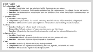
Body fluids and
blood
By- Samruddhi S. Khonde
Asst. Prof
P. R. Patil Institute of Pharmacy. Talegaon
UNIT III
 Body fluids refer to the various liquids or semi-liquids that are present in the human body.
These fluids play crucial roles in maintaining the body's homeostasis, transporting nutrients and waste
products, regulating temperature, and supporting various physiological functions.
The major types of body fluids include blood, lymph, cerebrospinal fluid, synovial fluid, saliva, gastric
juices, bile, sweat, and urine.
Blood:
 Composition: Blood is a connective tissue composed of plasma, red blood cells (erythrocytes),
white blood cells (leukocytes), and platelets. Plasma is the liquid component of blood, and it
contains water, electrolytes, proteins, hormones, and waste products.
 Function: Blood carries oxygen and nutrients to cells, removes waste products, transports
hormones, and plays a crucial role in the immune system.
1. Lymph:
 Composition: Lymph is a clear fluid that resembles plasma but lacks red blood cells. It contains
white blood cells, proteins, and fats.
 Function: Lymphatic fluid is part of the immune system, helping to fight infections and diseases. It
also plays a role in maintaining fluid balance in tissues.
Body fluids
BODY FLUIDS
1.Cerebrospinal Fluid:
 Location: Found in the brain and spinal cord within the central nervous system.
 Composition: Cerebrospinal fluid is a clear, colorless fluid that contains water, electrolytes, glucose, and proteins.
 Function: It acts as a cushion, protecting the brain and spinal cord from injury. It also helps transport nutrients and
waste products.
2.Synovial Fluid:
 Location: Found in joints.
 Composition: Synovial fluid is a viscous, lubricating fluid that contains water, electrolytes, and proteins.
 Function: It lubricates the joints, reducing friction between bones and facilitating smooth movement.
3.Saliva:
 Location: Produced by salivary glands in the mouth.
 Composition: Saliva is a mixture of water, enzymes, mucus, and electrolytes.
 Function: It helps in the digestion of food, moistens the mouth, and has antimicrobial properties.
4.Gastric Juices:
 Location: Found in the stomach.
 Composition: Gastric juices contain hydrochloric acid, enzymes, mucus, and water.
 Function: These fluids aid in the digestion of food in the stomach.
5.Bile:
 Location: Produced by the liver and stored in the gallbladder.
 Composition: Bile is a digestive fluid containing bile salts, pigments, cholesterol, and water.
 Function: Bile aids in the digestion and absorption of fats.
Blood – it’s Composition and Functions
• Blood may be described as a specialized liquid connective tissue that consists of cells surrounded by
a liquid extracellular matrix. The extracellular matrix is called blood plasma, and it suspends various
cells and cell fragments
• The specific gravity of whole blood varies from 1.055 to 1.060. The freshly shed, blood is red, thick,
opaque and slightly alkaline.
• Blood is denser and more viscous (thicker) than water and feels slightly sticky.
• The temperature of blood is 38°C (100.4°F), about 1°C higher than oral or rectal body temperature,
and it has a slightly alkaline pH ranging from 7.35 to 7.45 (average = 7.4).
• The color of blood varies with its oxygen content. When saturated with oxygen, it is bright red.
When unsaturated with oxygen, it is dark red.
• Blood constitutes about 20% of extracellular fluid, amounting to 8% of the total body mass. The
blood volume is 5 to 6 liters (1.5 gal) in an average-sized adult male and 4 to 5 liters (1.2 gal) in an
average-sized adult female.
• The gender difference in volume is due to differences in body size.
FUNCTIONS OF BLOOD
Blood has three general functions:
• 1. Transportation. Blood carries carbon dioxide from body cells back to the lungs for expiration and oxygen
from the lungs to the body's cells. It transports hormones from endocrine glands to other body cells and
nutrients from the digestive system to other body cells. Additionally, blood carries heat and waste materials to
different organs so they can be expelled from the body.
• 2. Regulation. All bodily fluids are kept in a state of equilibrium when blood is flowing. The water in blood
plasma has cooling and heat-absorbing qualities, and its varying rate of flow through the skin—where extra
heat can escape from the body—helps regulate body temperature as well. Additionally, the water content of
cells is influenced by blood osmotic pressure, primarily through interactions of dissolved.
• 3. Protection. Blood can clot (become gel-like), which protects against its excessive loss from the
cardiovascular system after an injury. In addition, its white blood cells protect against disease by carrying on
phagocytosis. Several types of blood proteins, including antibodies, interferons, and complement, help protect
against disease in a variety of ways.
COMPOSITION OF BLOOD
• Blood is a highly complex fluid which is composed of two parts-a liquid, called the plasma and
different types of cells which remain suspended in the plasma. The cells are called the blood
corpuscles. The plasma constitutes about 55%, and the cells about 45% of the total volume of human
blood.
• The blood is made up of more than 10 different cell types. Each of these cell types falls into one of
three broad categories: 1. Red blood cells (erythrocytes) 2. White blood cells (leukocytes) 3.
Platelets (thrombocytes)
The general composition of the whole blood is as follows-
• Whole blood cells: (a) Red blood corpuscles or erythrocytes (RBC).
(b) White blood corpuscles or leucocytes (WBC).
(c) Platelets or thrombocytes.
• Plasma: (a) Water, 91 to 92%, (b) Solids, 8 to 9%.
• Inorganic constituents: 0.9% Sodium, Potassium, Calcium, Magnésium, Phosphorus, Iron, Copper.
• Organic constituents
I. Proteins: 7.5% serum albumin, serum globulin, fibrinogen, prothrombin, etc.
II. Non-protein nitrogenous (NPN) substances: Urea, uric acid, xanthine, hypoxanthine, creatine,
creatinine, ammonia, amino acids, etc.
III. Carbohydrate: Glucose, etc.
IV. Fats: Neutral fat, phospholipid, cholesterol, cholesterides, etc.
V. Other substances: Internal secretions, antibodies and various enzymes (amylases, proteases, lipases,
phosphatases, etc.).
VI. Colouring matter: The yellow colour of plasma is due to small amounts of bilirubin, carotene and
xanthophyllin.
1. Red blood cells (erythrocytes)
• Erythrocytes are small, biconcave disc-shaped cells without a nucleus in humans
(nucleated in some animals). The biconcave shape provides a large surface area for the
efficient exchange of gases and allows the cells to be flexible, facilitating their
movement through narrow capillaries.
• The primary function of red blood cells is to transport oxygen, and they achieve this
through the presence of hemoglobin, a protein that binds to oxygen in the lungs and
releases it in the tissues. Hemoglobin also plays a role in carrying some carbon dioxide
back to the lungs.
• Erythrocytes are produced in the bone marrow through a process called
erythropoiesis. The production is regulated by the hormone erythropoietin, which is
released by the kidneys in response to low oxygen levels in the blood.
• Red blood cells have a finite lifespan, typically around 120 days in circulation. After this
period, they are removed from the bloodstream by the spleen and liver, and new red
blood cells are continually produced to replace them.
• The characteristic red color of blood comes from the iron-containing hemoglobin
molecules within red blood cells.
• Erythropoiesis begins in the bone marrow, where hematopoietic
stem cells differentiate into committed progenitor cells called
erythroblasts.
Stem Cell Stage:
• Erythroblasts undergo several stages of maturation. The
proerythroblast is the earliest recognizable stage committed to
becoming a red blood cell.
Proerythroblast Stage:
• The proerythroblast transforms into an early erythroblast, which
then undergoes a series of divisions, producing a population of
cells that commit to the erythroid lineage.
Early Erythroblast Stage:
• Late erythroblasts further mature, reducing their size and nucleus,
and begin to produce hemoglobin.
Late Erythroblast Stage:
• The late-stage erythroblasts eventually lose their nuclei, becoming
reticulocytes. These reticulocytes are still immature but can enter
circulation.
Reticulocyte Formation:
• Reticulocytes circulate in the bloodstream for about 1-2 days,
during which they lose residual organelles and complete their
maturation into fully functional erythrocytes.
Maturation in Circulation:
Erythropoiesis (Development)
Lifespan of
Erythrocytes
Circulation:
• Once fully mature, erythrocytes circulate in the bloodstream for an average of 120 days.
Removal from Circulation:
• As erythrocytes age, they become more fragile, and their membranes become less
flexible. This can lead to their removal from circulation, especially as they traverse
narrow capillaries.
Macrophage Phagocytosis:
• Aged or damaged erythrocytes are recognized and engulfed by macrophages, particularly in the spleen and
liver.
Hemoglobin Recycling:
• Once engulfed, hemoglobin is broken down, and its components (heme and globin) are recycled. Iron from
heme is salvaged and reused, while the globin portion is broken down into amino acids.
Bilirubin Production:
• The breakdown of heme also produces bilirubin, a yellow pigment. Bilirubin is eventually excreted by the
liver into bile, and then it passes out of the body in feces, giving stool its characteristic brown color.
White blood corpuscles or leucocytes (WBC).
• White blood cells, also known as leukocytes, are an essential component of the immune system.
Their primary function is to defend the body against infectious diseases and foreign substances.
These cells play a crucial role in identifying and eliminating pathogens, such as bacteria, viruses, and
fungi, through various mechanisms like phagocytosis or the production of antibodies. Additionally,
white blood cells also contribute to the regulation of inflammation and help in repairing
damaged tissues.
• White blood cells, or leukocytes, are vital for the proper functioning of the immune system. They act
as the body's defense mechanism by detecting and destroying harmful pathogens, preventing the
spread of infection. Without white blood cells, the body would be more susceptible to diseases and
unable to effectively fight off infections.
• There are several types of white blood cells, each with their own specific functions in the immune
system. These include
neutrophils, which are the most abundant and quickly respond to infections, as well as
lymphocytes, which play a key role in recognizing and destroying infected cells.
monocytes help to engulf and remove dead or damaged cells,
eosinophils and basophils are involved in allergic reactions and inflammation.
 Neutrophils are the most abundant type of white blood cell and are responsible for quickly responding
to infections. They are often the first cells to arrive at the site of an infection and release chemicals that
help to kill bacteria. Neutrophils have a short lifespan and are constantly being produced in the bone
marrow to replenish their numbers.
Monocytes are large white blood cells that can differentiate into macrophages, are the most abundant
type of white blood cell and are responsible for quickly responding to infections. They are often the first
cells to arrive at the site of an infection and release chemicals that help to kill bacteria. Neutrophils have a
short lifespan and are constantly being produced in the bone marrow to replenish their numbers.
Eosinophils primarily function in dealing with parasitic infections and allergic reactions. Eosinophils
have a bilobed nucleus and cytoplasmic granules that stain with acidic dyes, giving them a distinctive red
or pink color. The granules in eosinophils contain enzymes and proteins, such as major basic protein,
eosinophil peroxidase, and eosinophil cationic protein.
• Role in Immune Response:
• Parasitic Infections: Eosinophils are particularly effective against parasitic infections, as they
release toxic substances that can destroy the parasites.
• Allergic Reactions: Eosinophils are also involved in allergic responses, especially in the late phase.
They release substances that modulate the inflammatory response.
 Basophils:
• Basophils are involved in the inflammatory response and play a role in allergic
reactions. Basophils have a bilobed nucleus, but their cytoplasmic granules stain
with basic dyes, giving them a deep blue or purple colour. Basophil granules contain
histamine, heparin, and various other chemical mediators.
• Role in Immune Response:
• Inflammation: Basophils release histamine and other substances that promote
inflammation. Histamine increases blood vessel permeability, allowing immune cells to
reach the site of infection or injury.
• Allergic Reactions: Basophils are involved in immediate hypersensitivity reactions. When
triggered by an allergen, they release histamine, contributing to symptoms such as
itching and swelling.
Lymphocytes are another type of white blood cell that are crucial for the immune response.
There are two main types of lymphocytes: B cells and T cells. B cells produce antibodies that
can neutralize pathogens, while T cells directly attack infected cells. These cells work together
to provide long-term immunity against specific pathogens. The development and maturation of
T cells takes place in the thymus, while B cells mature in the bone marrow. Once matured,
both types of lymphocytes circulate throughout the body, constantly scanning for foreign
invaders. - T cells play a crucial role in cell-mediated immunity by recognizing and destroying
infected or abnormal cells, such as cancer cells.
• T cells mature in the thymus, a small organ located near the heart. There are several subtypes
of T cells, each with distinct functions. Some of the key types include:
• Helper T Cells: These cells assist other immune cells by releasing signals (cytokines) that
activate B cells and cytotoxic T cells. They play a central role in coordinating the immune
response.
• Cytotoxic T Cells: These cells directly attack and destroy infected or abnormal cells, such
as cells infected by viruses or cancer cells.
• Regulatory T Cells: These cells help regulate the immune response, preventing excessive
reactions that could harm the body's own tissues.
 B Cells (B Lymphocytes):
1. B cells mature in the bone marrow.
2. When B cells encounter specific pathogens, they can differentiate into plasma
cells, which produce antibodies. Antibodies are proteins that can bind to and
neutralize pathogens, marking them for destruction by other immune cells.
 Natural Killer (NK) Cells:
1. NK cells are part of the innate immune system and do not require prior
exposure to specific pathogens.
2. They are responsible for recognizing and destroying infected or abnormal cells,
such as those infected by viruses or cancer cells.
Platelets (thrombocytes)
• Platelets, also known as thrombocytes, are small, disc-shaped cell fragments found in the blood. They
play a crucial role in the process of blood clotting, which is essential for preventing excessive bleeding
when a blood vessel is injured. Platelets are one of the three main types of blood cells, along with red
blood cells and white blood cells.
• Platelets are formed in the bone marrow through a process called thrombopoiesis. Megakaryocytes,
which are large cells found in the bone marrow, undergo fragmentation to produce platelets. A single
megakaryocyte can give rise to thousands of platelets.
• Platelets are tiny cell fragments, much smaller than red or white blood cells, with a diameter ranging
from 1 to 3 micrometers. They lack a nucleus but contain cellular organelles such as mitochondria and
granules. The granules in platelets contain various substances that are released during the clotting
process.
Function: The primary function of platelets is to prevent bleeding by forming blood clots. When a blood
vessel is injured, platelets adhere to the site of injury and become activated. Activated platelets release
chemicals that make nearby platelets sticky, causing them to adhere to each other and the damaged area
of the blood vessel. This forms a temporary plug that helps to seal the breach in the vessel wall.
HEMOPOEISIS
• Simply, hematopoiesis is the process through which the body manufactures blood cells. It begins early in
the development of an embryo, well before birth, and continues for the life of an individual. It occurs
within the hematopoietic system, which includes organs and tissues such as the bone marrow, liver, and
spleen.
• All blood cells originate from pluripotent stem cells and go through several developmental stages before
entering the blood. Different types of blood cells follow separate lines of development. The process of
blood cell formation is called haemopoiesis takes place within red bone marrow.
• Hematopoiesis begins during the first weeks of embryonic development.
• Before birth, hemopoiesis first occurs in the yolk sac of an embryo and later in the liver, spleen, thymus,
and lymph nodes of a fetus. Red bone marrow becomes the primary site of hemopoiesis in the last 3
months before birth, and continues as the source of blood cells after birth and throughout life.
• Red bone marrow is a vascularized connective tissue found in the axial skeleton, pectoral and pelvic
girdles, and proximal epiphyses of the humerus and femur.
• About 0.05–0.1% of red bone marrow cells are called pluripotent stem cells. or hemocytoblasts and
are derived from mesenchyme (tissue from which almost all connective tissues develop). These cells
have the capacity to develop into many different types of cells.
• In order to form blood cells, pluripotent stem cells in red bone marrow produce two further types of stem cells,
which have the capacity to develop into several types of cells. These stem cells are called myeloid stem cells and
lymphoid stem cells.
• Myeloid stem cells begin their development in red bone marrow and give rise to red blood cells, platelets,
monocytes, neutrophils, eosinophils, basophils, and mast cells.
• Lymphoid stem cells, which give rise to lymphocytes, begin their development in red bone marrow but complete it
in lymphatic tissues Lymphoid stem cells also give rise to natural killer (NK) cells. Although the various stem cells
have distinctive cell identity markers in their plasma membranes, they cannot be distinguished histologically and
resemble lymphocytes.
• During hemopoiesis, some of the myeloid stem cells differentiate into progenitor cells (prō-JEN-i-tor). Other
myeloid stem cells and the lymphoid stem cells develop directly into precursor cells (described shortly). Progenitor
cells are no longer capable of reproducing themselves and are committed to giving rise to more specific elements
of blood. Some progenitor cells are known as colony-forming units (CFUs).
THANK YOU

Body fluids and blood.pptx

  • 2.
    Body fluids and blood By-Samruddhi S. Khonde Asst. Prof P. R. Patil Institute of Pharmacy. Talegaon UNIT III
  • 3.
     Body fluidsrefer to the various liquids or semi-liquids that are present in the human body. These fluids play crucial roles in maintaining the body's homeostasis, transporting nutrients and waste products, regulating temperature, and supporting various physiological functions. The major types of body fluids include blood, lymph, cerebrospinal fluid, synovial fluid, saliva, gastric juices, bile, sweat, and urine. Blood:  Composition: Blood is a connective tissue composed of plasma, red blood cells (erythrocytes), white blood cells (leukocytes), and platelets. Plasma is the liquid component of blood, and it contains water, electrolytes, proteins, hormones, and waste products.  Function: Blood carries oxygen and nutrients to cells, removes waste products, transports hormones, and plays a crucial role in the immune system. 1. Lymph:  Composition: Lymph is a clear fluid that resembles plasma but lacks red blood cells. It contains white blood cells, proteins, and fats.  Function: Lymphatic fluid is part of the immune system, helping to fight infections and diseases. It also plays a role in maintaining fluid balance in tissues. Body fluids
  • 4.
    BODY FLUIDS 1.Cerebrospinal Fluid: Location: Found in the brain and spinal cord within the central nervous system.  Composition: Cerebrospinal fluid is a clear, colorless fluid that contains water, electrolytes, glucose, and proteins.  Function: It acts as a cushion, protecting the brain and spinal cord from injury. It also helps transport nutrients and waste products. 2.Synovial Fluid:  Location: Found in joints.  Composition: Synovial fluid is a viscous, lubricating fluid that contains water, electrolytes, and proteins.  Function: It lubricates the joints, reducing friction between bones and facilitating smooth movement. 3.Saliva:  Location: Produced by salivary glands in the mouth.  Composition: Saliva is a mixture of water, enzymes, mucus, and electrolytes.  Function: It helps in the digestion of food, moistens the mouth, and has antimicrobial properties. 4.Gastric Juices:  Location: Found in the stomach.  Composition: Gastric juices contain hydrochloric acid, enzymes, mucus, and water.  Function: These fluids aid in the digestion of food in the stomach. 5.Bile:  Location: Produced by the liver and stored in the gallbladder.  Composition: Bile is a digestive fluid containing bile salts, pigments, cholesterol, and water.  Function: Bile aids in the digestion and absorption of fats.
  • 5.
    Blood – it’sComposition and Functions • Blood may be described as a specialized liquid connective tissue that consists of cells surrounded by a liquid extracellular matrix. The extracellular matrix is called blood plasma, and it suspends various cells and cell fragments • The specific gravity of whole blood varies from 1.055 to 1.060. The freshly shed, blood is red, thick, opaque and slightly alkaline. • Blood is denser and more viscous (thicker) than water and feels slightly sticky. • The temperature of blood is 38°C (100.4°F), about 1°C higher than oral or rectal body temperature, and it has a slightly alkaline pH ranging from 7.35 to 7.45 (average = 7.4). • The color of blood varies with its oxygen content. When saturated with oxygen, it is bright red. When unsaturated with oxygen, it is dark red. • Blood constitutes about 20% of extracellular fluid, amounting to 8% of the total body mass. The blood volume is 5 to 6 liters (1.5 gal) in an average-sized adult male and 4 to 5 liters (1.2 gal) in an average-sized adult female. • The gender difference in volume is due to differences in body size.
  • 6.
    FUNCTIONS OF BLOOD Bloodhas three general functions: • 1. Transportation. Blood carries carbon dioxide from body cells back to the lungs for expiration and oxygen from the lungs to the body's cells. It transports hormones from endocrine glands to other body cells and nutrients from the digestive system to other body cells. Additionally, blood carries heat and waste materials to different organs so they can be expelled from the body. • 2. Regulation. All bodily fluids are kept in a state of equilibrium when blood is flowing. The water in blood plasma has cooling and heat-absorbing qualities, and its varying rate of flow through the skin—where extra heat can escape from the body—helps regulate body temperature as well. Additionally, the water content of cells is influenced by blood osmotic pressure, primarily through interactions of dissolved. • 3. Protection. Blood can clot (become gel-like), which protects against its excessive loss from the cardiovascular system after an injury. In addition, its white blood cells protect against disease by carrying on phagocytosis. Several types of blood proteins, including antibodies, interferons, and complement, help protect against disease in a variety of ways.
  • 7.
    COMPOSITION OF BLOOD •Blood is a highly complex fluid which is composed of two parts-a liquid, called the plasma and different types of cells which remain suspended in the plasma. The cells are called the blood corpuscles. The plasma constitutes about 55%, and the cells about 45% of the total volume of human blood. • The blood is made up of more than 10 different cell types. Each of these cell types falls into one of three broad categories: 1. Red blood cells (erythrocytes) 2. White blood cells (leukocytes) 3. Platelets (thrombocytes) The general composition of the whole blood is as follows- • Whole blood cells: (a) Red blood corpuscles or erythrocytes (RBC). (b) White blood corpuscles or leucocytes (WBC). (c) Platelets or thrombocytes. • Plasma: (a) Water, 91 to 92%, (b) Solids, 8 to 9%. • Inorganic constituents: 0.9% Sodium, Potassium, Calcium, Magnésium, Phosphorus, Iron, Copper.
  • 8.
    • Organic constituents I.Proteins: 7.5% serum albumin, serum globulin, fibrinogen, prothrombin, etc. II. Non-protein nitrogenous (NPN) substances: Urea, uric acid, xanthine, hypoxanthine, creatine, creatinine, ammonia, amino acids, etc. III. Carbohydrate: Glucose, etc. IV. Fats: Neutral fat, phospholipid, cholesterol, cholesterides, etc. V. Other substances: Internal secretions, antibodies and various enzymes (amylases, proteases, lipases, phosphatases, etc.). VI. Colouring matter: The yellow colour of plasma is due to small amounts of bilirubin, carotene and xanthophyllin.
  • 9.
    1. Red bloodcells (erythrocytes) • Erythrocytes are small, biconcave disc-shaped cells without a nucleus in humans (nucleated in some animals). The biconcave shape provides a large surface area for the efficient exchange of gases and allows the cells to be flexible, facilitating their movement through narrow capillaries. • The primary function of red blood cells is to transport oxygen, and they achieve this through the presence of hemoglobin, a protein that binds to oxygen in the lungs and releases it in the tissues. Hemoglobin also plays a role in carrying some carbon dioxide back to the lungs. • Erythrocytes are produced in the bone marrow through a process called erythropoiesis. The production is regulated by the hormone erythropoietin, which is released by the kidneys in response to low oxygen levels in the blood. • Red blood cells have a finite lifespan, typically around 120 days in circulation. After this period, they are removed from the bloodstream by the spleen and liver, and new red blood cells are continually produced to replace them. • The characteristic red color of blood comes from the iron-containing hemoglobin molecules within red blood cells.
  • 10.
    • Erythropoiesis beginsin the bone marrow, where hematopoietic stem cells differentiate into committed progenitor cells called erythroblasts. Stem Cell Stage: • Erythroblasts undergo several stages of maturation. The proerythroblast is the earliest recognizable stage committed to becoming a red blood cell. Proerythroblast Stage: • The proerythroblast transforms into an early erythroblast, which then undergoes a series of divisions, producing a population of cells that commit to the erythroid lineage. Early Erythroblast Stage: • Late erythroblasts further mature, reducing their size and nucleus, and begin to produce hemoglobin. Late Erythroblast Stage: • The late-stage erythroblasts eventually lose their nuclei, becoming reticulocytes. These reticulocytes are still immature but can enter circulation. Reticulocyte Formation: • Reticulocytes circulate in the bloodstream for about 1-2 days, during which they lose residual organelles and complete their maturation into fully functional erythrocytes. Maturation in Circulation: Erythropoiesis (Development)
  • 11.
    Lifespan of Erythrocytes Circulation: • Oncefully mature, erythrocytes circulate in the bloodstream for an average of 120 days. Removal from Circulation: • As erythrocytes age, they become more fragile, and their membranes become less flexible. This can lead to their removal from circulation, especially as they traverse narrow capillaries. Macrophage Phagocytosis: • Aged or damaged erythrocytes are recognized and engulfed by macrophages, particularly in the spleen and liver. Hemoglobin Recycling: • Once engulfed, hemoglobin is broken down, and its components (heme and globin) are recycled. Iron from heme is salvaged and reused, while the globin portion is broken down into amino acids. Bilirubin Production: • The breakdown of heme also produces bilirubin, a yellow pigment. Bilirubin is eventually excreted by the liver into bile, and then it passes out of the body in feces, giving stool its characteristic brown color.
  • 12.
    White blood corpusclesor leucocytes (WBC). • White blood cells, also known as leukocytes, are an essential component of the immune system. Their primary function is to defend the body against infectious diseases and foreign substances. These cells play a crucial role in identifying and eliminating pathogens, such as bacteria, viruses, and fungi, through various mechanisms like phagocytosis or the production of antibodies. Additionally, white blood cells also contribute to the regulation of inflammation and help in repairing damaged tissues. • White blood cells, or leukocytes, are vital for the proper functioning of the immune system. They act as the body's defense mechanism by detecting and destroying harmful pathogens, preventing the spread of infection. Without white blood cells, the body would be more susceptible to diseases and unable to effectively fight off infections. • There are several types of white blood cells, each with their own specific functions in the immune system. These include neutrophils, which are the most abundant and quickly respond to infections, as well as lymphocytes, which play a key role in recognizing and destroying infected cells. monocytes help to engulf and remove dead or damaged cells, eosinophils and basophils are involved in allergic reactions and inflammation.
  • 14.
     Neutrophils arethe most abundant type of white blood cell and are responsible for quickly responding to infections. They are often the first cells to arrive at the site of an infection and release chemicals that help to kill bacteria. Neutrophils have a short lifespan and are constantly being produced in the bone marrow to replenish their numbers. Monocytes are large white blood cells that can differentiate into macrophages, are the most abundant type of white blood cell and are responsible for quickly responding to infections. They are often the first cells to arrive at the site of an infection and release chemicals that help to kill bacteria. Neutrophils have a short lifespan and are constantly being produced in the bone marrow to replenish their numbers. Eosinophils primarily function in dealing with parasitic infections and allergic reactions. Eosinophils have a bilobed nucleus and cytoplasmic granules that stain with acidic dyes, giving them a distinctive red or pink color. The granules in eosinophils contain enzymes and proteins, such as major basic protein, eosinophil peroxidase, and eosinophil cationic protein. • Role in Immune Response: • Parasitic Infections: Eosinophils are particularly effective against parasitic infections, as they release toxic substances that can destroy the parasites. • Allergic Reactions: Eosinophils are also involved in allergic responses, especially in the late phase. They release substances that modulate the inflammatory response.
  • 15.
     Basophils: • Basophilsare involved in the inflammatory response and play a role in allergic reactions. Basophils have a bilobed nucleus, but their cytoplasmic granules stain with basic dyes, giving them a deep blue or purple colour. Basophil granules contain histamine, heparin, and various other chemical mediators. • Role in Immune Response: • Inflammation: Basophils release histamine and other substances that promote inflammation. Histamine increases blood vessel permeability, allowing immune cells to reach the site of infection or injury. • Allergic Reactions: Basophils are involved in immediate hypersensitivity reactions. When triggered by an allergen, they release histamine, contributing to symptoms such as itching and swelling.
  • 16.
    Lymphocytes are anothertype of white blood cell that are crucial for the immune response. There are two main types of lymphocytes: B cells and T cells. B cells produce antibodies that can neutralize pathogens, while T cells directly attack infected cells. These cells work together to provide long-term immunity against specific pathogens. The development and maturation of T cells takes place in the thymus, while B cells mature in the bone marrow. Once matured, both types of lymphocytes circulate throughout the body, constantly scanning for foreign invaders. - T cells play a crucial role in cell-mediated immunity by recognizing and destroying infected or abnormal cells, such as cancer cells. • T cells mature in the thymus, a small organ located near the heart. There are several subtypes of T cells, each with distinct functions. Some of the key types include: • Helper T Cells: These cells assist other immune cells by releasing signals (cytokines) that activate B cells and cytotoxic T cells. They play a central role in coordinating the immune response. • Cytotoxic T Cells: These cells directly attack and destroy infected or abnormal cells, such as cells infected by viruses or cancer cells. • Regulatory T Cells: These cells help regulate the immune response, preventing excessive reactions that could harm the body's own tissues.
  • 17.
     B Cells(B Lymphocytes): 1. B cells mature in the bone marrow. 2. When B cells encounter specific pathogens, they can differentiate into plasma cells, which produce antibodies. Antibodies are proteins that can bind to and neutralize pathogens, marking them for destruction by other immune cells.  Natural Killer (NK) Cells: 1. NK cells are part of the innate immune system and do not require prior exposure to specific pathogens. 2. They are responsible for recognizing and destroying infected or abnormal cells, such as those infected by viruses or cancer cells.
  • 18.
    Platelets (thrombocytes) • Platelets,also known as thrombocytes, are small, disc-shaped cell fragments found in the blood. They play a crucial role in the process of blood clotting, which is essential for preventing excessive bleeding when a blood vessel is injured. Platelets are one of the three main types of blood cells, along with red blood cells and white blood cells. • Platelets are formed in the bone marrow through a process called thrombopoiesis. Megakaryocytes, which are large cells found in the bone marrow, undergo fragmentation to produce platelets. A single megakaryocyte can give rise to thousands of platelets. • Platelets are tiny cell fragments, much smaller than red or white blood cells, with a diameter ranging from 1 to 3 micrometers. They lack a nucleus but contain cellular organelles such as mitochondria and granules. The granules in platelets contain various substances that are released during the clotting process. Function: The primary function of platelets is to prevent bleeding by forming blood clots. When a blood vessel is injured, platelets adhere to the site of injury and become activated. Activated platelets release chemicals that make nearby platelets sticky, causing them to adhere to each other and the damaged area of the blood vessel. This forms a temporary plug that helps to seal the breach in the vessel wall.
  • 19.
    HEMOPOEISIS • Simply, hematopoiesisis the process through which the body manufactures blood cells. It begins early in the development of an embryo, well before birth, and continues for the life of an individual. It occurs within the hematopoietic system, which includes organs and tissues such as the bone marrow, liver, and spleen. • All blood cells originate from pluripotent stem cells and go through several developmental stages before entering the blood. Different types of blood cells follow separate lines of development. The process of blood cell formation is called haemopoiesis takes place within red bone marrow. • Hematopoiesis begins during the first weeks of embryonic development. • Before birth, hemopoiesis first occurs in the yolk sac of an embryo and later in the liver, spleen, thymus, and lymph nodes of a fetus. Red bone marrow becomes the primary site of hemopoiesis in the last 3 months before birth, and continues as the source of blood cells after birth and throughout life. • Red bone marrow is a vascularized connective tissue found in the axial skeleton, pectoral and pelvic girdles, and proximal epiphyses of the humerus and femur.
  • 21.
    • About 0.05–0.1%of red bone marrow cells are called pluripotent stem cells. or hemocytoblasts and are derived from mesenchyme (tissue from which almost all connective tissues develop). These cells have the capacity to develop into many different types of cells. • In order to form blood cells, pluripotent stem cells in red bone marrow produce two further types of stem cells, which have the capacity to develop into several types of cells. These stem cells are called myeloid stem cells and lymphoid stem cells. • Myeloid stem cells begin their development in red bone marrow and give rise to red blood cells, platelets, monocytes, neutrophils, eosinophils, basophils, and mast cells. • Lymphoid stem cells, which give rise to lymphocytes, begin their development in red bone marrow but complete it in lymphatic tissues Lymphoid stem cells also give rise to natural killer (NK) cells. Although the various stem cells have distinctive cell identity markers in their plasma membranes, they cannot be distinguished histologically and resemble lymphocytes. • During hemopoiesis, some of the myeloid stem cells differentiate into progenitor cells (prō-JEN-i-tor). Other myeloid stem cells and the lymphoid stem cells develop directly into precursor cells (described shortly). Progenitor cells are no longer capable of reproducing themselves and are committed to giving rise to more specific elements of blood. Some progenitor cells are known as colony-forming units (CFUs).
  • 22.