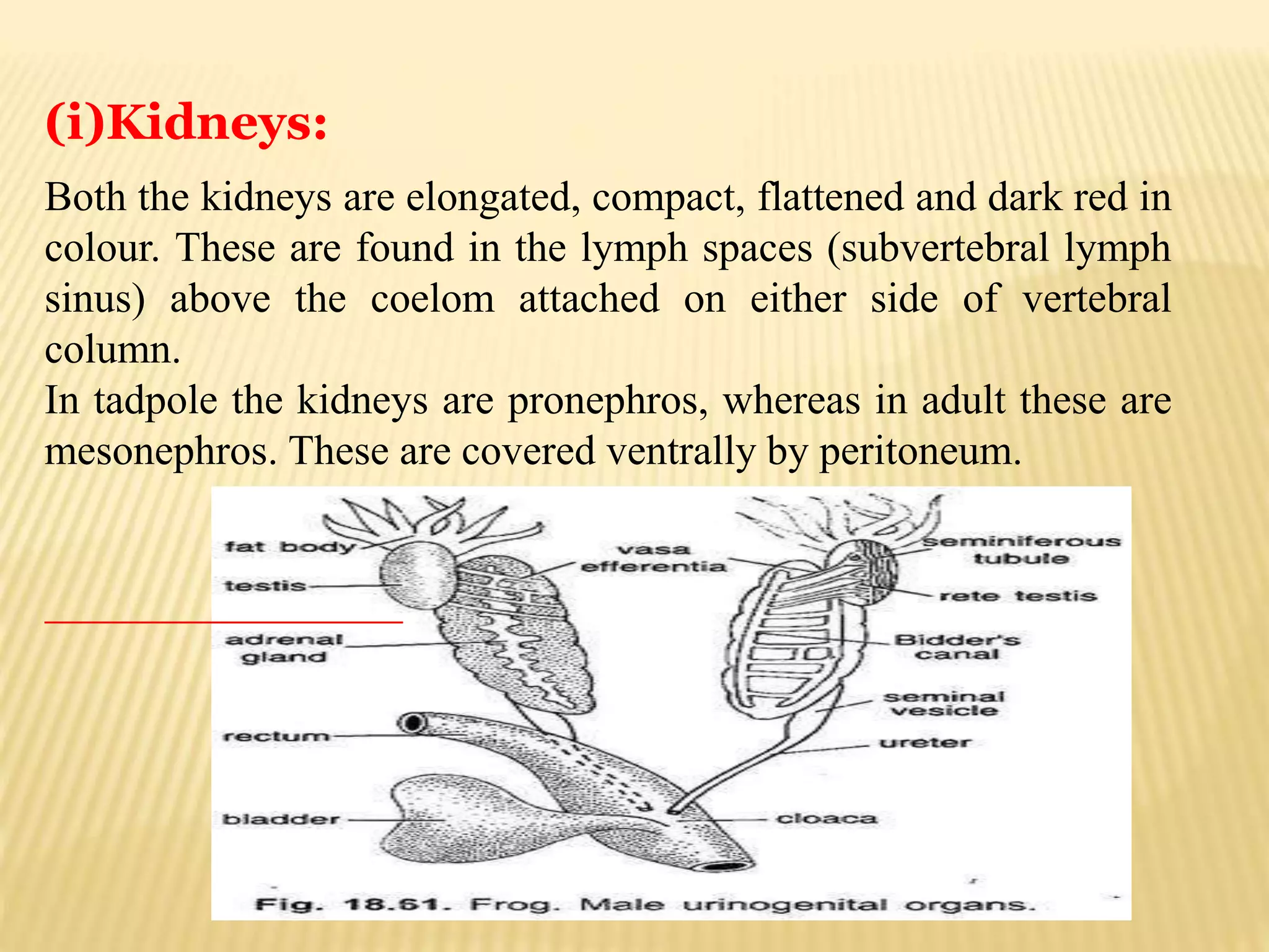
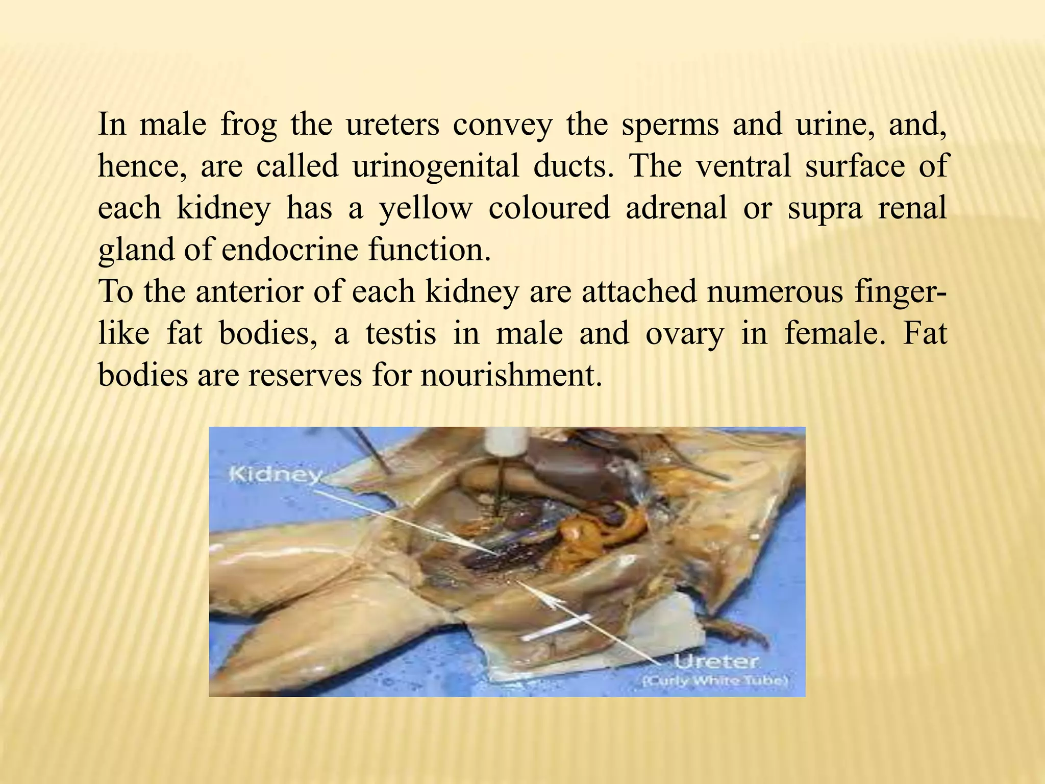
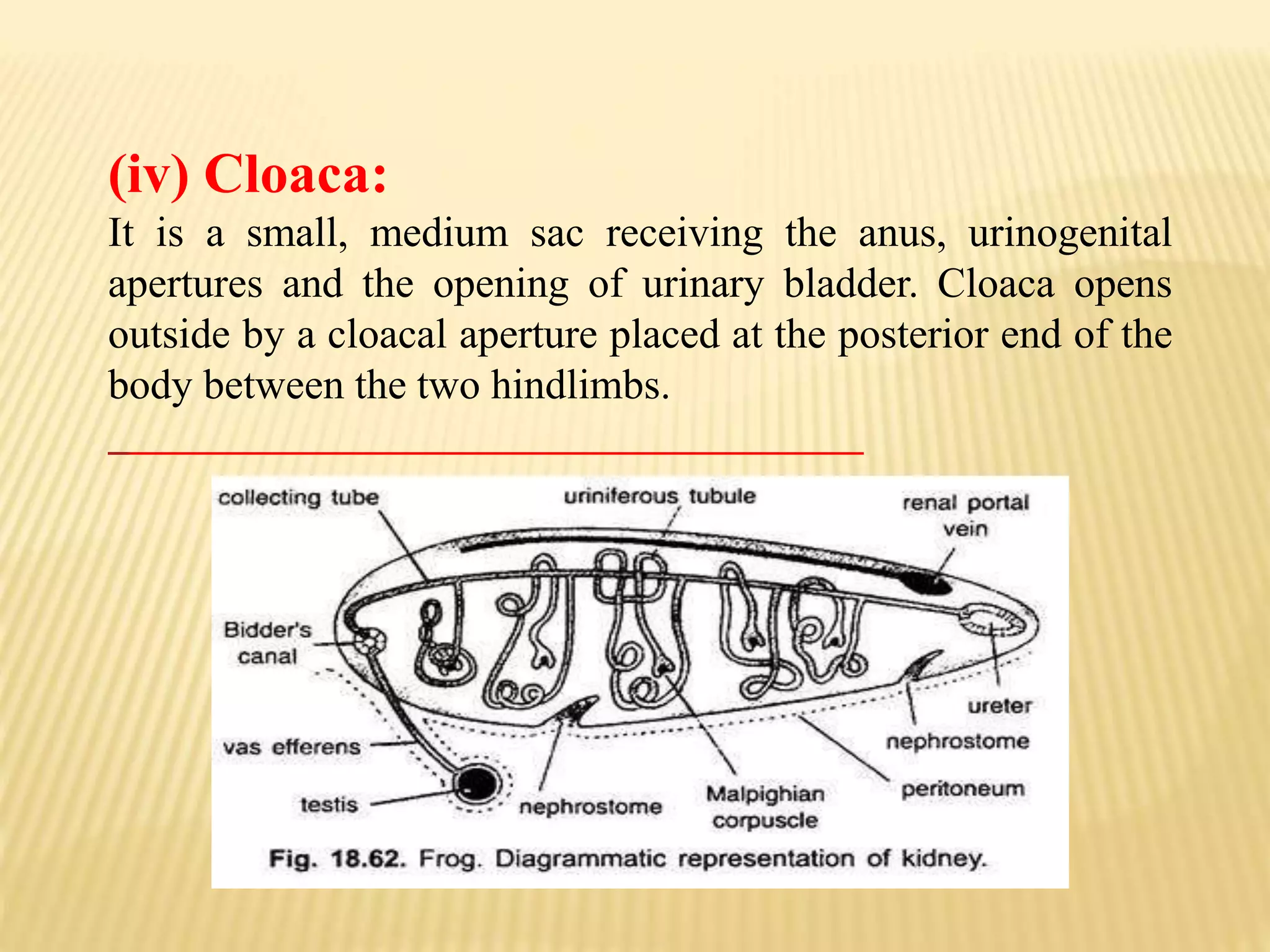
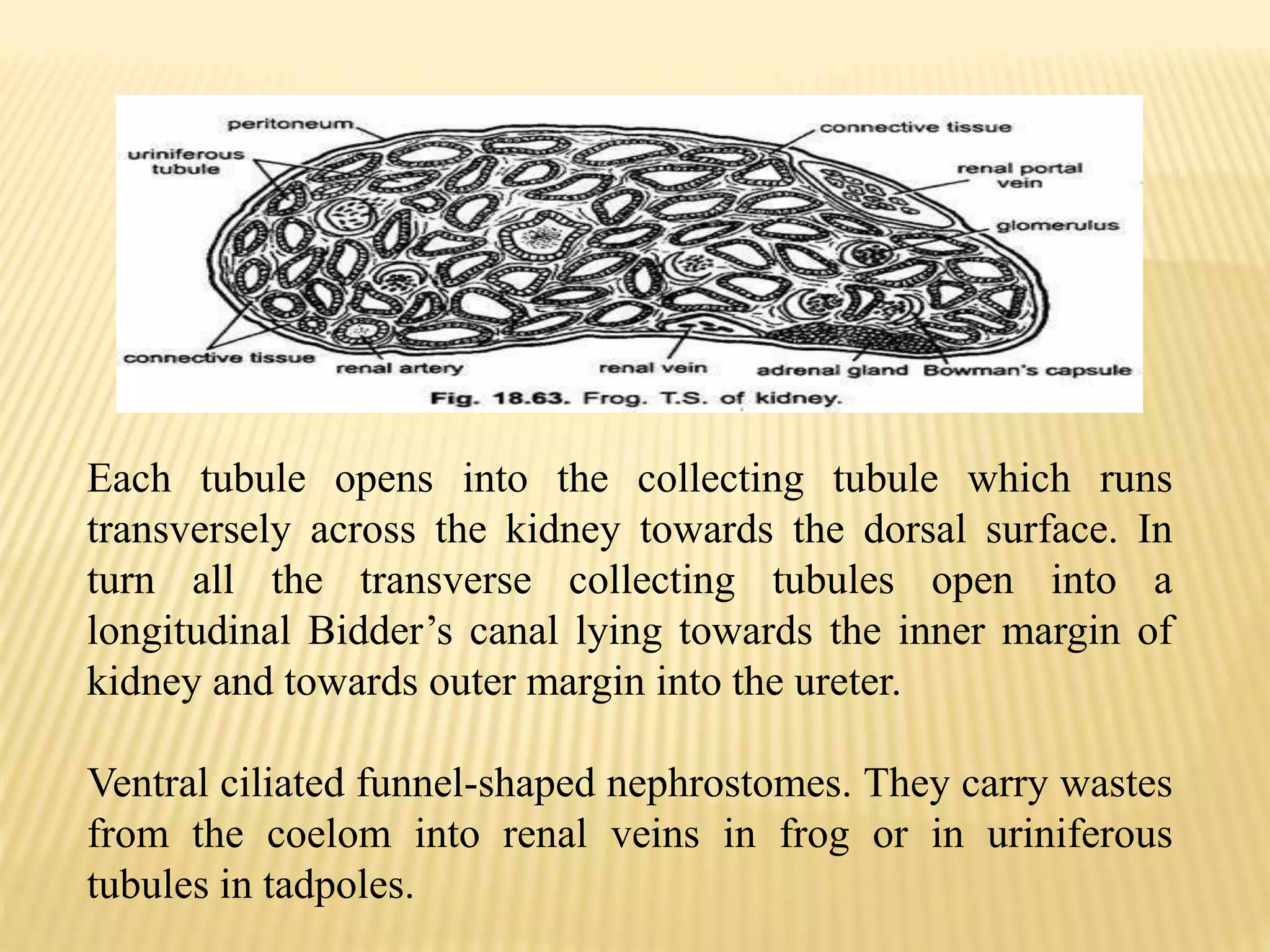
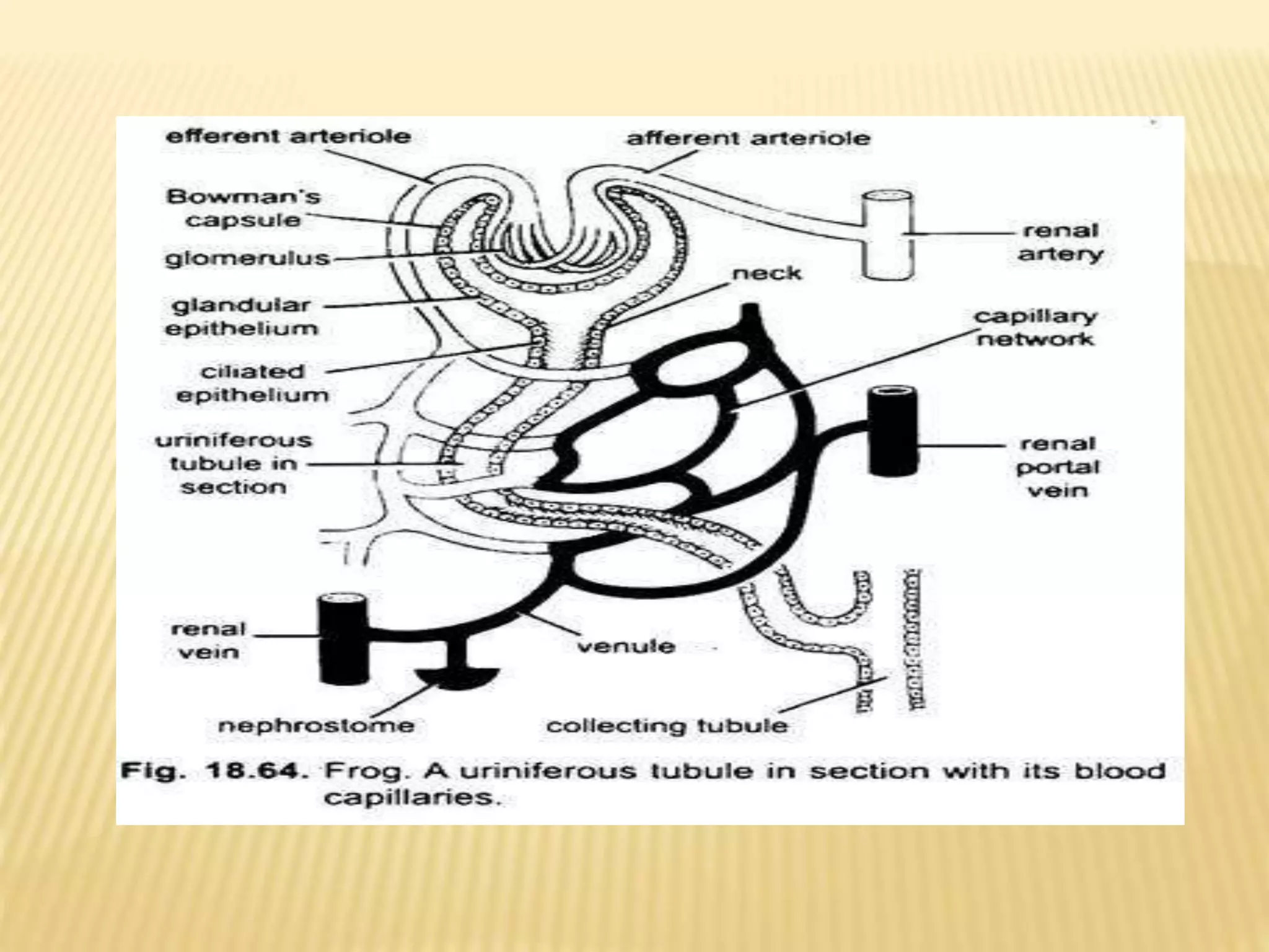
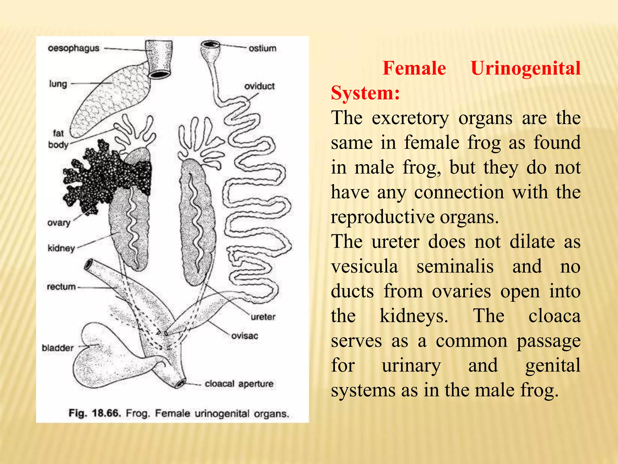
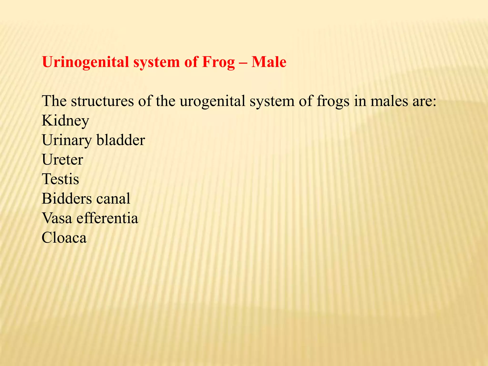
DR. G.D. HANDE
Urinogenital System in Frog
In vertebrates, the organs concerned with reproduction
and urinary excretion.
The major structures of the reproductive system in males
are the testes, sperm ducts, urethra, and penis; in females,
they are the ovaries, fallopian tubes, uterus, and vagina.
The urogenital system is responsible for the
formation, storage and elimination of urine and for
reproductive activity.
Many functions of the urogenital system are controlled
by autonomic (parasympathetic and sympathetic) and
somatic efferent pathways originating in the
lumbosacral spinal cord.
The urinary and reproductive systems are associated
closely in frogs and are jointly referred to as the
Urogenital system.
It is primarily involved in assisting the frog in carrying
out urine excretion and reproduction.
The main excretory product of frogs is urea and hence
frogs are ureotelic animals.
The urinogenital organs can be studied under the following
heads:
1. Excretory System:
The excretory system in both male and female frog is similar.
The excretion is mainly carried out with the help of a pair of
kidneys, a pair of ureters, a urinary bladder and cloaca.
The term urogenital refers to something that has both
urinary and genital origins.
The word urogenital is used because the urinary and
reproductive systems in males merge.
(i)Kidneys:
Both the kidneys are elongated, compact, flattened and dark red in
colour. These are found in the lymph spaces (subvertebral lymph
sinus) above the coelom attached on either side of vertebral
column.
In tadpole the kidneys are pronephros, whereas in adult these are
mesonephros. These are covered ventrally by peritoneum.
(ii) Ureters:
From the outer smooth convex posterior side of each
kidney arises a mesonephric or Wolffian duct or ureter which
passes backwards to open into dorsal side of the cloaca.
The openings of the ureters are placed over a separate
papilla on the dorsal side of cloaca. In male frog the ureters dilate
just posterior to the kidney to form a vesicula seminalis in which
sperms are stored.
In male frog the ureters convey the sperms and urine, and,
hence, are called urinogenital ducts. The ventral surface of
each kidney has a yellow coloured adrenal or supra renal
gland of endocrine function.
To the anterior of each kidney are attached numerous finger-
like fat bodies, a testis in male and ovary in female. Fat
bodies are reserves for nourishment.
(iii) Urinary Bladder:
It is large, thin-walled bilobed distensible
structure. It also opens into the ventral wall
of cloaca by a sphinctered aperture. Its
aperture lies below and opposite to the
openings of ureters.
The inner surface of bladder is lined with
a layer of epithelium about three cells
thick. The middle layer of the bladder
consists of a network of smooth muscle
fibres and outside this layer is a thin sheet
of connective tissue covered externally by
the peritoneum.
(iv) Cloaca:
It is a small, medium sac receiving the anus, urinogenital
apertures and the opening of urinary bladder. Cloaca opens
outside by a cloacal aperture placed at the posterior end of the
body between the two hindlimbs.
Histology of Kidney:
The kidneys are most of the red blood corpuscles.
Each kidney is composed of a number of (about 2000) twisted
renal or uriniferous tubules held together by connective tissue
and richly supplied with blood vessels and their capillaries.
Each renal or uriniferous tubule starts as a thin,
double-walled ciliated cup, the Bowman’s capsule enclosing a
bunch of blood capillaries, the glomerulus which receives blood
from an afferent arteriole of the renal artery.
Uriniferous tubules are the functional units of kidney. These
are lined with a glandular and at places with ciliated
epithelium.
From the glomerulus blood is collected by efferent
arteriole which joins with a renal vein.
The tubule gets the blood supply from the capillaries
of efferent arteriole and renal portal vein.
The Bowman’s capsule along with its glomerulus is
called Malpighian body or corpuscle tubules.
Each tubule opens into the collecting tubule which runs
transversely across the kidney towards the dorsal surface. In
turn all the transverse collecting tubules open into a
longitudinal Bidder’s canal lying towards the inner margin of
kidney and towards outer margin into the ureter.
Ventral ciliated funnel-shaped nephrostomes. They carry wastes
from the coelom into renal veins in frog or in uriniferous
tubules in tadpoles.
Physiology of Excretion:
Due to continuous catabolic activities certain substances
are formed which are harmful to body, hence, their
elimination is very essential which is done by the kidneys.
The blood brings these substances into the kidneys through
the renal portal veins.
Blood comes to glomeruli from afferent arterioles which
have a wider lumen then the capillaries so that the blood in
glomeruli is under high pressure which causes filtration of
blood.
The glomerular filtrate does not contain corpuscles
nor plasma proteins, it has plasma, wastes and crystalloids
consisting of urea, inorganic salts, glucose and large
quantities of water.
The glomerular filtrate passes through the thin wall
of the Bowman’s capsules into the lumen of uriniferous
tubules and the filtered blood goes into renal vein through
the efferent arteriole.
Female Urinogenital
System:
The excretory organs are the
same in female frog as found
in male frog, but they do not
have any connection with the
reproductive organs.
The ureter does not dilate as
vesicula seminalis and no
ducts from ovaries open into
the kidneys. The cloaca
serves as a common passage
for urinary and genital
systems as in the male frog.
Urinogenital system of Frog – Male
The structures of the urogenital system of frogs in males are:
Kidney
Urinary bladder
Ureter
Testis
Bidders canal
Vasa efferentia
Cloaca
Kidney – Kidneys are dark red and bean like structures, compact,
occurring in a pair located posteriorly in the body cavity on both
sides of the vertebral column. Each kidney consists of many
functional and structural units – nephrons or uriniferous tubules.
Urinary Bladder – it is a thin walled structure present ventral to
the rectum. It stores urine.
Ureter – the ureter or the urinogenital duct occur in pairs, projects
from the kidney in the form of ureters in males.
Testis – These structures occur in a pair and are yellowish in
appearance. These ovoid structures are located in the upper section
of the kidneys by a double fold of the peritoneum known as
mesorchium.
Bidder’s canal – It is a canal into which the vasa efferentia
opens into the kidneys. Eventually this canal meets the
ureter. This structure is responsible for passing urine, ova,
water and sperm.
Vasa efferentia – It emerges from the testis and are 10-12
in number. They pass through mesorchium and move into
the kidneys.
Cloaca – It is a small chamber mediating to pass urine,
faeces and sperms to the outside. The ureter and urinary
bladder opens into the cloaca.
Urinogenital system of Frog – Female
The structures of the urogenital system of frogs in females
are:
Kidney
Urinary bladder
Ureter
Ovaries
Oviduct
Cloaca
The common structures in both male and female
urogenital systems in frogs are kidneys, ureters and
urinary bladder, their functionalities remain the same.
Ovary – It is a paired structure present close to kidneys.
These yellow-colored structures are present on the outer
side of the kidneys by a fold of peritoneum known as the
mesovarium. Through the process of oogenesis, they
produce ova. A mature female releases close to 2500-
3000 ova at once.
Oviduct – These structures are paired and found on each
side of the ovary. They are thin, large and coiled
structures appearing white in color. These ducts open
separately into the cloaca.
How is the blood supply to the Kidneys?
Blood supply to the kidneys is through the renal portal veins
and renal arteries. Majorly blood constitutes urea as the
excretory product. The renal artery branches in the kidney
and redivides forming the afferent arteriole having a
diameter larger than the efferent branch. The renal veins on
the other side carries away the detoxified blood from the
kidneys.
The renal portal veins branch into the kidney and these
branches and the efferent arteriole unites forming a mesh of
capillaries near the uriniferous tubules and ultimately
together they form the renal venule.
Urinogenital System in Frog 1.pptx

Urinogenital System in Frog 1.pptx

  • 1.
  • 2.
    In vertebrates, theorgans concerned with reproduction and urinary excretion. The major structures of the reproductive system in males are the testes, sperm ducts, urethra, and penis; in females, they are the ovaries, fallopian tubes, uterus, and vagina. The urogenital system is responsible for the formation, storage and elimination of urine and for reproductive activity. Many functions of the urogenital system are controlled by autonomic (parasympathetic and sympathetic) and somatic efferent pathways originating in the lumbosacral spinal cord.
  • 3.
    The urinary andreproductive systems are associated closely in frogs and are jointly referred to as the Urogenital system. It is primarily involved in assisting the frog in carrying out urine excretion and reproduction. The main excretory product of frogs is urea and hence frogs are ureotelic animals.
  • 4.
    The urinogenital organscan be studied under the following heads: 1. Excretory System: The excretory system in both male and female frog is similar. The excretion is mainly carried out with the help of a pair of kidneys, a pair of ureters, a urinary bladder and cloaca. The term urogenital refers to something that has both urinary and genital origins. The word urogenital is used because the urinary and reproductive systems in males merge.
  • 5.
    (i)Kidneys: Both the kidneysare elongated, compact, flattened and dark red in colour. These are found in the lymph spaces (subvertebral lymph sinus) above the coelom attached on either side of vertebral column. In tadpole the kidneys are pronephros, whereas in adult these are mesonephros. These are covered ventrally by peritoneum.
  • 7.
    (ii) Ureters: From theouter smooth convex posterior side of each kidney arises a mesonephric or Wolffian duct or ureter which passes backwards to open into dorsal side of the cloaca. The openings of the ureters are placed over a separate papilla on the dorsal side of cloaca. In male frog the ureters dilate just posterior to the kidney to form a vesicula seminalis in which sperms are stored.
  • 8.
    In male frogthe ureters convey the sperms and urine, and, hence, are called urinogenital ducts. The ventral surface of each kidney has a yellow coloured adrenal or supra renal gland of endocrine function. To the anterior of each kidney are attached numerous finger- like fat bodies, a testis in male and ovary in female. Fat bodies are reserves for nourishment.
  • 9.
    (iii) Urinary Bladder: Itis large, thin-walled bilobed distensible structure. It also opens into the ventral wall of cloaca by a sphinctered aperture. Its aperture lies below and opposite to the openings of ureters. The inner surface of bladder is lined with a layer of epithelium about three cells thick. The middle layer of the bladder consists of a network of smooth muscle fibres and outside this layer is a thin sheet of connective tissue covered externally by the peritoneum.
  • 10.
    (iv) Cloaca: It isa small, medium sac receiving the anus, urinogenital apertures and the opening of urinary bladder. Cloaca opens outside by a cloacal aperture placed at the posterior end of the body between the two hindlimbs.
  • 11.
    Histology of Kidney: Thekidneys are most of the red blood corpuscles. Each kidney is composed of a number of (about 2000) twisted renal or uriniferous tubules held together by connective tissue and richly supplied with blood vessels and their capillaries. Each renal or uriniferous tubule starts as a thin, double-walled ciliated cup, the Bowman’s capsule enclosing a bunch of blood capillaries, the glomerulus which receives blood from an afferent arteriole of the renal artery.
  • 12.
    Uriniferous tubules arethe functional units of kidney. These are lined with a glandular and at places with ciliated epithelium. From the glomerulus blood is collected by efferent arteriole which joins with a renal vein. The tubule gets the blood supply from the capillaries of efferent arteriole and renal portal vein. The Bowman’s capsule along with its glomerulus is called Malpighian body or corpuscle tubules.
  • 13.
    Each tubule opensinto the collecting tubule which runs transversely across the kidney towards the dorsal surface. In turn all the transverse collecting tubules open into a longitudinal Bidder’s canal lying towards the inner margin of kidney and towards outer margin into the ureter. Ventral ciliated funnel-shaped nephrostomes. They carry wastes from the coelom into renal veins in frog or in uriniferous tubules in tadpoles.
  • 14.
    Physiology of Excretion: Dueto continuous catabolic activities certain substances are formed which are harmful to body, hence, their elimination is very essential which is done by the kidneys. The blood brings these substances into the kidneys through the renal portal veins. Blood comes to glomeruli from afferent arterioles which have a wider lumen then the capillaries so that the blood in glomeruli is under high pressure which causes filtration of blood.
  • 16.
    The glomerular filtratedoes not contain corpuscles nor plasma proteins, it has plasma, wastes and crystalloids consisting of urea, inorganic salts, glucose and large quantities of water. The glomerular filtrate passes through the thin wall of the Bowman’s capsules into the lumen of uriniferous tubules and the filtered blood goes into renal vein through the efferent arteriole.
  • 17.
    Female Urinogenital System: The excretoryorgans are the same in female frog as found in male frog, but they do not have any connection with the reproductive organs. The ureter does not dilate as vesicula seminalis and no ducts from ovaries open into the kidneys. The cloaca serves as a common passage for urinary and genital systems as in the male frog.
  • 18.
    Urinogenital system ofFrog – Male The structures of the urogenital system of frogs in males are: Kidney Urinary bladder Ureter Testis Bidders canal Vasa efferentia Cloaca
  • 19.
    Kidney – Kidneysare dark red and bean like structures, compact, occurring in a pair located posteriorly in the body cavity on both sides of the vertebral column. Each kidney consists of many functional and structural units – nephrons or uriniferous tubules. Urinary Bladder – it is a thin walled structure present ventral to the rectum. It stores urine. Ureter – the ureter or the urinogenital duct occur in pairs, projects from the kidney in the form of ureters in males. Testis – These structures occur in a pair and are yellowish in appearance. These ovoid structures are located in the upper section of the kidneys by a double fold of the peritoneum known as mesorchium.
  • 20.
    Bidder’s canal –It is a canal into which the vasa efferentia opens into the kidneys. Eventually this canal meets the ureter. This structure is responsible for passing urine, ova, water and sperm. Vasa efferentia – It emerges from the testis and are 10-12 in number. They pass through mesorchium and move into the kidneys. Cloaca – It is a small chamber mediating to pass urine, faeces and sperms to the outside. The ureter and urinary bladder opens into the cloaca.
  • 21.
    Urinogenital system ofFrog – Female The structures of the urogenital system of frogs in females are: Kidney Urinary bladder Ureter Ovaries Oviduct Cloaca
  • 22.
    The common structuresin both male and female urogenital systems in frogs are kidneys, ureters and urinary bladder, their functionalities remain the same. Ovary – It is a paired structure present close to kidneys. These yellow-colored structures are present on the outer side of the kidneys by a fold of peritoneum known as the mesovarium. Through the process of oogenesis, they produce ova. A mature female releases close to 2500- 3000 ova at once. Oviduct – These structures are paired and found on each side of the ovary. They are thin, large and coiled structures appearing white in color. These ducts open separately into the cloaca.
  • 23.
    How is theblood supply to the Kidneys? Blood supply to the kidneys is through the renal portal veins and renal arteries. Majorly blood constitutes urea as the excretory product. The renal artery branches in the kidney and redivides forming the afferent arteriole having a diameter larger than the efferent branch. The renal veins on the other side carries away the detoxified blood from the kidneys. The renal portal veins branch into the kidney and these branches and the efferent arteriole unites forming a mesh of capillaries near the uriniferous tubules and ultimately together they form the renal venule.