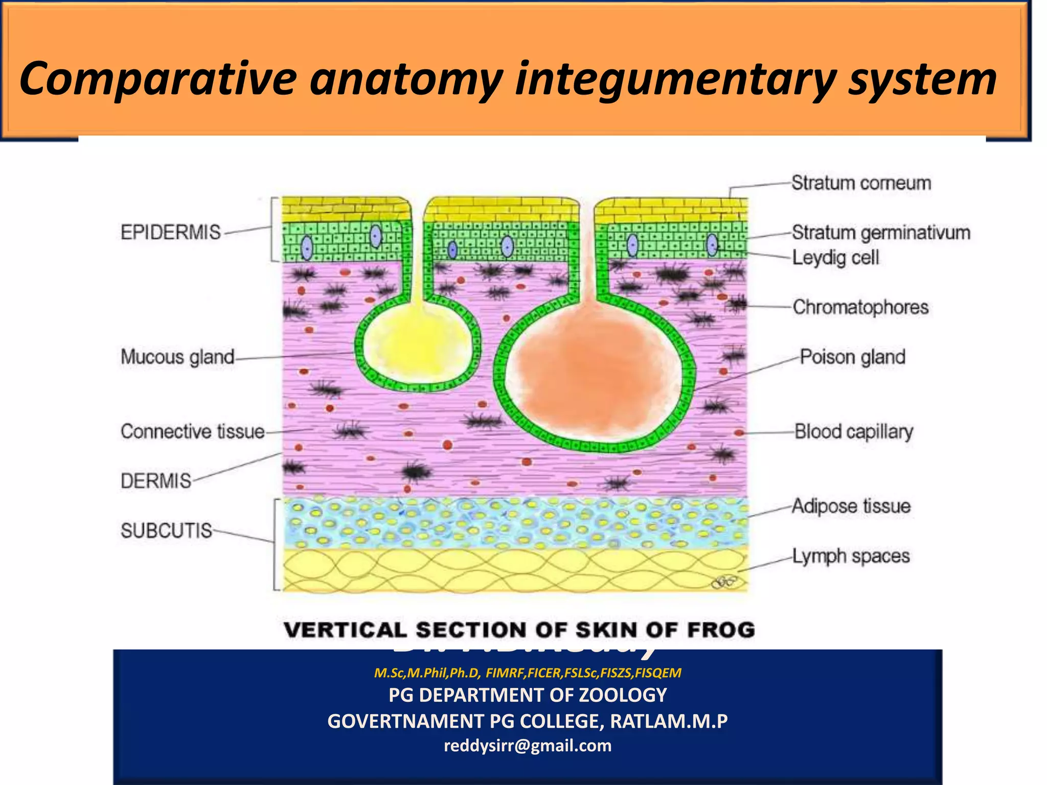
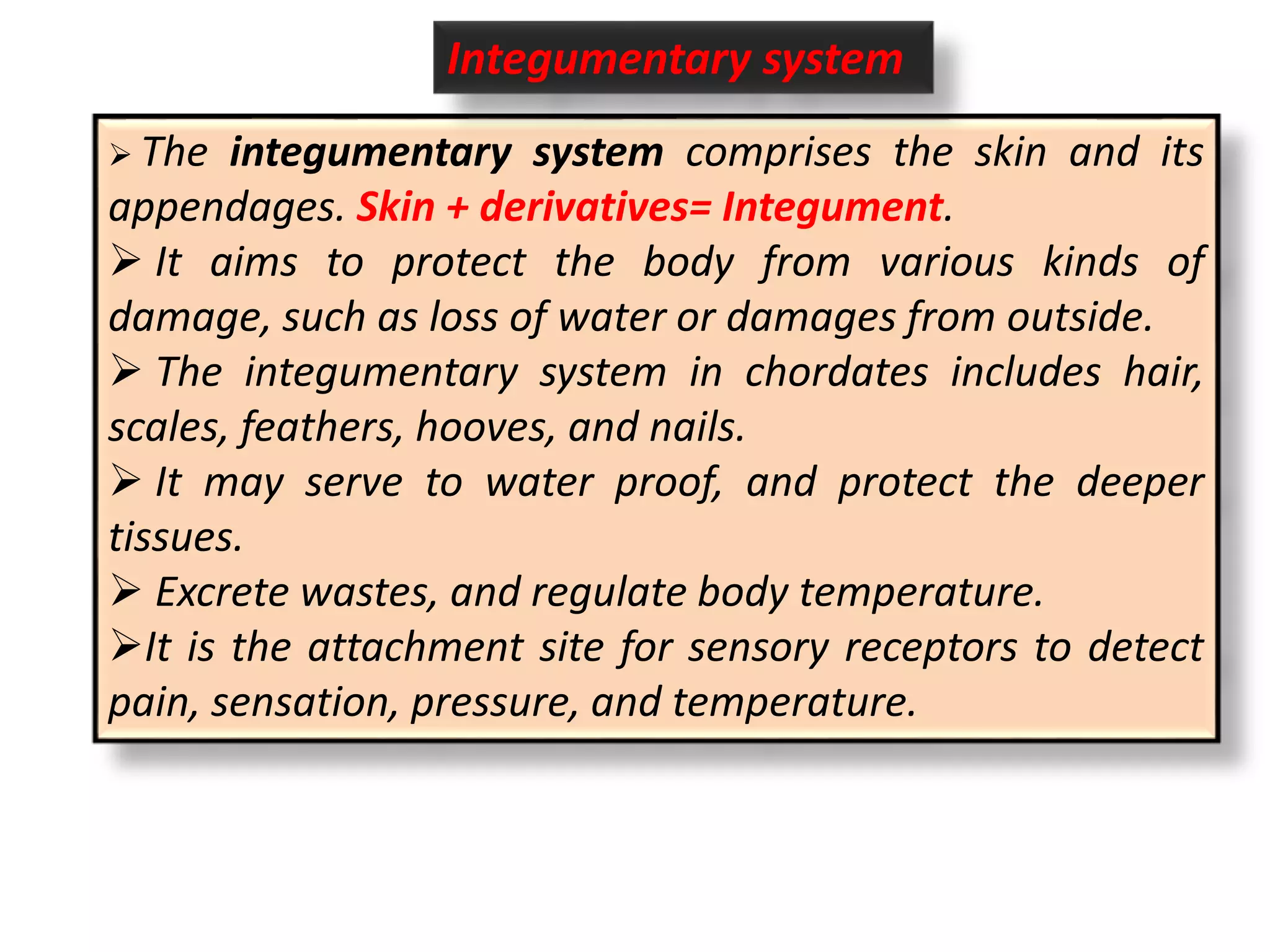
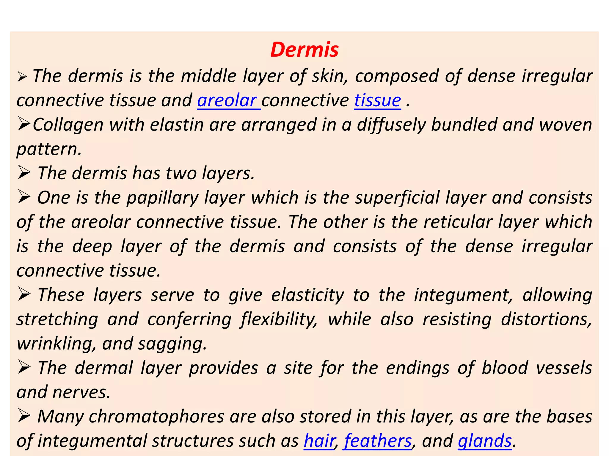
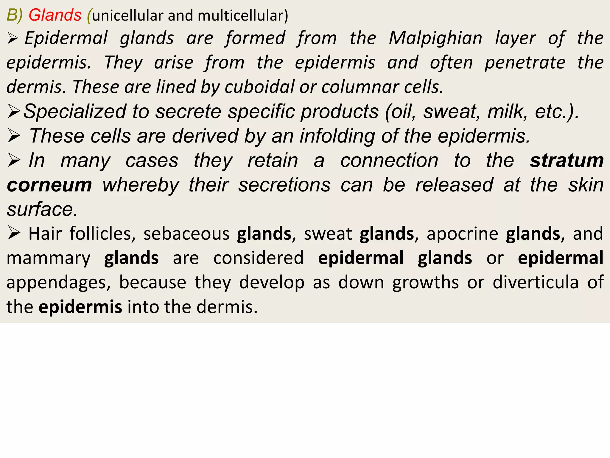
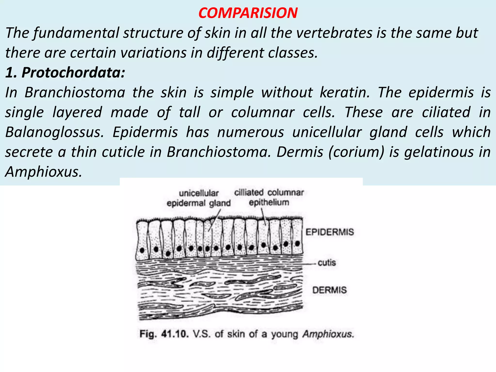
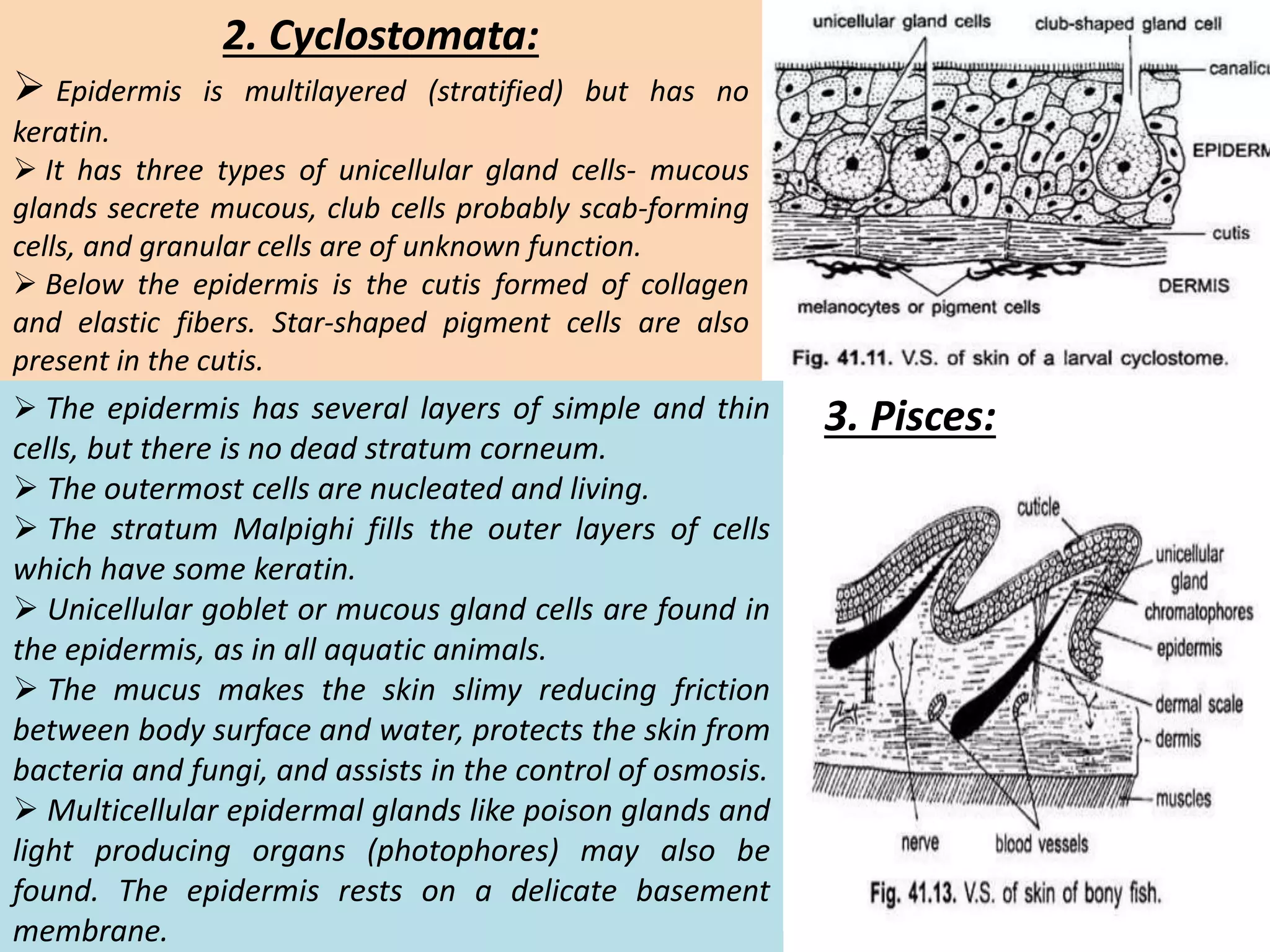
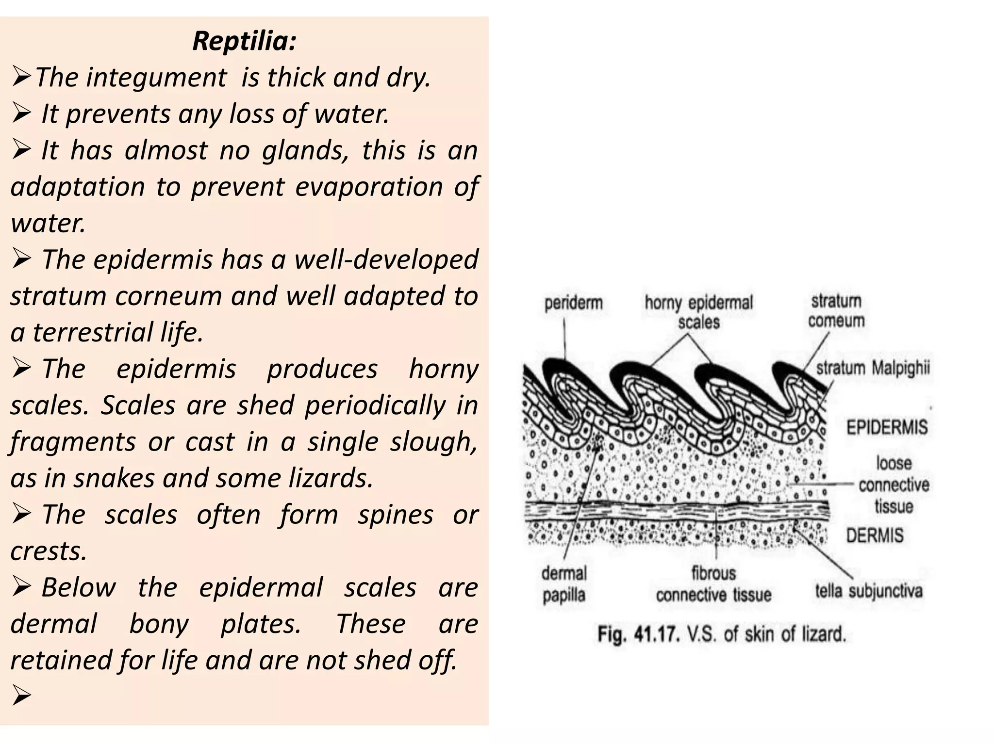
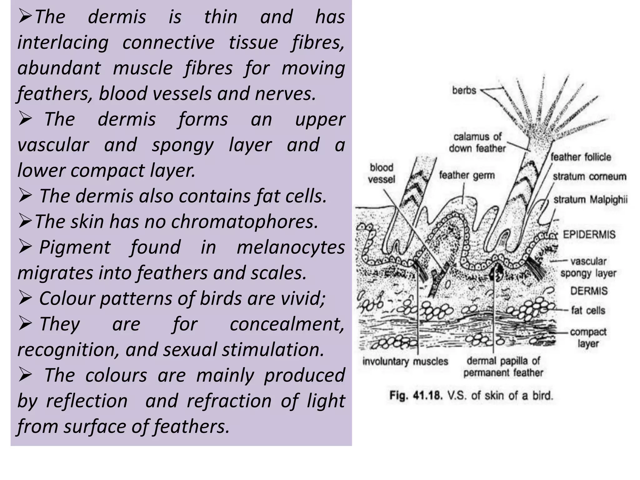
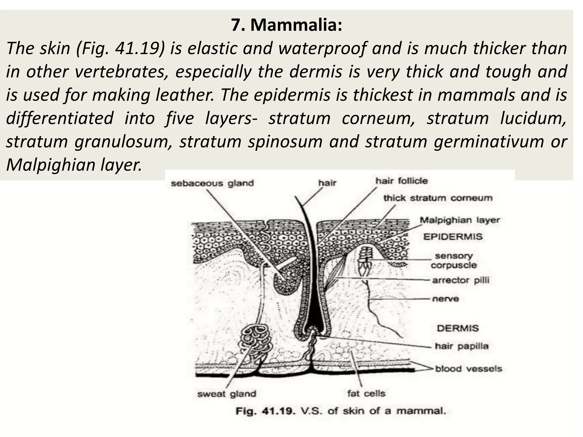
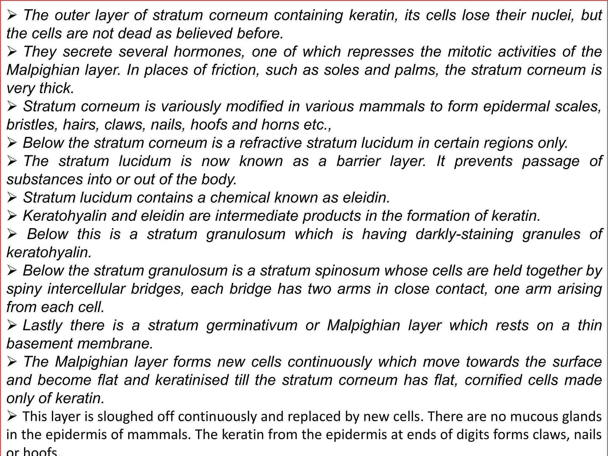
SlideShare a Scribd company logo
1 of 24
Dr. P.B.Reddy
M.Sc,M.Phil,Ph.D, FIMRF,FICER,FSLSc,FISZS,FISQEM
PG DEPARTMENT OF ZOOLOGY
GOVERTNAMENT PG COLLEGE, RATLAM.M.P
reddysirr@gmail.com
Comparative anatomy integumentary system
 The integumentary system comprises the skin and its
appendages. Skin + derivatives= Integument.
 It aims to protect the body from various kinds of
damage, such as loss of water or damages from outside.
 The integumentary system in chordates includes hair,
scales, feathers, hooves, and nails.
 It may serve to water proof, and protect the deeper
tissues.
 Excrete wastes, and regulate body temperature.
It is the attachment site for sensory receptors to detect
pain, sensation, pressure, and temperature.
Integumentary system
Functions (Jack of all trades and Master of none)
 Maintains the body's equilibrium.
 The acts as the body's first line of defense.
 Regulates the body temperature change, and homeostasis.
 Protect the body's internal living tissues and organs.
 Protect against invasion by infectious organisms.
 Protect the body from dehydration.
 Help excrete waste materials through perspiration.
 Act as a receptor for touch, pressure, pain, heat, and cold (Tactile
receptors)
 Protect the body against sunburns by secreting melanin.
 Generate vitamin D through exposure to ultraviolet light.
 Store water, fat, glucose, vitamin D.
 Maintenance of the body shape and outline.
 Formation of new cells from stratum germinativum to repair minor injuries
Protect from UV rays.
 It distinguishes, separates, and protects the organism from its
surroundings.
 Some invertebrates and frogs respire (cutaneous) using the outer layer
Skin
 The skin is the largest organ of the body.
 In humans, it accounts for about 12 to 15 percent of total body weight
and covers 1.5-2m2 of surface area.
 The is composed of two major layers of tissue: the epidermis and dermis.
(The hypodermis or subcutaneous layer is not part of the skin)
 The epidermis is the outermost layer, providing the initial barrier to the
external environment.
 It is separated from the dermis by the basement membrane.
 The epidermis contains melanocytes and gives color to the skin.
 The deepest layer of epidermis also contains nerve endings.
 Beneath this, the dermis comprises two sections, the papillary and
reticular layers, and contains connective tissues, vessels, glands, follicles,
hair roots, sensory nerve endings, and muscular tissue.
 The deepest layer, the hypodermis, is primarily made up of adipose
tissue. Substantial collagen bundles anchor the dermis to the hypodermis in
a way that permits most areas of the skin to move freely over the deeper
tissue layers.
 The epidermis, derived from somatic ectoderm,
The epidermis is the top layer of skin and made up of
epithelial cells.
 It does not contain blood vessels.
 Its main functions are protection, absorption of
nutrients, and homeostasis.
 It provides a barrier to infection from
environmental pathogens and regulates the amount
of water.
 In structure, it consists of a keratinized stratified
squamous epithelium; four types of cells:
keratinocytes, melanocytes, Merkel cells, and
Langerhans cells.
Keratin is a waterproofing protein. Millions of dead
keratinocytes rub off daily (ecdysis or moulting).
 The layers of the epidermis include the stratum
basale (the deepest portion of the epidermis),
stratum spinosum, stratum granulosum, stratum
lucidum, and stratum corneum (the most superficial
portion of the epidermis).
 Fishes and amphibians have a mucus layer for
bacterial and mechanical
Epidermis
The epidermis of fish and of most
amphibians consists entirely of live
cells, with only minimal quantities of
keratin in the cells of the superficial
layer. It is generally permeable, and
in the case of many amphibians, may
actually be a major respiratory
organ.
Dermis
 The dermis is the middle layer of skin, composed of dense irregular
connective tissue and areolar connective tissue .
Collagen with elastin are arranged in a diffusely bundled and woven
pattern.
 The dermis has two layers.
 One is the papillary layer which is the superficial layer and consists
of the areolar connective tissue. The other is the reticular layer which
is the deep layer of the dermis and consists of the dense irregular
connective tissue.
 These layers serve to give elasticity to the integument, allowing
stretching and conferring flexibility, while also resisting distortions,
wrinkling, and sagging.
 The dermal layer provides a site for the endings of blood vessels
and nerves.
 Many chromatophores are also stored in this layer, as are the bases
of integumental structures such as hair, feathers, and glands.
Epidermal Derivatives of the Integument
A) Keratin Structures
New epidermal cells are formed continuously in the lower layers of the epidermis. In terrestrial vertebrates, new
epidermal cells push more superficial ones to the stratum corneum, the outer-most epithelial layer. In the
process of self-destruction, these exterior epidermal cells accumulate protein products called keratin. Keratinized
or cornified skin serves to prevent water escape and to protect against friction and direct mechanical stimulation
(e.g. calluses in humans). The production of all of the following structures involves keratinization:
Epidermal Scales: a continuous layer of repetitious thickenings of the stratum corneum; These scales
may be shed entirely or in small flakes. Examine preserved specimen of snake and dried specimens of
bird legs and feet.
Claws and Talons: curved, laterally compressed keratinized projections from the tips of digits. See dried
specimen of cat claws and bird talons.
The possible functions of these structures
Hooves: enlarged keratinized plates found on the ends of ungulate digits. Examine the hooves of pig and
horse.
Nails: keratinized epithelial cells produced at the name base by pushing the existing nail forward. Protect
from mechanical injury and stabilize skin for better grasping. Found only in primates.
Horns: a tough, cornified layer of the integument covers horns. Their core, however, is bone, of dermal
origin. Horns are found in bovines (cattle, antelope, sheep, goats, bison, wildebeest). They are retained
year-round and grow throughout the animal’s lifetime.
Baleen: found in some whales, is a series of keratinized plates that arise from oral epithelium. These
sheets hang from the palate along its length. use would the sieve-like action of these plates be.
Beaks: epidermal structures, jaws are covered by keratinized sheaths in birds.
Feathers: are believed to have evolved from reptilian scales. Columns of epidermal cells project into the
skin initially to form an invagination called the feather follicle.
Hair: just as in feathers, there is an initial in growth of epidermal cells to form the hair follicle, followed by
an outward growth of keratinized cells to form the hair shaft. Both feather and hair possess dermal
papillae, shafts, an inner pulp and columns of specialized keratinized cells. Hair is characteristic of
mammals.
B) Glands (unicellular and multicellular)
 Epidermal glands are formed from the Malpighian layer of the
epidermis. They arise from the epidermis and often penetrate the
dermis. These are lined by cuboidal or columnar cells.
Specialized to secrete specific products (oil, sweat, milk, etc.).
 These cells are derived by an infolding of the epidermis.
 In many cases they retain a connection to the stratum
corneum whereby their secretions can be released at the skin
surface.
 Hair follicles, sebaceous glands, sweat glands, apocrine glands, and
mammary glands are considered epidermal glands or epidermal
appendages, because they develop as down growths or diverticula of
the epidermis into the dermis.
COMPARISION
The fundamental structure of skin in all the vertebrates is the same but
there are certain variations in different classes.
1. Protochordata:
In Branchiostoma the skin is simple without keratin. The epidermis is
single layered made of tall or columnar cells. These are ciliated in
Balanoglossus. Epidermis has numerous unicellular gland cells which
secrete a thin cuticle in Branchiostoma. Dermis (corium) is gelatinous in
Amphioxus.
2. Cyclostomata:
 Epidermis is multilayered (stratified) but has no
keratin.
 It has three types of unicellular gland cells- mucous
glands secrete mucous, club cells probably scab-forming
cells, and granular cells are of unknown function.
 Below the epidermis is the cutis formed of collagen
and elastic fibers. Star-shaped pigment cells are also
present in the cutis.
3. Pisces: The epidermis has several layers of simple and thin
cells, but there is no dead stratum corneum.
 The outermost cells are nucleated and living.
 The stratum Malpighi fills the outer layers of cells
which have some keratin.
 Unicellular goblet or mucous gland cells are found in
the epidermis, as in all aquatic animals.
 The mucus makes the skin slimy reducing friction
between body surface and water, protects the skin from
bacteria and fungi, and assists in the control of osmosis.
 Multicellular epidermal glands like poison glands and
light producing organs (photophores) may also be
found. The epidermis rests on a delicate basement
membrane.
 The mucus makes the skin slimy reducing friction between body surface and water, protects
the skin from bacteria and fungi, and assists in the control of osmosis.
 Multicellular epidermal glands like poison glands and light producing organs (photophores)
may also be found. The epidermis rests on a delicate basement membrane.
 The dermis contains connective tissue, smooth muscles, blood vessels, nerves, lymph
vessels, and collagen fibers. The connective tissue fibers are generally not arranged at right
angles, but run parallel to the surface. Scales are embedded in the dermis and projected above
the epidermal surface.
These are of five types. The elasmobranches have placoid scales, Chondrostei and Holostei
have ganoid scales, while most Teleostei have cycloid and ctenoid scales lodged in pouches of
the dermis. Extinct Crossopterygii had cosmoid scales.
Many bony fishes show more brilliant colours than any other group of animals. The colours of
fishes are due to chromatophores and iridocytes.
(a) Chromatophores in the dermis are derived from neural crest cells. They contain pigments
which not only produce colours but also cause variations of colours. Chromatophores
containing brown or black pigment are known as melanophores and those containing red,
yellow, or orange pigment are collectively called lipophores.
(b) Iridocytes or guanophores are reflecting cells. They have no pigment but contain crystals
of guanin. They lie in the dermis and cause iridescence. Iridocytes reflect light from guanin
crystals to produce white or silvery colours if the iridocytes are below the scales, if the
iridocytes are above the scales they cause iridescent hues. By combinations of
chromatophores and iridocytes various colours are produced, e.g., blue is produced by
reflection from iridocytes, the blue combines with yellow pigment to produce green.
4. Amphibia:
 The epidermis is multilayered.
 The stratum corneum made of flattened, highly keratinized cells. Such a
dead layer appears first in amphibians. The stratum corneum is an
adaptation to terrestrial life. It not only protects the body but prevents any
excessive loss of moisture.
In ecdysis, the stratum corneum is cast off in fragments or as a whole in
some.
 The dermis is relatively thin, it is made of two layers, an upper loose
stratum spongiosum and a lower dense and compact stratum compactum.
Connective tissue fibers run both vertically and horizontally.
 There are two kinds of glands, they are multicellular mucous glands and
poison glands in the dermis, but they are derivatives of the epidermis.
 The mucous glands produce mucus which not only forms a slimy
protective covering but also helps in respiration.
 The poison glands found in toads as parotid glands produce a mild but
unpleasant poison which is protective, keep the enemies away.
 In the upper part of the dermis are chromatophores which have black
melanophores and yellow lipophores, these produce the colour of the skin.

 The ability of the skin for changing colour to blend with the environment is well
developed.
 Skin of labyrinthodontia, the stem Amphibia had a armour of dermal scales.
 Bony dermal scales are found embedded in the skin of some Gymnophiona (Apoda)
and a few tropical toads. These scales are absent in modern Amphibia.
The skin is sensitive to light in amphibians, especially in cave-dwelling forms.
 It is an important organ of respiration, and also enables the frog to respire under
water for long periods, during hibernation or aestivation it is the only organ of
respiration.
 The skin is loose being attached to muscles only at certain places by connective
tissue septa which mark the boundaries of subcutaneous lymph spaces.
Reptilia:
The integument is thick and dry.
 It prevents any loss of water.
 It has almost no glands, this is an
adaptation to prevent evaporation of
water.
 The epidermis has a well-developed
stratum corneum and well adapted to
a terrestrial life.
 The epidermis produces horny
scales. Scales are shed periodically in
fragments or cast in a single slough,
as in snakes and some lizards.
 The scales often form spines or
crests.
 Below the epidermal scales are
dermal bony plates. These are
retained for life and are not shed off.

The dermis is thick and has an upper loose connective tissue layer and
a lower layer tella subjunctiva and separating the two is a horizontal
layer of fibrous connective tissue.
 The upper layer has an abundance of chromatophores in snakes and
lizards like fishes and amphibians.
 Many lizards and snakes have elaborate colour patterns, for
concealment or as warning colours. In Calotes, the chromatophores have
no nerve fibers, they are controlled by hormones of the posterior lobe of
the pituitary. In Chameleon, chromatophores are controlled by the
autonomic nervous system.
 Reptiles essentially lack skin glands.
 Many lizards have glands near the cloaca.
 Musk glands in the throat and cloacal opening of crocodilians function
during courtship.
 Generation glands found recently are associated with periodic
shedding of the skin.

6. Aves:
 The integument is thin, loose, dry and devoid of glands except a
uropygial gland at the base of the tail whose secreted oil is used for
preening the feathers, especially in aquatic birds.
 The stratified epidermis is delicate, except on shanks and feet where
it is thick and forms epidermal scales.
 The claws, spurs and horny sheaths of beaks are also the
modifications of stratum corneum of epidermis.
 Claws and beaks are variously modified in birds according to habitat.
 The rest of the body has a protective covering of epidermal feathers
which are evolved from epidermal scales.
 Feathers protect and insulate the body, i.e, keep the body warm.

The dermis is thin and has
interlacing connective tissue fibres,
abundant muscle fibres for moving
feathers, blood vessels and nerves.
 The dermis forms an upper
vascular and spongy layer and a
lower compact layer.
 The dermis also contains fat cells.
The skin has no chromatophores.
 Pigment found in melanocytes
migrates into feathers and scales.
 Colour patterns of birds are vivid;
 They are for concealment,
recognition, and sexual stimulation.
 The colours are mainly produced
by reflection and refraction of light
from surface of feathers.
7. Mammalia:
The skin (Fig. 41.19) is elastic and waterproof and is much thicker than
in other vertebrates, especially the dermis is very thick and tough and
is used for making leather. The epidermis is thickest in mammals and is
differentiated into five layers- stratum corneum, stratum lucidum,
stratum granulosum, stratum spinosum and stratum germinativum or
Malpighian layer.
 The outer layer of stratum corneum containing keratin, its cells lose their nuclei, but
the cells are not dead as believed before.
 They secrete several hormones, one of which represses the mitotic activities of the
Malpighian layer. In places of friction, such as soles and palms, the stratum corneum is
very thick.
 Stratum corneum is variously modified in various mammals to form epidermal scales,
bristles, hairs, claws, nails, hoofs and horns etc.,
 Below the stratum corneum is a refractive stratum lucidum in certain regions only.
 The stratum lucidum is now known as a barrier layer. It prevents passage of
substances into or out of the body.
 Stratum lucidum contains a chemical known as eleidin.
 Keratohyalin and eleidin are intermediate products in the formation of keratin.
 Below this is a stratum granulosum which is having darkly-staining granules of
keratohyalin.
 Below the stratum granulosum is a stratum spinosum whose cells are held together by
spiny intercellular bridges, each bridge has two arms in close contact, one arm arising
from each cell.
 Lastly there is a stratum germinativum or Malpighian layer which rests on a thin
basement membrane.
 The Malpighian layer forms new cells continuously which move towards the surface
and become flat and keratinised till the stratum corneum has flat, cornified cells made
only of keratin.
 This layer is sloughed off continuously and replaced by new cells. There are no mucous glands
in the epidermis of mammals. The keratin from the epidermis at ends of digits forms claws, nails
 The dermis is best developed in mammals.
 The upper part of the dermis in contact with the epidermis is the papillary
layer which is made of elastic and collagen fibers with capillaries in between
and lower part of the dermis is a reticular layer having elastic and collagen
fibers.
 In both layers there are blood vessels, nerves, smooth muscles, certain
glands, tactile corpuscles, and connective tissue fibers extending in all
directions.
 Below the dermis the subcutaneous tissue has a layer of fat cells forming
adipose tissue which helps to maintain body heat.
 no pigment-bearing chromatophores in mammals.
 In man some branching dendritic cells or melanoblasts lie between the
epidermis and dermis, they contain pigment.
 The epidermis forms hairs, sudorific glands, sebaceous glands and
mammary glands.

Sebaceous glands are out
pushings of the wall of hair
follicle and produce an oily
substance which keeps the hair
supple and prevents its wetting in
water.
 It also lubricates the skin.
 In the dermis are present
coiled sudorific or sweat glands,
which occur all over except lips
and glans penis.
 Mammary glands are modified
sebaceous glands, but in
monotremes they are modified
sudorific glands.
 They are functional only in
females for producing milk for the
young. Mucous glands are not
found in mammals.
Integument of vertebrates

More Related Content

What's hot

Comparative Anatomy - Respiratory System
Comparative Anatomy - Respiratory SystemComparative Anatomy - Respiratory System
Comparative Anatomy - Respiratory System
Emsi Onairpic
 
Jaw suspension in vertebrates [autosaved]
Jaw suspension in vertebrates [autosaved]Jaw suspension in vertebrates [autosaved]
Jaw suspension in vertebrates [autosaved]
Govt.college,Nagda, ujjain.M.P
 
block to polyspermy.pptx
block to polyspermy.pptxblock to polyspermy.pptx
block to polyspermy.pptx
College
 

What's hot (20)

Protochordates
Protochordates Protochordates
Protochordates
 
Integumentary system vertebrates
Integumentary system vertebratesIntegumentary system vertebrates
Integumentary system vertebrates
 
Comparative Anatomy - Respiratory System
Comparative Anatomy - Respiratory SystemComparative Anatomy - Respiratory System
Comparative Anatomy - Respiratory System
 
Origin of chordata
Origin of chordataOrigin of chordata
Origin of chordata
 
Visceral arches
Visceral archesVisceral arches
Visceral arches
 
Jaw suspension in vertebrates [autosaved]
Jaw suspension in vertebrates [autosaved]Jaw suspension in vertebrates [autosaved]
Jaw suspension in vertebrates [autosaved]
 
Adaptive radiation in reptiles copy
Adaptive radiation in reptiles copyAdaptive radiation in reptiles copy
Adaptive radiation in reptiles copy
 
Comparative anatomy integumentary system
Comparative anatomy integumentary systemComparative anatomy integumentary system
Comparative anatomy integumentary system
 
Skull typpes
Skull typpesSkull typpes
Skull typpes
 
larval forms of crustaceans
larval forms of crustaceans larval forms of crustaceans
larval forms of crustaceans
 
Development of chick embryo- structure of egg,cleavages,fate map and primitiv...
Development of chick embryo- structure of egg,cleavages,fate map and primitiv...Development of chick embryo- structure of egg,cleavages,fate map and primitiv...
Development of chick embryo- structure of egg,cleavages,fate map and primitiv...
 
Heart in vetrebrates
Heart in vetrebratesHeart in vetrebrates
Heart in vetrebrates
 
Comparative account of jaw suspension
Comparative account of jaw suspensionComparative account of jaw suspension
Comparative account of jaw suspension
 
Phylum annelida- excretion in annelida
Phylum annelida- excretion in annelidaPhylum annelida- excretion in annelida
Phylum annelida- excretion in annelida
 
block to polyspermy.pptx
block to polyspermy.pptxblock to polyspermy.pptx
block to polyspermy.pptx
 
Metamorphosis in Insects
Metamorphosis in InsectsMetamorphosis in Insects
Metamorphosis in Insects
 
Flight adaptations in birds
Flight adaptations in birdsFlight adaptations in birds
Flight adaptations in birds
 
Physiology of Respiration in Invertebrates
Physiology of Respiration in InvertebratesPhysiology of Respiration in Invertebrates
Physiology of Respiration in Invertebrates
 
Origin and Organization of Coelom_MS.pptx
Origin and Organization of Coelom_MS.pptxOrigin and Organization of Coelom_MS.pptx
Origin and Organization of Coelom_MS.pptx
 
Parental care in Amphibians
Parental care in AmphibiansParental care in Amphibians
Parental care in Amphibians
 

Similar to Integument of vertebrates

Lesson-2-Integumentary-System.pptx
Lesson-2-Integumentary-System.pptxLesson-2-Integumentary-System.pptx
Lesson-2-Integumentary-System.pptx
Amielle2
 
Powerpoint of integumentary syytem
Powerpoint of  integumentary syytemPowerpoint of  integumentary syytem
Powerpoint of integumentary syytem
none
 
Powerpoint of integumentary system of vertebrates
Powerpoint of  integumentary system of vertebratesPowerpoint of  integumentary system of vertebrates
Powerpoint of integumentary system of vertebrates
none
 

Similar to Integument of vertebrates (20)

Skin structure and development
Skin structure and developmentSkin structure and development
Skin structure and development
 
3. Integumentary system (Skin).pptx
3. Integumentary system (Skin).pptx3. Integumentary system (Skin).pptx
3. Integumentary system (Skin).pptx
 
TAM1
TAM1TAM1
TAM1
 
General structure and function of Integument by Dr. Jayvardhan V Balkhande
General structure and function of Integument by Dr. Jayvardhan V BalkhandeGeneral structure and function of Integument by Dr. Jayvardhan V Balkhande
General structure and function of Integument by Dr. Jayvardhan V Balkhande
 
Skin structures and functions ppt Dr.Mumux
Skin structures and functions ppt Dr.MumuxSkin structures and functions ppt Dr.Mumux
Skin structures and functions ppt Dr.Mumux
 
INTEGUMENTARY SYSTEM BY EVA KIPTOO.pptx
INTEGUMENTARY SYSTEM BY EVA KIPTOO.pptxINTEGUMENTARY SYSTEM BY EVA KIPTOO.pptx
INTEGUMENTARY SYSTEM BY EVA KIPTOO.pptx
 
Anatomy 1
 Anatomy 1 Anatomy 1
Anatomy 1
 
Skin presentation
Skin presentationSkin presentation
Skin presentation
 
Lesson-2-Integumentary-System.pptx
Lesson-2-Integumentary-System.pptxLesson-2-Integumentary-System.pptx
Lesson-2-Integumentary-System.pptx
 
Integumnetary system
Integumnetary systemIntegumnetary system
Integumnetary system
 
Survey of integumentary system
Survey of integumentary systemSurvey of integumentary system
Survey of integumentary system
 
Integumnetary system
Integumnetary systemIntegumnetary system
Integumnetary system
 
Protection, support and movement in animals
Protection, support and movement in animals Protection, support and movement in animals
Protection, support and movement in animals
 
Powerpoint of integumentary syytem
Powerpoint of  integumentary syytemPowerpoint of  integumentary syytem
Powerpoint of integumentary syytem
 
Powerpoint of integumentary system of vertebrates
Powerpoint of  integumentary system of vertebratesPowerpoint of  integumentary system of vertebrates
Powerpoint of integumentary system of vertebrates
 
EVA KIPTOO DENTAL PRESENTATION - Copy.pptx
EVA KIPTOO DENTAL PRESENTATION - Copy.pptxEVA KIPTOO DENTAL PRESENTATION - Copy.pptx
EVA KIPTOO DENTAL PRESENTATION - Copy.pptx
 
Final-ANIMAL FORM AND FUNCTION.pptx
Final-ANIMAL FORM AND FUNCTION.pptxFinal-ANIMAL FORM AND FUNCTION.pptx
Final-ANIMAL FORM AND FUNCTION.pptx
 
Basic Skin Structure
Basic Skin StructureBasic Skin Structure
Basic Skin Structure
 
The Integumentary System
The Integumentary SystemThe Integumentary System
The Integumentary System
 
Integumentary system
Integumentary systemIntegumentary system
Integumentary system
 

More from Govt.college,Nagda, ujjain.M.P

Climate change and economic growth.pptx
Climate change and economic growth.pptxClimate change and economic growth.pptx
Climate change and economic growth.pptx
Govt.college,Nagda, ujjain.M.P
 
Antibiotic Resistance Scenario in India
Antibiotic Resistance Scenario in IndiaAntibiotic Resistance Scenario in India
Antibiotic Resistance Scenario in India
Govt.college,Nagda, ujjain.M.P
 
Physiological and histopathological effects of Bisphenol A.pptx
Physiological and histopathological effects of Bisphenol A.pptxPhysiological and histopathological effects of Bisphenol A.pptx
Physiological and histopathological effects of Bisphenol A.pptx
Govt.college,Nagda, ujjain.M.P
 
Concepts of toxicology
Concepts of toxicologyConcepts of toxicology
Concepts of toxicology
Govt.college,Nagda, ujjain.M.P
 
Gnathostomata
GnathostomataGnathostomata
Origin and evolution of ostracoderms
Origin and evolution of ostracodermsOrigin and evolution of ostracoderms
Origin and evolution of ostracoderms
Govt.college,Nagda, ujjain.M.P
 
Aquatic adaptations
Aquatic adaptationsAquatic adaptations
Aquatic adaptations
Govt.college,Nagda, ujjain.M.P
 
Peripheral and autonomic nervous system of vertebrates
Peripheral and autonomic nervous system of vertebratesPeripheral and autonomic nervous system of vertebrates
Peripheral and autonomic nervous system of vertebrates
Govt.college,Nagda, ujjain.M.P
 
Comparision of brain in vertebrates
Comparision of brain in vertebratesComparision of brain in vertebrates
Comparision of brain in vertebrates
Govt.college,Nagda, ujjain.M.P
 

More from Govt.college,Nagda, ujjain.M.P (20)

Biomarker assessments in Mystus tengara against pollutin stress.pptx
Biomarker assessments in Mystus tengara against pollutin stress.pptxBiomarker assessments in Mystus tengara against pollutin stress.pptx
Biomarker assessments in Mystus tengara against pollutin stress.pptx
 
Assessment of with reference to water quality in Shivna River at Mandsaur, I...
Assessment of  with reference to water quality in Shivna River at Mandsaur, I...Assessment of  with reference to water quality in Shivna River at Mandsaur, I...
Assessment of with reference to water quality in Shivna River at Mandsaur, I...
 
Climate change and economic growth.pptx
Climate change and economic growth.pptxClimate change and economic growth.pptx
Climate change and economic growth.pptx
 
Climate change 2-3-21.pptx
Climate change 2-3-21.pptxClimate change 2-3-21.pptx
Climate change 2-3-21.pptx
 
Antibiotic Resistance Scenario in India
Antibiotic Resistance Scenario in IndiaAntibiotic Resistance Scenario in India
Antibiotic Resistance Scenario in India
 
Sagar presentation.pptx
Sagar presentation.pptxSagar presentation.pptx
Sagar presentation.pptx
 
Physiological and histopathological effects of Bisphenol A.pptx
Physiological and histopathological effects of Bisphenol A.pptxPhysiological and histopathological effects of Bisphenol A.pptx
Physiological and histopathological effects of Bisphenol A.pptx
 
water pollution.pptx
water pollution.pptxwater pollution.pptx
water pollution.pptx
 
Reddy 4-0.pptx
Reddy 4-0.pptxReddy 4-0.pptx
Reddy 4-0.pptx
 
Good health and well being SDG3.pptx
Good health and well being SDG3.pptxGood health and well being SDG3.pptx
Good health and well being SDG3.pptx
 
Bhawna presentation.pptx
Bhawna presentation.pptxBhawna presentation.pptx
Bhawna presentation.pptx
 
PIPARIYA.pptx
PIPARIYA.pptxPIPARIYA.pptx
PIPARIYA.pptx
 
Country rankings.pptx
Country rankings.pptxCountry rankings.pptx
Country rankings.pptx
 
Career in Zoology.ppt
Career in Zoology.pptCareer in Zoology.ppt
Career in Zoology.ppt
 
Concepts of toxicology
Concepts of toxicologyConcepts of toxicology
Concepts of toxicology
 
Gnathostomata
GnathostomataGnathostomata
Gnathostomata
 
Origin and evolution of ostracoderms
Origin and evolution of ostracodermsOrigin and evolution of ostracoderms
Origin and evolution of ostracoderms
 
Aquatic adaptations
Aquatic adaptationsAquatic adaptations
Aquatic adaptations
 
Peripheral and autonomic nervous system of vertebrates
Peripheral and autonomic nervous system of vertebratesPeripheral and autonomic nervous system of vertebrates
Peripheral and autonomic nervous system of vertebrates
 
Comparision of brain in vertebrates
Comparision of brain in vertebratesComparision of brain in vertebrates
Comparision of brain in vertebrates
 

Recently uploaded

Reboulia: features, anatomy, morphology etc.
Reboulia: features, anatomy, morphology etc.Reboulia: features, anatomy, morphology etc.
Reboulia: features, anatomy, morphology etc.
Cherry
 
PODOCARPUS...........................pptx
PODOCARPUS...........................pptxPODOCARPUS...........................pptx
PODOCARPUS...........................pptx
Cherry
 
Asymmetry in the atmosphere of the ultra-hot Jupiter WASP-76 b
Asymmetry in the atmosphere of the ultra-hot Jupiter WASP-76 bAsymmetry in the atmosphere of the ultra-hot Jupiter WASP-76 b
Asymmetry in the atmosphere of the ultra-hot Jupiter WASP-76 b
Sérgio Sacani
 
(May 9, 2024) Enhanced Ultrafast Vector Flow Imaging (VFI) Using Multi-Angle ...
(May 9, 2024) Enhanced Ultrafast Vector Flow Imaging (VFI) Using Multi-Angle ...(May 9, 2024) Enhanced Ultrafast Vector Flow Imaging (VFI) Using Multi-Angle ...
(May 9, 2024) Enhanced Ultrafast Vector Flow Imaging (VFI) Using Multi-Angle ...
Scintica Instrumentation
 
CYTOGENETIC MAP................ ppt.pptx
CYTOGENETIC MAP................ ppt.pptxCYTOGENETIC MAP................ ppt.pptx
CYTOGENETIC MAP................ ppt.pptx
Cherry
 
development of diagnostic enzyme assay to detect leuser virus
development of diagnostic enzyme assay to detect leuser virusdevelopment of diagnostic enzyme assay to detect leuser virus
development of diagnostic enzyme assay to detect leuser virus
NazaninKarimi6
 
biology HL practice questions IB BIOLOGY
biology HL practice questions IB BIOLOGYbiology HL practice questions IB BIOLOGY
biology HL practice questions IB BIOLOGY
1301aanya
 

Recently uploaded (20)

Genetics and epigenetics of ADHD and comorbid conditions
Genetics and epigenetics of ADHD and comorbid conditionsGenetics and epigenetics of ADHD and comorbid conditions
Genetics and epigenetics of ADHD and comorbid conditions
 
Cyanide resistant respiration pathway.pptx
Cyanide resistant respiration pathway.pptxCyanide resistant respiration pathway.pptx
Cyanide resistant respiration pathway.pptx
 
Reboulia: features, anatomy, morphology etc.
Reboulia: features, anatomy, morphology etc.Reboulia: features, anatomy, morphology etc.
Reboulia: features, anatomy, morphology etc.
 
Role of AI in seed science Predictive modelling and Beyond.pptx
Role of AI in seed science  Predictive modelling and  Beyond.pptxRole of AI in seed science  Predictive modelling and  Beyond.pptx
Role of AI in seed science Predictive modelling and Beyond.pptx
 
Selaginella: features, morphology ,anatomy and reproduction.
Selaginella: features, morphology ,anatomy and reproduction.Selaginella: features, morphology ,anatomy and reproduction.
Selaginella: features, morphology ,anatomy and reproduction.
 
GBSN - Microbiology (Unit 3)Defense Mechanism of the body
GBSN - Microbiology (Unit 3)Defense Mechanism of the body GBSN - Microbiology (Unit 3)Defense Mechanism of the body
GBSN - Microbiology (Unit 3)Defense Mechanism of the body
 
PODOCARPUS...........................pptx
PODOCARPUS...........................pptxPODOCARPUS...........................pptx
PODOCARPUS...........................pptx
 
Asymmetry in the atmosphere of the ultra-hot Jupiter WASP-76 b
Asymmetry in the atmosphere of the ultra-hot Jupiter WASP-76 bAsymmetry in the atmosphere of the ultra-hot Jupiter WASP-76 b
Asymmetry in the atmosphere of the ultra-hot Jupiter WASP-76 b
 
(May 9, 2024) Enhanced Ultrafast Vector Flow Imaging (VFI) Using Multi-Angle ...
(May 9, 2024) Enhanced Ultrafast Vector Flow Imaging (VFI) Using Multi-Angle ...(May 9, 2024) Enhanced Ultrafast Vector Flow Imaging (VFI) Using Multi-Angle ...
(May 9, 2024) Enhanced Ultrafast Vector Flow Imaging (VFI) Using Multi-Angle ...
 
Cot curve, melting temperature, unique and repetitive DNA
Cot curve, melting temperature, unique and repetitive DNACot curve, melting temperature, unique and repetitive DNA
Cot curve, melting temperature, unique and repetitive DNA
 
Climate Change Impacts on Terrestrial and Aquatic Ecosystems.pptx
Climate Change Impacts on Terrestrial and Aquatic Ecosystems.pptxClimate Change Impacts on Terrestrial and Aquatic Ecosystems.pptx
Climate Change Impacts on Terrestrial and Aquatic Ecosystems.pptx
 
Clean In Place(CIP).pptx .
Clean In Place(CIP).pptx                 .Clean In Place(CIP).pptx                 .
Clean In Place(CIP).pptx .
 
Call Girls Ahmedabad +917728919243 call me Independent Escort Service
Call Girls Ahmedabad +917728919243 call me Independent Escort ServiceCall Girls Ahmedabad +917728919243 call me Independent Escort Service
Call Girls Ahmedabad +917728919243 call me Independent Escort Service
 
CYTOGENETIC MAP................ ppt.pptx
CYTOGENETIC MAP................ ppt.pptxCYTOGENETIC MAP................ ppt.pptx
CYTOGENETIC MAP................ ppt.pptx
 
module for grade 9 for distance learning
module for grade 9 for distance learningmodule for grade 9 for distance learning
module for grade 9 for distance learning
 
development of diagnostic enzyme assay to detect leuser virus
development of diagnostic enzyme assay to detect leuser virusdevelopment of diagnostic enzyme assay to detect leuser virus
development of diagnostic enzyme assay to detect leuser virus
 
Gwalior ❤CALL GIRL 84099*07087 ❤CALL GIRLS IN Gwalior ESCORT SERVICE❤CALL GIRL
Gwalior ❤CALL GIRL 84099*07087 ❤CALL GIRLS IN Gwalior ESCORT SERVICE❤CALL GIRLGwalior ❤CALL GIRL 84099*07087 ❤CALL GIRLS IN Gwalior ESCORT SERVICE❤CALL GIRL
Gwalior ❤CALL GIRL 84099*07087 ❤CALL GIRLS IN Gwalior ESCORT SERVICE❤CALL GIRL
 
Terpineol and it's characterization pptx
Terpineol and it's characterization pptxTerpineol and it's characterization pptx
Terpineol and it's characterization pptx
 
biology HL practice questions IB BIOLOGY
biology HL practice questions IB BIOLOGYbiology HL practice questions IB BIOLOGY
biology HL practice questions IB BIOLOGY
 
Factory Acceptance Test( FAT).pptx .
Factory Acceptance Test( FAT).pptx       .Factory Acceptance Test( FAT).pptx       .
Factory Acceptance Test( FAT).pptx .
 

Integument of vertebrates

  • 1. Dr. P.B.Reddy M.Sc,M.Phil,Ph.D, FIMRF,FICER,FSLSc,FISZS,FISQEM PG DEPARTMENT OF ZOOLOGY GOVERTNAMENT PG COLLEGE, RATLAM.M.P reddysirr@gmail.com Comparative anatomy integumentary system
  • 2.
  • 3.
  • 4.  The integumentary system comprises the skin and its appendages. Skin + derivatives= Integument.  It aims to protect the body from various kinds of damage, such as loss of water or damages from outside.  The integumentary system in chordates includes hair, scales, feathers, hooves, and nails.  It may serve to water proof, and protect the deeper tissues.  Excrete wastes, and regulate body temperature. It is the attachment site for sensory receptors to detect pain, sensation, pressure, and temperature. Integumentary system
  • 5. Functions (Jack of all trades and Master of none)  Maintains the body's equilibrium.  The acts as the body's first line of defense.  Regulates the body temperature change, and homeostasis.  Protect the body's internal living tissues and organs.  Protect against invasion by infectious organisms.  Protect the body from dehydration.  Help excrete waste materials through perspiration.  Act as a receptor for touch, pressure, pain, heat, and cold (Tactile receptors)  Protect the body against sunburns by secreting melanin.  Generate vitamin D through exposure to ultraviolet light.  Store water, fat, glucose, vitamin D.  Maintenance of the body shape and outline.  Formation of new cells from stratum germinativum to repair minor injuries Protect from UV rays.  It distinguishes, separates, and protects the organism from its surroundings.  Some invertebrates and frogs respire (cutaneous) using the outer layer
  • 6. Skin  The skin is the largest organ of the body.  In humans, it accounts for about 12 to 15 percent of total body weight and covers 1.5-2m2 of surface area.  The is composed of two major layers of tissue: the epidermis and dermis. (The hypodermis or subcutaneous layer is not part of the skin)  The epidermis is the outermost layer, providing the initial barrier to the external environment.  It is separated from the dermis by the basement membrane.  The epidermis contains melanocytes and gives color to the skin.  The deepest layer of epidermis also contains nerve endings.  Beneath this, the dermis comprises two sections, the papillary and reticular layers, and contains connective tissues, vessels, glands, follicles, hair roots, sensory nerve endings, and muscular tissue.  The deepest layer, the hypodermis, is primarily made up of adipose tissue. Substantial collagen bundles anchor the dermis to the hypodermis in a way that permits most areas of the skin to move freely over the deeper tissue layers.
  • 7.  The epidermis, derived from somatic ectoderm, The epidermis is the top layer of skin and made up of epithelial cells.  It does not contain blood vessels.  Its main functions are protection, absorption of nutrients, and homeostasis.  It provides a barrier to infection from environmental pathogens and regulates the amount of water.  In structure, it consists of a keratinized stratified squamous epithelium; four types of cells: keratinocytes, melanocytes, Merkel cells, and Langerhans cells. Keratin is a waterproofing protein. Millions of dead keratinocytes rub off daily (ecdysis or moulting).  The layers of the epidermis include the stratum basale (the deepest portion of the epidermis), stratum spinosum, stratum granulosum, stratum lucidum, and stratum corneum (the most superficial portion of the epidermis).  Fishes and amphibians have a mucus layer for bacterial and mechanical Epidermis The epidermis of fish and of most amphibians consists entirely of live cells, with only minimal quantities of keratin in the cells of the superficial layer. It is generally permeable, and in the case of many amphibians, may actually be a major respiratory organ.
  • 8. Dermis  The dermis is the middle layer of skin, composed of dense irregular connective tissue and areolar connective tissue . Collagen with elastin are arranged in a diffusely bundled and woven pattern.  The dermis has two layers.  One is the papillary layer which is the superficial layer and consists of the areolar connective tissue. The other is the reticular layer which is the deep layer of the dermis and consists of the dense irregular connective tissue.  These layers serve to give elasticity to the integument, allowing stretching and conferring flexibility, while also resisting distortions, wrinkling, and sagging.  The dermal layer provides a site for the endings of blood vessels and nerves.  Many chromatophores are also stored in this layer, as are the bases of integumental structures such as hair, feathers, and glands.
  • 9. Epidermal Derivatives of the Integument A) Keratin Structures New epidermal cells are formed continuously in the lower layers of the epidermis. In terrestrial vertebrates, new epidermal cells push more superficial ones to the stratum corneum, the outer-most epithelial layer. In the process of self-destruction, these exterior epidermal cells accumulate protein products called keratin. Keratinized or cornified skin serves to prevent water escape and to protect against friction and direct mechanical stimulation (e.g. calluses in humans). The production of all of the following structures involves keratinization: Epidermal Scales: a continuous layer of repetitious thickenings of the stratum corneum; These scales may be shed entirely or in small flakes. Examine preserved specimen of snake and dried specimens of bird legs and feet. Claws and Talons: curved, laterally compressed keratinized projections from the tips of digits. See dried specimen of cat claws and bird talons. The possible functions of these structures Hooves: enlarged keratinized plates found on the ends of ungulate digits. Examine the hooves of pig and horse. Nails: keratinized epithelial cells produced at the name base by pushing the existing nail forward. Protect from mechanical injury and stabilize skin for better grasping. Found only in primates. Horns: a tough, cornified layer of the integument covers horns. Their core, however, is bone, of dermal origin. Horns are found in bovines (cattle, antelope, sheep, goats, bison, wildebeest). They are retained year-round and grow throughout the animal’s lifetime. Baleen: found in some whales, is a series of keratinized plates that arise from oral epithelium. These sheets hang from the palate along its length. use would the sieve-like action of these plates be. Beaks: epidermal structures, jaws are covered by keratinized sheaths in birds. Feathers: are believed to have evolved from reptilian scales. Columns of epidermal cells project into the skin initially to form an invagination called the feather follicle. Hair: just as in feathers, there is an initial in growth of epidermal cells to form the hair follicle, followed by an outward growth of keratinized cells to form the hair shaft. Both feather and hair possess dermal papillae, shafts, an inner pulp and columns of specialized keratinized cells. Hair is characteristic of mammals.
  • 10. B) Glands (unicellular and multicellular)  Epidermal glands are formed from the Malpighian layer of the epidermis. They arise from the epidermis and often penetrate the dermis. These are lined by cuboidal or columnar cells. Specialized to secrete specific products (oil, sweat, milk, etc.).  These cells are derived by an infolding of the epidermis.  In many cases they retain a connection to the stratum corneum whereby their secretions can be released at the skin surface.  Hair follicles, sebaceous glands, sweat glands, apocrine glands, and mammary glands are considered epidermal glands or epidermal appendages, because they develop as down growths or diverticula of the epidermis into the dermis.
  • 11. COMPARISION The fundamental structure of skin in all the vertebrates is the same but there are certain variations in different classes. 1. Protochordata: In Branchiostoma the skin is simple without keratin. The epidermis is single layered made of tall or columnar cells. These are ciliated in Balanoglossus. Epidermis has numerous unicellular gland cells which secrete a thin cuticle in Branchiostoma. Dermis (corium) is gelatinous in Amphioxus.
  • 12. 2. Cyclostomata:  Epidermis is multilayered (stratified) but has no keratin.  It has three types of unicellular gland cells- mucous glands secrete mucous, club cells probably scab-forming cells, and granular cells are of unknown function.  Below the epidermis is the cutis formed of collagen and elastic fibers. Star-shaped pigment cells are also present in the cutis. 3. Pisces: The epidermis has several layers of simple and thin cells, but there is no dead stratum corneum.  The outermost cells are nucleated and living.  The stratum Malpighi fills the outer layers of cells which have some keratin.  Unicellular goblet or mucous gland cells are found in the epidermis, as in all aquatic animals.  The mucus makes the skin slimy reducing friction between body surface and water, protects the skin from bacteria and fungi, and assists in the control of osmosis.  Multicellular epidermal glands like poison glands and light producing organs (photophores) may also be found. The epidermis rests on a delicate basement membrane.
  • 13.  The mucus makes the skin slimy reducing friction between body surface and water, protects the skin from bacteria and fungi, and assists in the control of osmosis.  Multicellular epidermal glands like poison glands and light producing organs (photophores) may also be found. The epidermis rests on a delicate basement membrane.  The dermis contains connective tissue, smooth muscles, blood vessels, nerves, lymph vessels, and collagen fibers. The connective tissue fibers are generally not arranged at right angles, but run parallel to the surface. Scales are embedded in the dermis and projected above the epidermal surface. These are of five types. The elasmobranches have placoid scales, Chondrostei and Holostei have ganoid scales, while most Teleostei have cycloid and ctenoid scales lodged in pouches of the dermis. Extinct Crossopterygii had cosmoid scales. Many bony fishes show more brilliant colours than any other group of animals. The colours of fishes are due to chromatophores and iridocytes. (a) Chromatophores in the dermis are derived from neural crest cells. They contain pigments which not only produce colours but also cause variations of colours. Chromatophores containing brown or black pigment are known as melanophores and those containing red, yellow, or orange pigment are collectively called lipophores. (b) Iridocytes or guanophores are reflecting cells. They have no pigment but contain crystals of guanin. They lie in the dermis and cause iridescence. Iridocytes reflect light from guanin crystals to produce white or silvery colours if the iridocytes are below the scales, if the iridocytes are above the scales they cause iridescent hues. By combinations of chromatophores and iridocytes various colours are produced, e.g., blue is produced by reflection from iridocytes, the blue combines with yellow pigment to produce green.
  • 14. 4. Amphibia:  The epidermis is multilayered.  The stratum corneum made of flattened, highly keratinized cells. Such a dead layer appears first in amphibians. The stratum corneum is an adaptation to terrestrial life. It not only protects the body but prevents any excessive loss of moisture. In ecdysis, the stratum corneum is cast off in fragments or as a whole in some.  The dermis is relatively thin, it is made of two layers, an upper loose stratum spongiosum and a lower dense and compact stratum compactum. Connective tissue fibers run both vertically and horizontally.  There are two kinds of glands, they are multicellular mucous glands and poison glands in the dermis, but they are derivatives of the epidermis.  The mucous glands produce mucus which not only forms a slimy protective covering but also helps in respiration.  The poison glands found in toads as parotid glands produce a mild but unpleasant poison which is protective, keep the enemies away.  In the upper part of the dermis are chromatophores which have black melanophores and yellow lipophores, these produce the colour of the skin. 
  • 15.  The ability of the skin for changing colour to blend with the environment is well developed.  Skin of labyrinthodontia, the stem Amphibia had a armour of dermal scales.  Bony dermal scales are found embedded in the skin of some Gymnophiona (Apoda) and a few tropical toads. These scales are absent in modern Amphibia. The skin is sensitive to light in amphibians, especially in cave-dwelling forms.  It is an important organ of respiration, and also enables the frog to respire under water for long periods, during hibernation or aestivation it is the only organ of respiration.  The skin is loose being attached to muscles only at certain places by connective tissue septa which mark the boundaries of subcutaneous lymph spaces.
  • 16. Reptilia: The integument is thick and dry.  It prevents any loss of water.  It has almost no glands, this is an adaptation to prevent evaporation of water.  The epidermis has a well-developed stratum corneum and well adapted to a terrestrial life.  The epidermis produces horny scales. Scales are shed periodically in fragments or cast in a single slough, as in snakes and some lizards.  The scales often form spines or crests.  Below the epidermal scales are dermal bony plates. These are retained for life and are not shed off. 
  • 17. The dermis is thick and has an upper loose connective tissue layer and a lower layer tella subjunctiva and separating the two is a horizontal layer of fibrous connective tissue.  The upper layer has an abundance of chromatophores in snakes and lizards like fishes and amphibians.  Many lizards and snakes have elaborate colour patterns, for concealment or as warning colours. In Calotes, the chromatophores have no nerve fibers, they are controlled by hormones of the posterior lobe of the pituitary. In Chameleon, chromatophores are controlled by the autonomic nervous system.  Reptiles essentially lack skin glands.  Many lizards have glands near the cloaca.  Musk glands in the throat and cloacal opening of crocodilians function during courtship.  Generation glands found recently are associated with periodic shedding of the skin. 
  • 18. 6. Aves:  The integument is thin, loose, dry and devoid of glands except a uropygial gland at the base of the tail whose secreted oil is used for preening the feathers, especially in aquatic birds.  The stratified epidermis is delicate, except on shanks and feet where it is thick and forms epidermal scales.  The claws, spurs and horny sheaths of beaks are also the modifications of stratum corneum of epidermis.  Claws and beaks are variously modified in birds according to habitat.  The rest of the body has a protective covering of epidermal feathers which are evolved from epidermal scales.  Feathers protect and insulate the body, i.e, keep the body warm. 
  • 19. The dermis is thin and has interlacing connective tissue fibres, abundant muscle fibres for moving feathers, blood vessels and nerves.  The dermis forms an upper vascular and spongy layer and a lower compact layer.  The dermis also contains fat cells. The skin has no chromatophores.  Pigment found in melanocytes migrates into feathers and scales.  Colour patterns of birds are vivid;  They are for concealment, recognition, and sexual stimulation.  The colours are mainly produced by reflection and refraction of light from surface of feathers.
  • 20. 7. Mammalia: The skin (Fig. 41.19) is elastic and waterproof and is much thicker than in other vertebrates, especially the dermis is very thick and tough and is used for making leather. The epidermis is thickest in mammals and is differentiated into five layers- stratum corneum, stratum lucidum, stratum granulosum, stratum spinosum and stratum germinativum or Malpighian layer.
  • 21.  The outer layer of stratum corneum containing keratin, its cells lose their nuclei, but the cells are not dead as believed before.  They secrete several hormones, one of which represses the mitotic activities of the Malpighian layer. In places of friction, such as soles and palms, the stratum corneum is very thick.  Stratum corneum is variously modified in various mammals to form epidermal scales, bristles, hairs, claws, nails, hoofs and horns etc.,  Below the stratum corneum is a refractive stratum lucidum in certain regions only.  The stratum lucidum is now known as a barrier layer. It prevents passage of substances into or out of the body.  Stratum lucidum contains a chemical known as eleidin.  Keratohyalin and eleidin are intermediate products in the formation of keratin.  Below this is a stratum granulosum which is having darkly-staining granules of keratohyalin.  Below the stratum granulosum is a stratum spinosum whose cells are held together by spiny intercellular bridges, each bridge has two arms in close contact, one arm arising from each cell.  Lastly there is a stratum germinativum or Malpighian layer which rests on a thin basement membrane.  The Malpighian layer forms new cells continuously which move towards the surface and become flat and keratinised till the stratum corneum has flat, cornified cells made only of keratin.  This layer is sloughed off continuously and replaced by new cells. There are no mucous glands in the epidermis of mammals. The keratin from the epidermis at ends of digits forms claws, nails
  • 22.  The dermis is best developed in mammals.  The upper part of the dermis in contact with the epidermis is the papillary layer which is made of elastic and collagen fibers with capillaries in between and lower part of the dermis is a reticular layer having elastic and collagen fibers.  In both layers there are blood vessels, nerves, smooth muscles, certain glands, tactile corpuscles, and connective tissue fibers extending in all directions.  Below the dermis the subcutaneous tissue has a layer of fat cells forming adipose tissue which helps to maintain body heat.  no pigment-bearing chromatophores in mammals.  In man some branching dendritic cells or melanoblasts lie between the epidermis and dermis, they contain pigment.  The epidermis forms hairs, sudorific glands, sebaceous glands and mammary glands. 
  • 23. Sebaceous glands are out pushings of the wall of hair follicle and produce an oily substance which keeps the hair supple and prevents its wetting in water.  It also lubricates the skin.  In the dermis are present coiled sudorific or sweat glands, which occur all over except lips and glans penis.  Mammary glands are modified sebaceous glands, but in monotremes they are modified sudorific glands.  They are functional only in females for producing milk for the young. Mucous glands are not found in mammals.