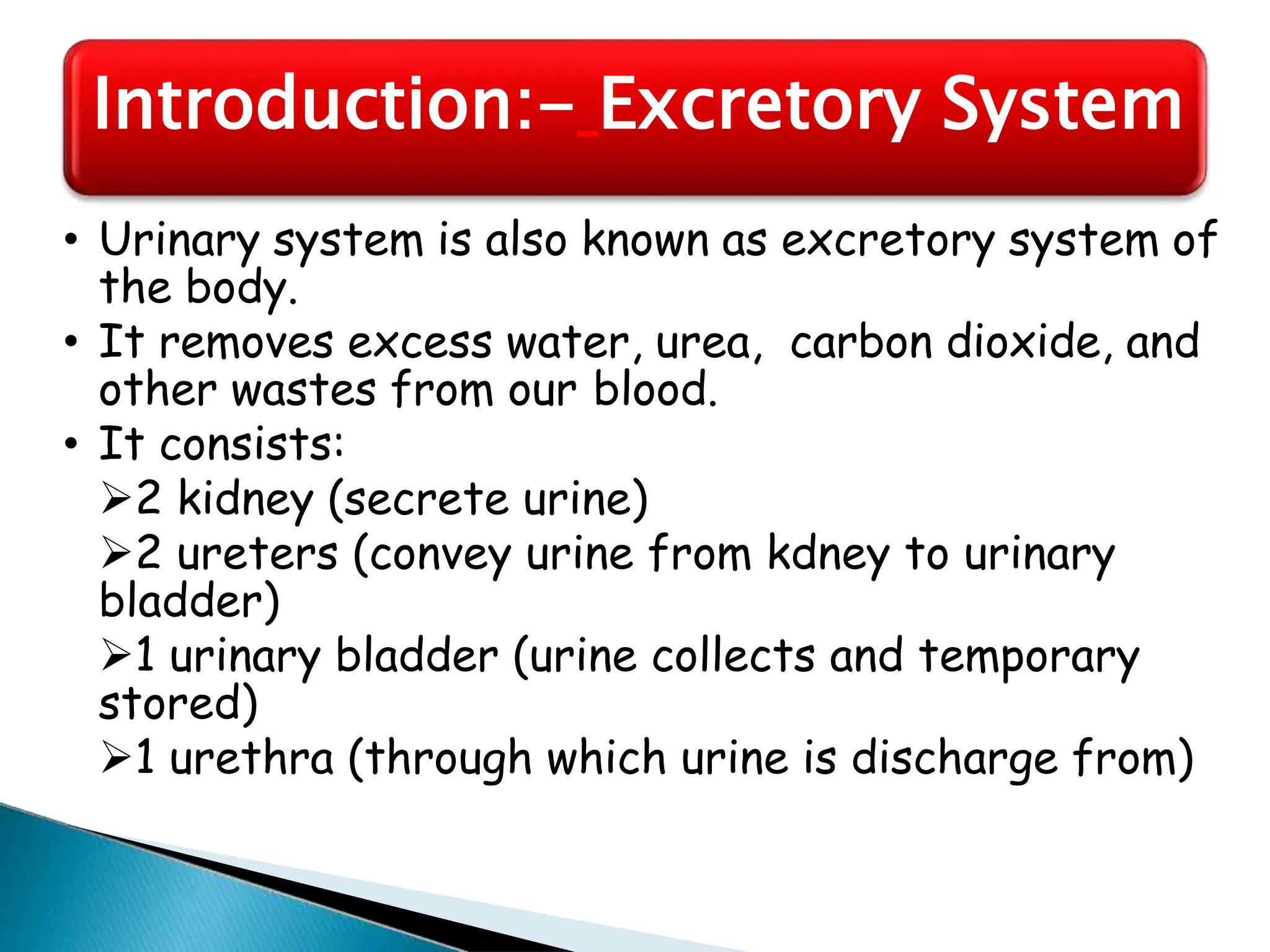
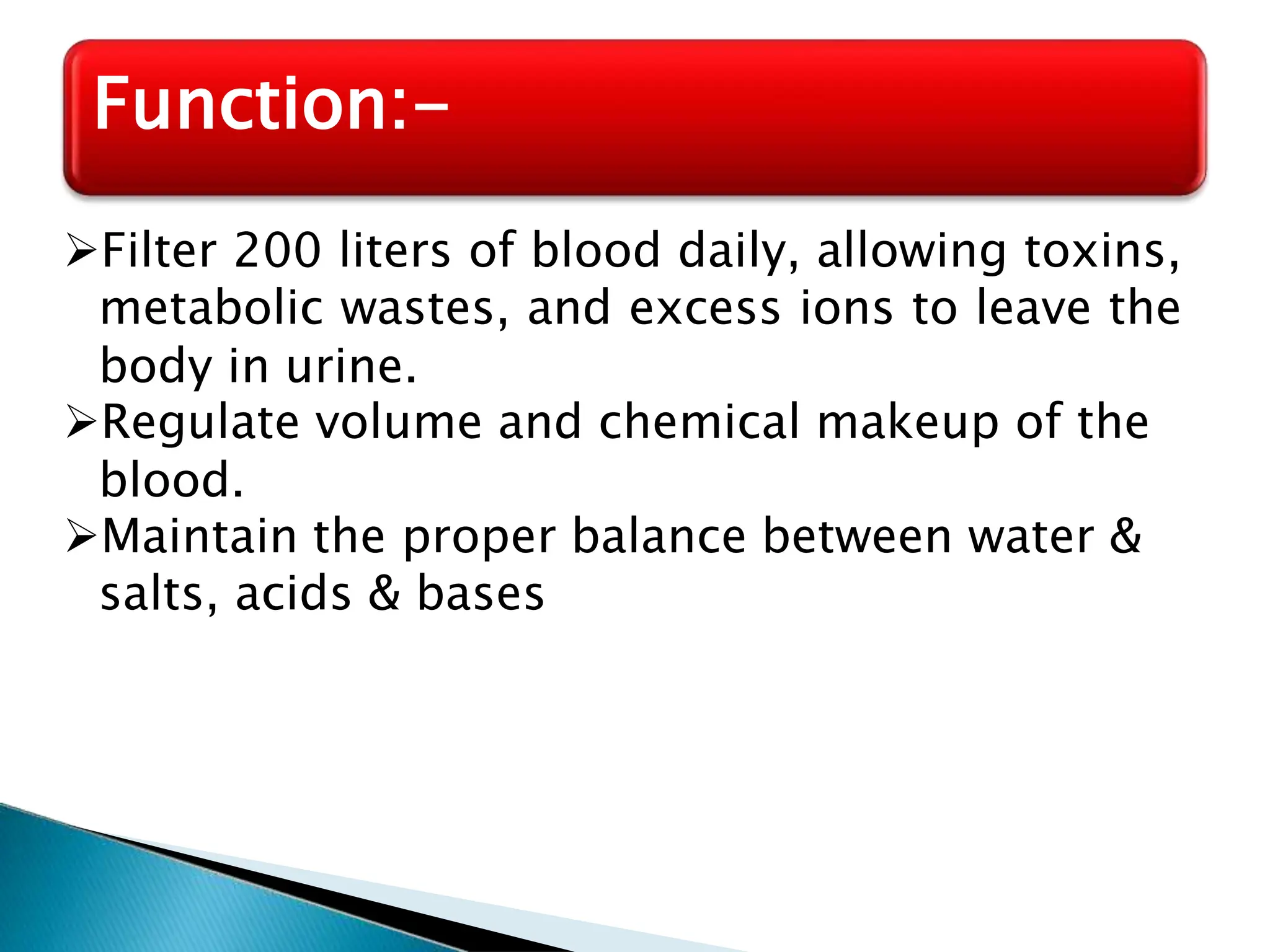
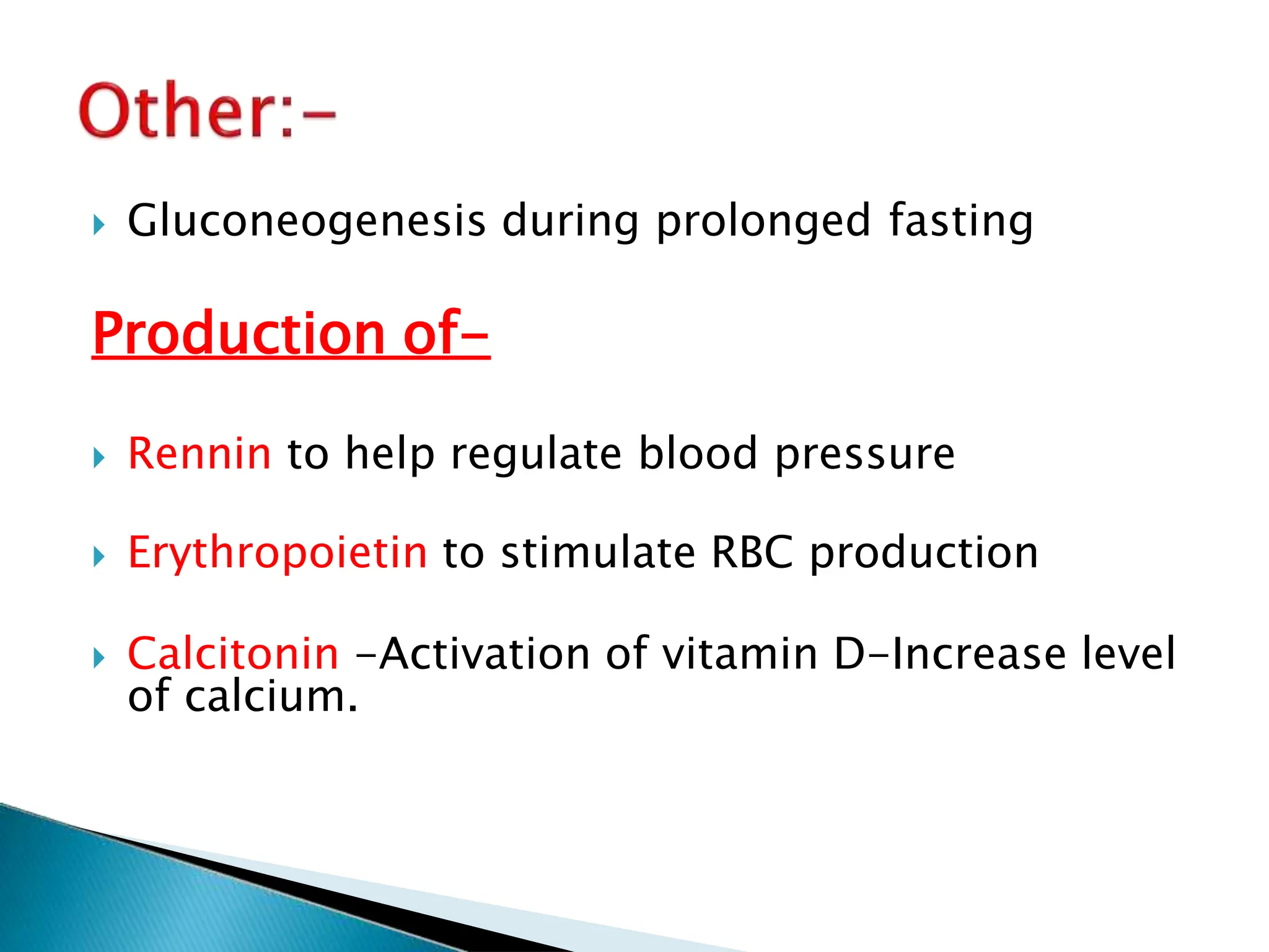
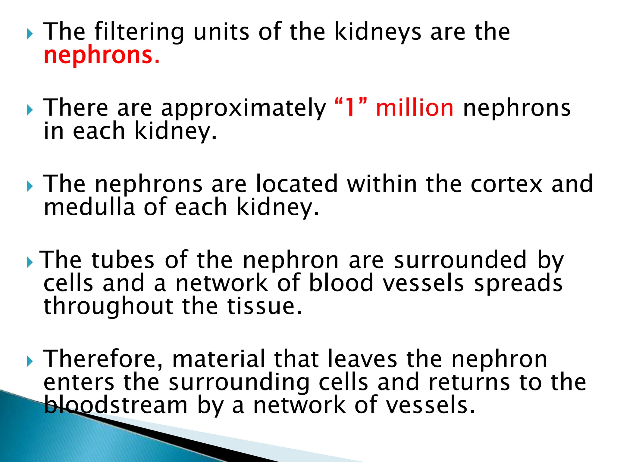
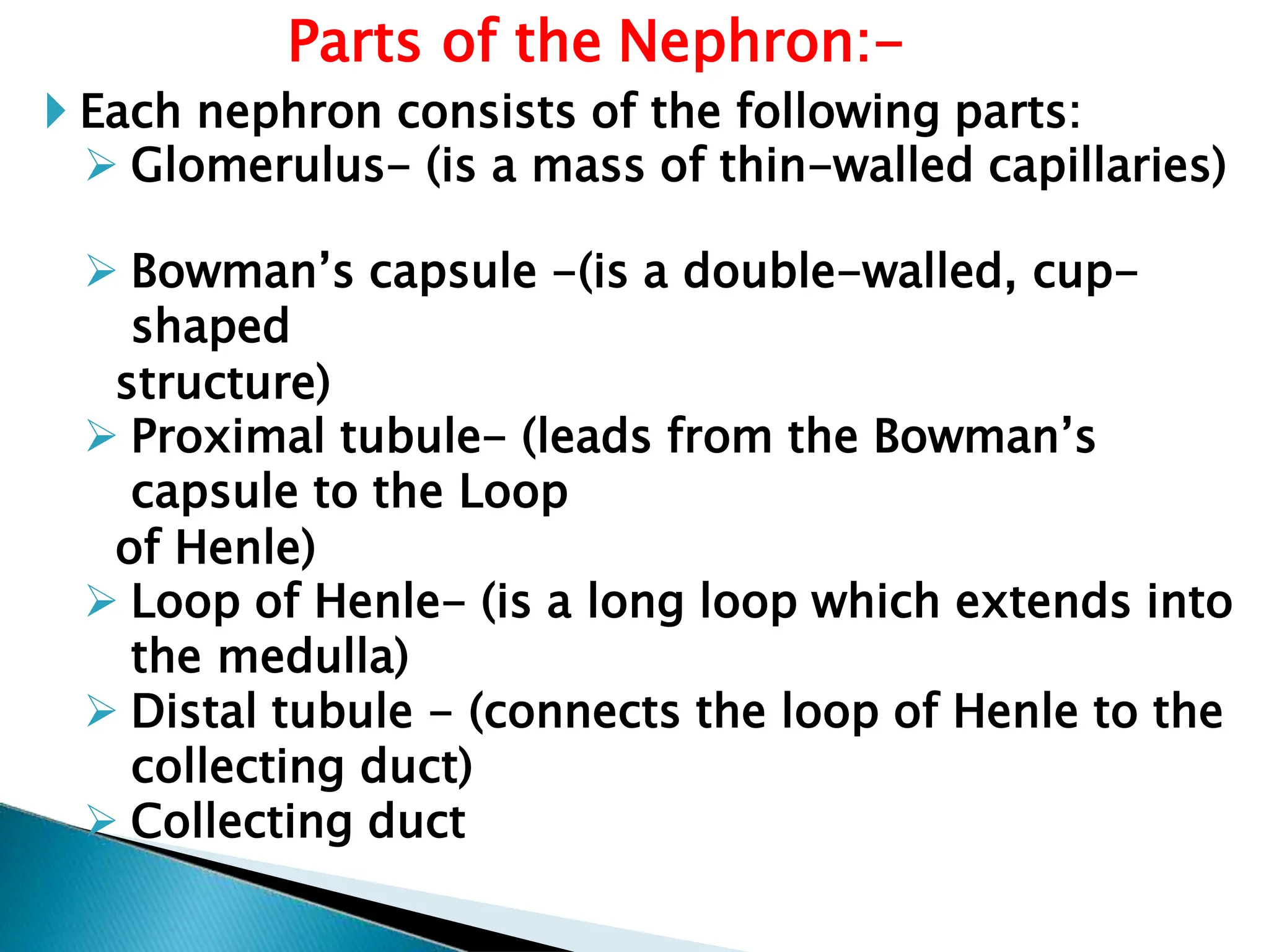
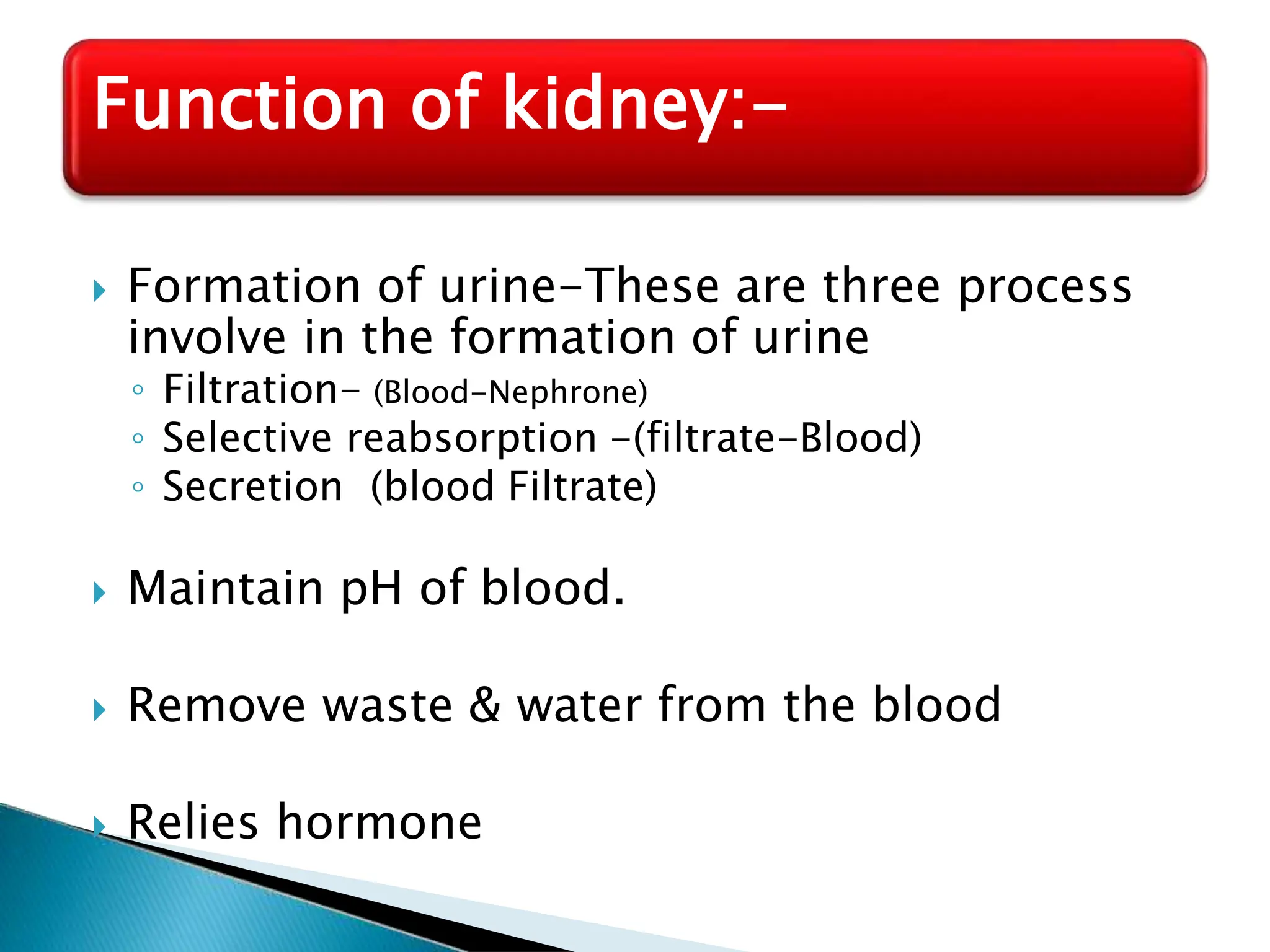
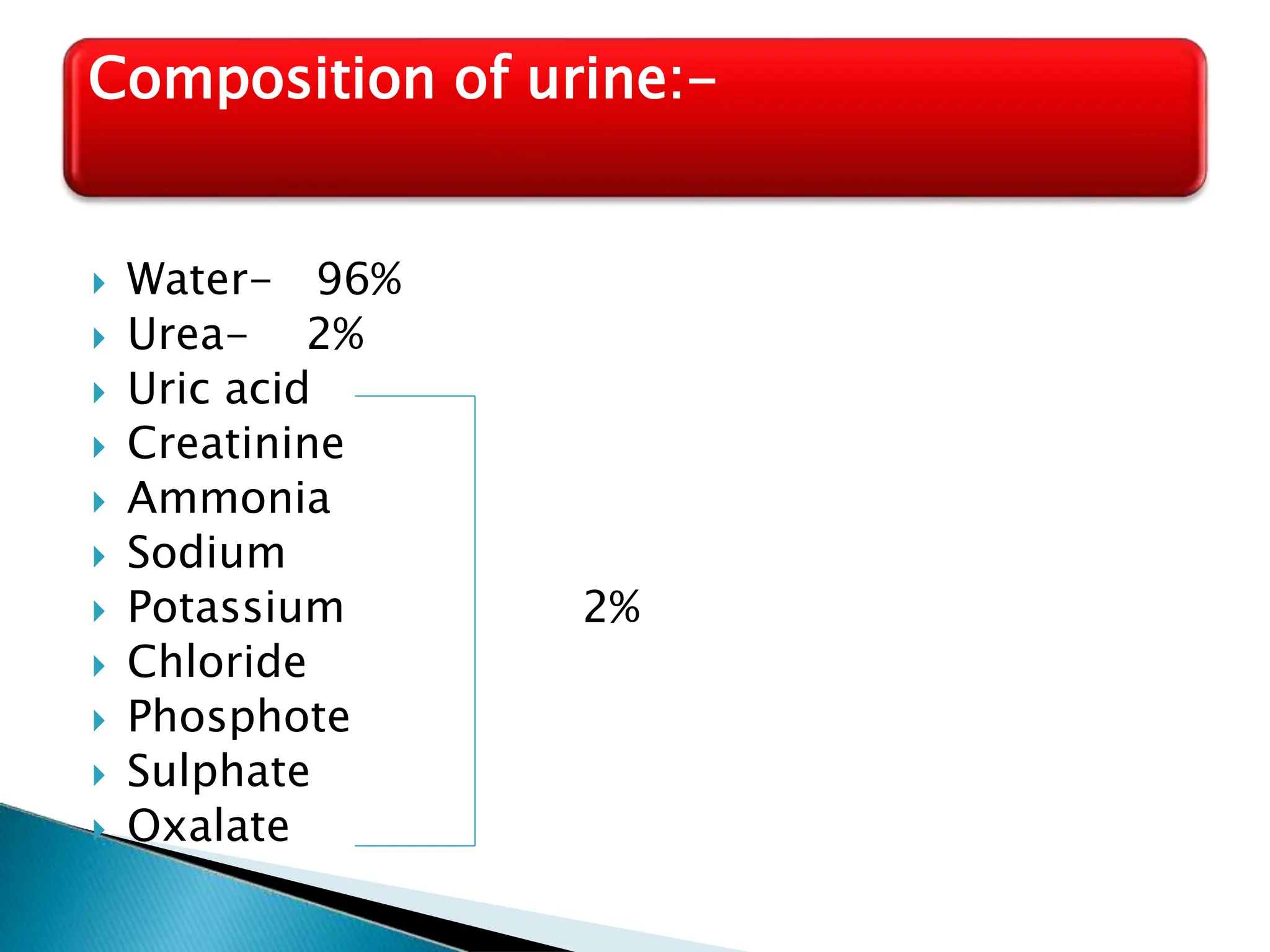
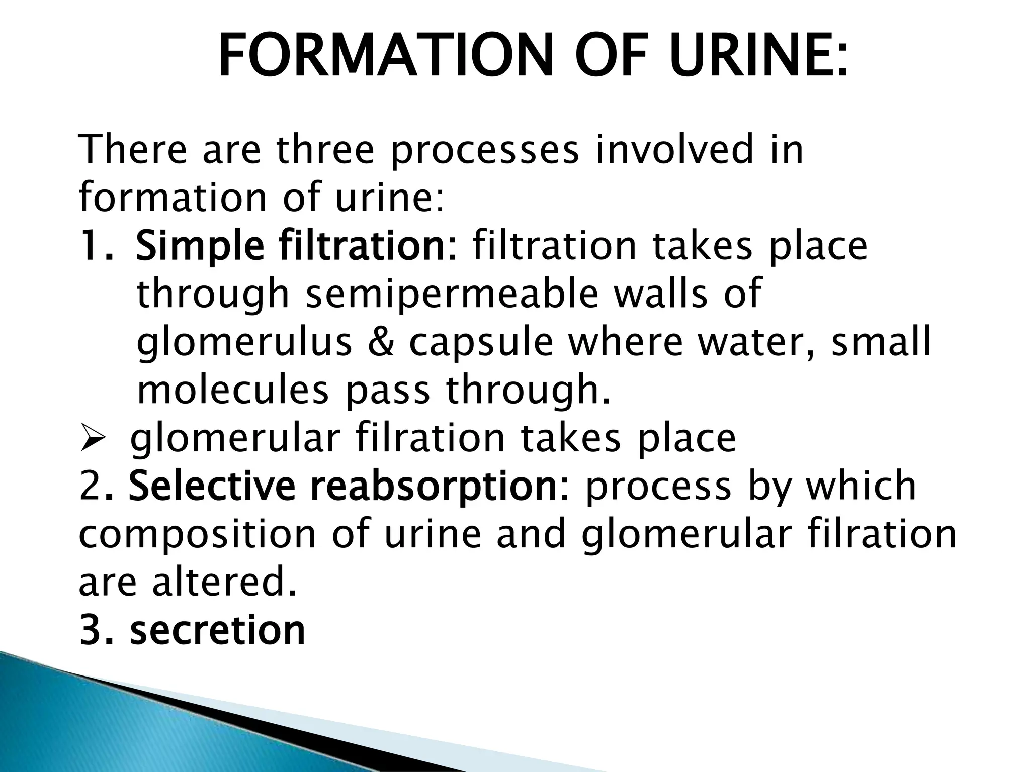
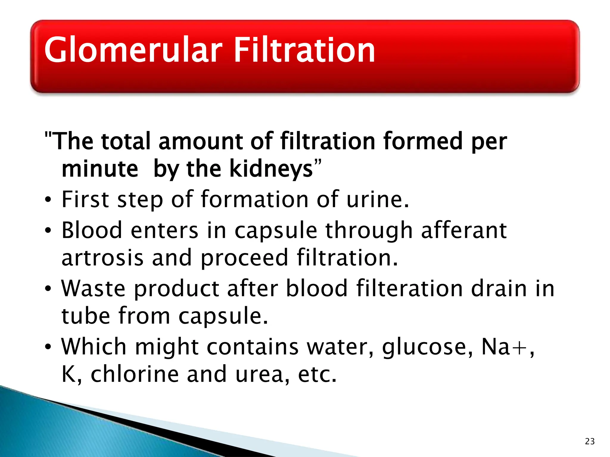
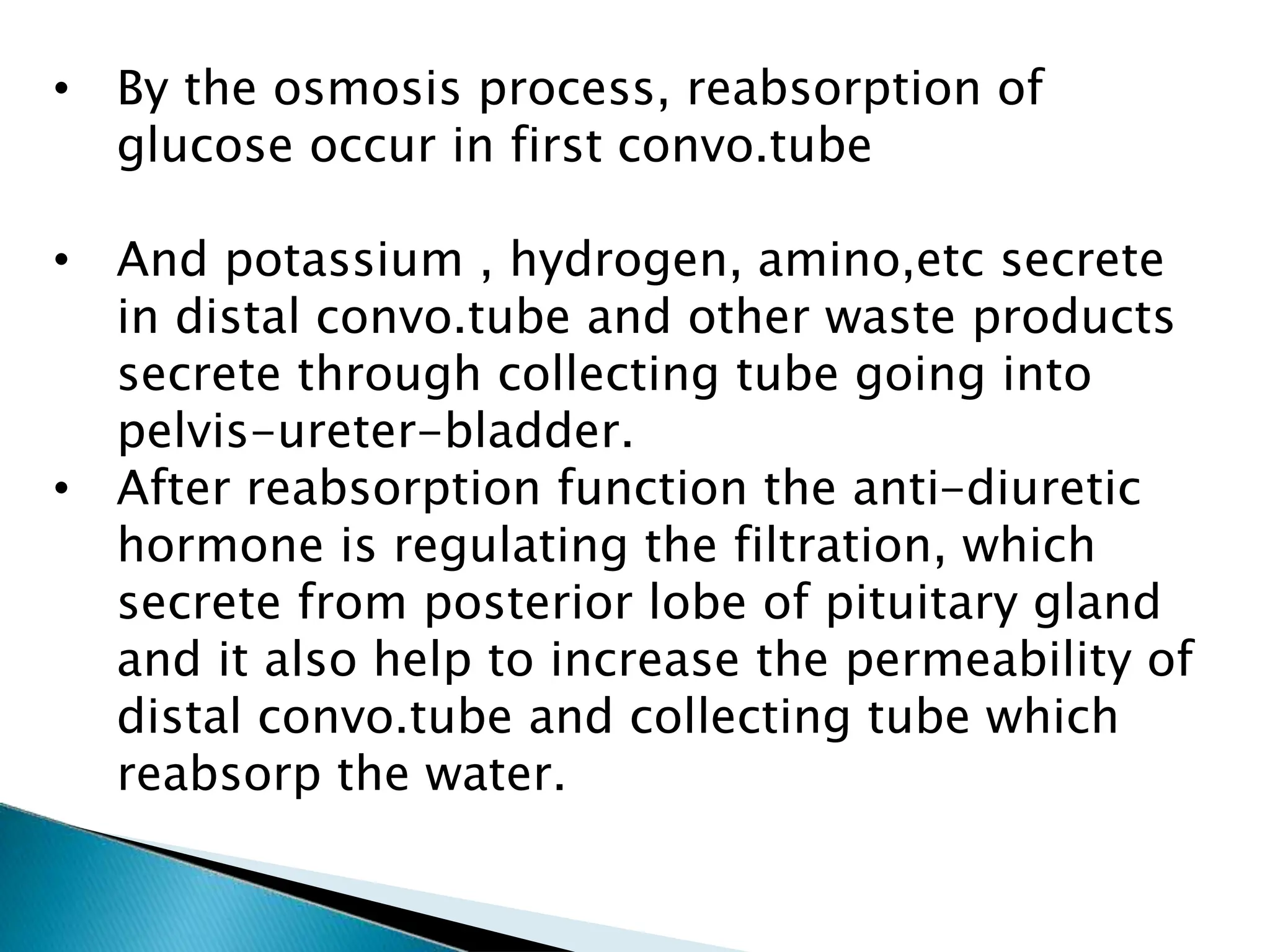
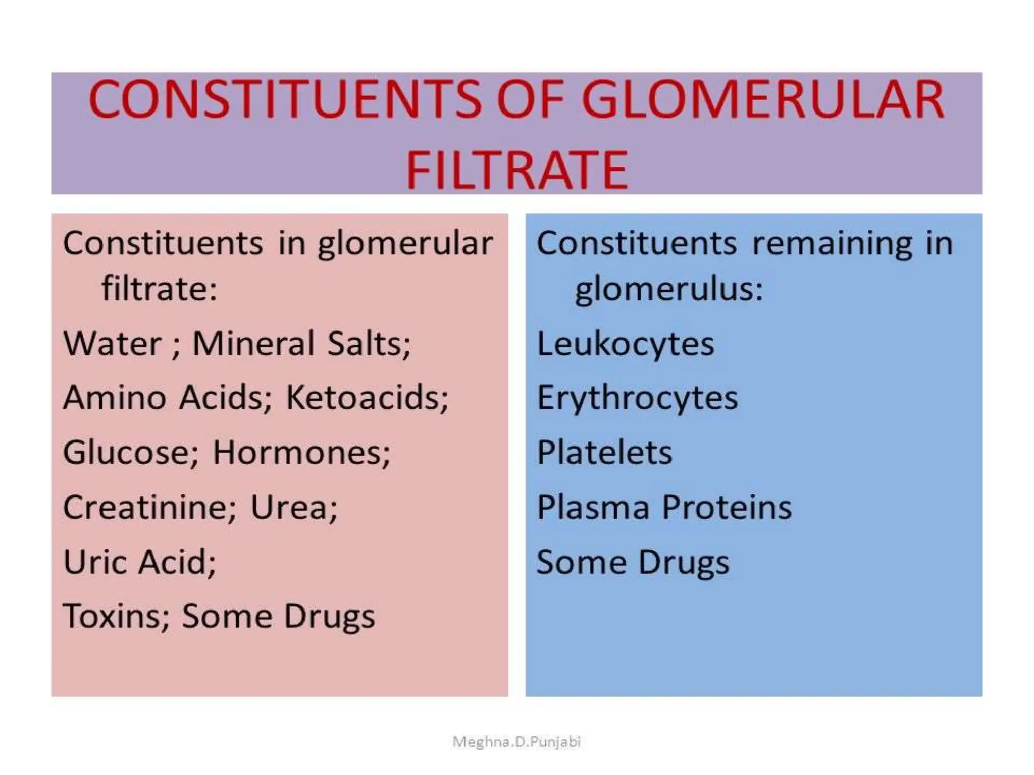
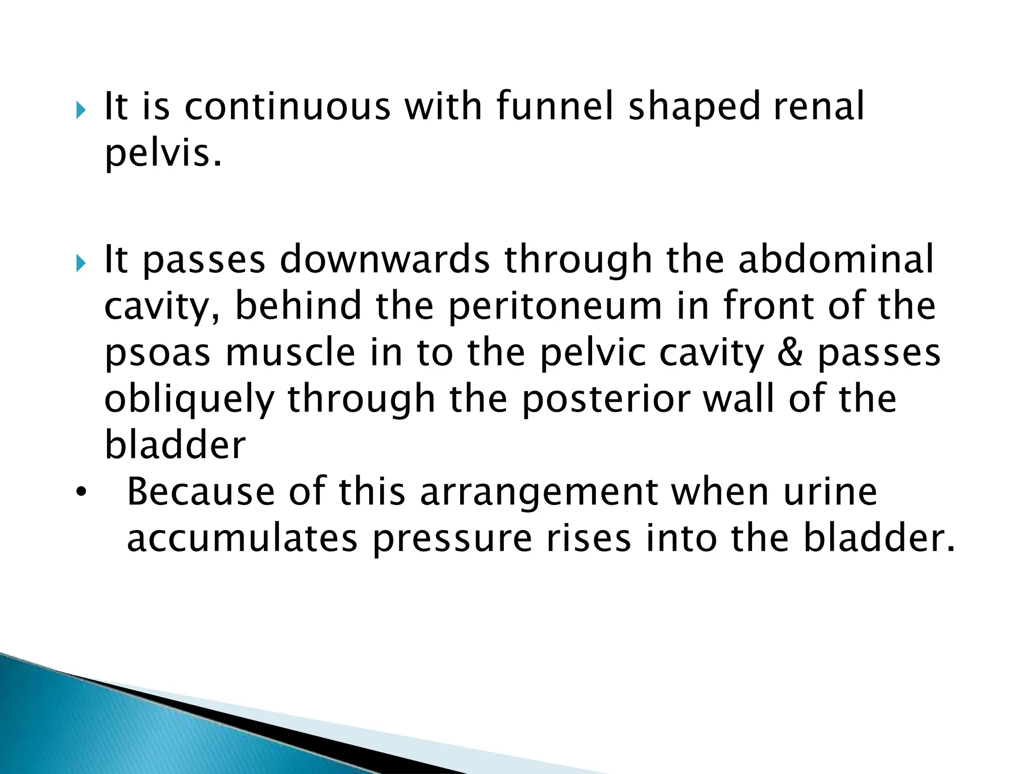
• Urinary system is also known as excretory system of
the body.
• It removes excess water, urea, carbon dioxide, and
other wastes from our blood.
• It consists:
2 kidney (secrete urine)
2 ureters (convey urine from kdney to urinary
bladder)
1 urinary bladder (urine collects and temporary
stored)
1 urethra (through which urine is discharge from)
Introduction:- Excretory System
Main functions of the kidney:
•Formation and secretion of urine
•Production and secretion of erythropoietin,
the hormone responsible for controling the
rate of formation of red blood cell
•Production and secretion of renin, an
important enzyme in the control of blood
pressure.
Types of metabolic wastes:-
Waste
Carbon Dioxide
Water
Salts
Nitrogenous wastes
Produced from
Aerobic Respiration
Aerobic Respiration
Metabolic activities
Breakdown of excess
Amino Acids & Proteins
Types of nitrogenous wastes
Ammonia (NH3)
Urea
Uric Acid Crystals
toxicity
Highly Toxic
Moderately Toxic
Minimally Toxic
The urinary system is the main excretory system
& consist of following organs...
 2 Kidneys:-Which secrete urine.
 2 Ureters:- Which convey urine from the kidney
to urinary bladder.
 1 Urinary bladder:- Where urine collect &
temporary stored.
 1 Urethra:- Through which the urine is
discharge from the urinary bladder to the
exterior.
Filter 200 liters of blood daily, allowing toxins,
metabolic wastes, and excess ions to leave the
body in urine.
Regulate volume and chemical makeup of the
blood.
Maintain the proper balance between water &
salts, acids & bases
Function:-
 Gluconeogenesis during prolonged fasting
Production of-
 Rennin to help regulate blood pressure
 Erythropoietin to stimulate RBC production
 Calcitonin -Activation of vitamin D-Increase level
of calcium.
Location:-
It occupy the Epigastric, Hypochondriac,
lumber & umbilical regions.
Vertically they extend from the upper boarder
of 12th thoracic vertebra to the centre of the
body of 3rd lumber vertebra
The right kidney is lower than the left because
of liver
There is adrenal glands are placed above both
of the kidneys.
Kidney (2)-Renal, Nephron:-
Shape:-
 It is Bean shaped organ
Size:-
 11cm long
 6 cm wide
 3 cm thick
Weight:-
 150gm in male
 135gm in female
 Colour:- Raddish brown in colour
The Parts of the Kidney:-
GROSS STRUCTURE OF KIDNEY:
 The cortex (cone-shaped) is where the blood is
filtered, Red-brown color, madeup of fibrous
tissue.
 The medulla inner most layer, pink color, consist 15-
16 pyramid conical shaped mass called renal pyramid.
 The HILUM: it is middle concave border of
kidney,which provide way to renalblood vessels,
lymphatic vessals, nerves and ureters to enter & exits.
 The Renal pelvis (funnel shaped) is a hollow cavity
where urine accumulates and drains into the ureter.
Walls of pelvis contains smooth muscles.
Nephron
 The filtering units of the kidneys are the
nephrons.
 There are approximately “1” million nephrons
in each kidney.
 The nephrons are located within the cortex and
medulla of each kidney.
 The tubes of the nephron are surrounded by
cells and a network of blood vessels spreads
throughout the tissue.
 Therefore, material that leaves the nephron
enters the surrounding cells and returns to the
bloodstream by a network of vessels.
Parts of the Nephron:-
 Each nephron consists of the following parts:
 Glomerulus- (is a mass of thin-walled capillaries)
 Bowman’s capsule -(is a double-walled, cup-
shaped
structure)
 Proximal tubule- (leads from the Bowman’s
capsule to the Loop
of Henle)
 Loop of Henle- (is a long loop which extends into
the medulla)
 Distal tubule - (connects the loop of Henle to the
collecting duct)
 Collecting duct
Microscopic structure of the kidney (Nephron):-
Flow of fluid through nephrone
Glomerulus (Bowman’s capsule)
Proximal convoluted tubule
Descending limb of loop of henle
Ascending limb of loop of henle
Distal convoluted tubule
Drain in to collecting duct
Blood
supply in
the kidney
Renal artery
Segment artery
Inter lobular artery
Afferent arteriole
Glomerular capillaries
Efferent arterioles
Inter lobule vein
Segmental vein
Renal vein
19
Blood and Nerve Supply:-
 Approximately one-fourth (1200 ml) of systemic
cardiac output flows through the kidneys each
minute.
 Arterial flow into and venous flow out of the kidneys
follow similar paths
 The nerve supply is via the renal plexus
 Formation of urine-These are three process
involve in the formation of urine
◦ Filtration- (Blood-Nephrone)
◦ Selective reabsorption -(filtrate-Blood)
◦ Secretion (blood Filtrate)
 Maintain pH of blood.
 Remove waste & water from the blood
 Relies hormone
Function of kidney:-
 Water- 96%
 Urea- 2%
 Uric acid
2%
 Creatinine
 Ammonia
 Sodium
 Potassium
 Chloride
 Phosphote
 Sulphate
 Oxalate
Composition of urine:-
FORMATION OF URINE:
There are three processes involved in
formation of urine:
1. Simple filtration: filtration takes place
through semipermeable walls of
glomerulus & capsule where water, small
molecules pass through.
 glomerular filration takes place
2. Selective reabsorption: process by which
composition of urine and glomerular filration
are altered.
3. secretion
23
Glomerular Filtration
"The total amount of filtration formed per
minute by the kidneys”
• First step of formation of urine.
• Blood enters in capsule through afferant
artrosis and proceed filtration.
• Waste product after blood filteration drain in
tube from capsule.
• Which might contains water, glucose, Na+,
K, chlorine and urea, etc.
• By the osmosis process, reabsorption of
glucose occur in first convo.tube
• And potassium , hydrogen, amino,etc secrete
in distal convo.tube and other waste products
secrete through collecting tube going into
pelvis-ureter-bladder.
• After reabsorption function the anti-diuretic
hormone is regulating the filtration, which
secrete from posterior lobe of pituitary gland
and it also help to increase the permeability of
distal convo.tube and collecting tube which
reabsorp the water.
• The para thyroid hormone of the para thyroid gand
along with calsitonin secrete from thyroid gland
together helps in reabsorption of calcium and
phosphate.
• Aldesterone hormone from the adrenal gland helps
in reabsorption of sodium and excretion of
potassium.
• Along with this it also excrete the nitrogenous
waste like urea and uric acid.
• Secretion: some of the substances cleared and
than excrete from the body in the form of urine.
• Normal GFR (Glomerular filration rate): 125ml/min
Ureters (2)
25
Introduction:-
 Slender tubes that convey urine from the
kidneys to the urinary bladder
 Ureters enter the base of the bladder
through the posterior wall.
 It is about 25-30 cm long
 It is about 3mm in diameter
 It is continuous with funnel shaped renal
pelvis.
 It passes downwards through the abdominal
cavity, behind the peritoneum in front of the
psoas muscle in to the pelvic cavity & passes
obliquely through the posterior wall of the
bladder
• Because of this arrangement when urine
accumulates pressure rises into the bladder.
Structure:-
28
Wall of ureters is consist of three layers;-
 Outer layer-Adventitia- of fibrous tissue
continuous with the fibrous capsule of the
kidney.
 Middle layer –Muscular-consisting of smooth
muscles fiber
 Inner layer - Mucosa -composed of transitional
epithelium.
Blood supply by:-Ureter receives its arterial blood
supply in three different parts, as explained
below.
 Upper part receives its blood supply from renal
artery
 Middle part receives its blood supply
from testicular or ovarian artery
 Pelvic part receives its blood supply from
the superior vesical artery
Venous drainage by:- The venous blood is
drained by veins that correspond to the arteries
explained above.
Lymph drainage by:-
 Lymph from the ureters drains into the lateral
aortic nodes and the iliac nodes.
Nerve supply by:-
 sympathetic nerves
 Propel urine to the bladder via response to
Peristaltic contraction of smooth muscle
layer.
Function of ureter:-
Urinary bladder (1)
 It is reservoir of urine
 It is pear shaped but become more oval as it
fills with the urine.
 It is a Smooth, collapsible, muscular sac that
temporarily stores urine
 It lies in the Pelvic cavity
 Total capacity is about 600ml
Introduction:-
 It lies retroperitoneal on the pelvic floor
posterior to the symphysis pubis
◦ Males – prostate gland surrounds the neck
inferiorly
◦ Females – Anterior to the vagina and
uterus
Structure:-
36
The bladder wall composed of 3 layers.
 Outer layer -of loose connective tissue-
containing blood, lymphatic vessels & nerve
covered on the upper surface by the peritoneum.
 Middle layer -Consisting of the interlacing
smooth muscle fiber & elastic tissue loosely
arranged in 3 layer is called Ditrusor muscle.
 Inner layer - Mucosa composed of transitional
epithelium
“3” Orifice of bladder wall form a Triangle or
trigone.
 The two orifice on the posterior wall are the
opening of the ureters.
 The lower orifice is opening in to the urethra.
 The bladder is distensible and collapses when
empty
 As urine accumulates, the bladder expands
without significant rise in internal pressure
Blood Supply by:-Superior & inferior vesical
arteries
Venous drainage by : Veins from the vesical
venous plexus that drain into the internal iliac
vein
Lymphatic drainage by : Into internal & external
iliac lymph nodes.
Nerve supply by:- Sympathetic & parasympathetic
nerve
Urethra (1)
 It is a canal extending from the neck of the
bladder to the exterior, at the external urethral
orifice.
 It is a longer in male then the female
 The male urethra has three named regions
◦ Prostatic urethra – runs within the prostate gland
◦ Membranous urethra – runs through the urogenital
diaphragm
◦ Spongy (penile) urethra – passes through the penis
and opens via the external urethral orifice
Introduction:-
Structure:-
 Male :- It is about “19-20” cm long
 Female :- it is about “4” cm long & “6” mm in
diameter.
 To transport urine from the bladder.
 To transport the semen (sperm cells and fluid
from the seminal vesicles and the prostate)
out the tip of the penis
Function in male urethra:-
26-46
Disorders of Urinary System
 Renal calculi
 Urinary tract infections
 Glomerular disease
 Renal failure
 Polycystic kidney disease
 The kidney has other functions but it
is usually associated with the
excretion of cellular waste such as :
 1) urea (a nitrogenous waste
produced in the liver from the
breakdown of protein. It is the main
component of urine) ;
 2) uric acid (usually produced from
breakdown of DNA or RNA) and
 3) creatinine (waste product of
muscle action).
 All of these compounds have nitrogen as a
major component.
 The kidneys are more than excretory organs.
 They are one of the major homeostatic
organs of the body.
 They control water pH, secrete erythropoietin
(a hormone that stimulates red blood cell
production) and activate vitamin D production
in the skin.
 That is why a doctor can tell so much from a
urine sample.

URINARY/ excretory SYSTEM anatomy & physiology

  • 2.
    • Urinary systemis also known as excretory system of the body. • It removes excess water, urea, carbon dioxide, and other wastes from our blood. • It consists: 2 kidney (secrete urine) 2 ureters (convey urine from kdney to urinary bladder) 1 urinary bladder (urine collects and temporary stored) 1 urethra (through which urine is discharge from) Introduction:- Excretory System
  • 3.
    Main functions ofthe kidney: •Formation and secretion of urine •Production and secretion of erythropoietin, the hormone responsible for controling the rate of formation of red blood cell •Production and secretion of renin, an important enzyme in the control of blood pressure.
  • 4.
    Types of metabolicwastes:- Waste Carbon Dioxide Water Salts Nitrogenous wastes Produced from Aerobic Respiration Aerobic Respiration Metabolic activities Breakdown of excess Amino Acids & Proteins Types of nitrogenous wastes Ammonia (NH3) Urea Uric Acid Crystals toxicity Highly Toxic Moderately Toxic Minimally Toxic
  • 6.
    The urinary systemis the main excretory system & consist of following organs...  2 Kidneys:-Which secrete urine.  2 Ureters:- Which convey urine from the kidney to urinary bladder.  1 Urinary bladder:- Where urine collect & temporary stored.  1 Urethra:- Through which the urine is discharge from the urinary bladder to the exterior.
  • 7.
    Filter 200 litersof blood daily, allowing toxins, metabolic wastes, and excess ions to leave the body in urine. Regulate volume and chemical makeup of the blood. Maintain the proper balance between water & salts, acids & bases Function:-
  • 8.
     Gluconeogenesis duringprolonged fasting Production of-  Rennin to help regulate blood pressure  Erythropoietin to stimulate RBC production  Calcitonin -Activation of vitamin D-Increase level of calcium.
  • 9.
    Location:- It occupy theEpigastric, Hypochondriac, lumber & umbilical regions. Vertically they extend from the upper boarder of 12th thoracic vertebra to the centre of the body of 3rd lumber vertebra The right kidney is lower than the left because of liver There is adrenal glands are placed above both of the kidneys. Kidney (2)-Renal, Nephron:-
  • 10.
    Shape:-  It isBean shaped organ Size:-  11cm long  6 cm wide  3 cm thick Weight:-  150gm in male  135gm in female  Colour:- Raddish brown in colour
  • 11.
    The Parts ofthe Kidney:-
  • 12.
    GROSS STRUCTURE OFKIDNEY:  The cortex (cone-shaped) is where the blood is filtered, Red-brown color, madeup of fibrous tissue.  The medulla inner most layer, pink color, consist 15- 16 pyramid conical shaped mass called renal pyramid.  The HILUM: it is middle concave border of kidney,which provide way to renalblood vessels, lymphatic vessals, nerves and ureters to enter & exits.  The Renal pelvis (funnel shaped) is a hollow cavity where urine accumulates and drains into the ureter. Walls of pelvis contains smooth muscles.
  • 13.
  • 14.
     The filteringunits of the kidneys are the nephrons.  There are approximately “1” million nephrons in each kidney.  The nephrons are located within the cortex and medulla of each kidney.  The tubes of the nephron are surrounded by cells and a network of blood vessels spreads throughout the tissue.  Therefore, material that leaves the nephron enters the surrounding cells and returns to the bloodstream by a network of vessels.
  • 15.
    Parts of theNephron:-  Each nephron consists of the following parts:  Glomerulus- (is a mass of thin-walled capillaries)  Bowman’s capsule -(is a double-walled, cup- shaped structure)  Proximal tubule- (leads from the Bowman’s capsule to the Loop of Henle)  Loop of Henle- (is a long loop which extends into the medulla)  Distal tubule - (connects the loop of Henle to the collecting duct)  Collecting duct
  • 16.
    Microscopic structure ofthe kidney (Nephron):-
  • 18.
    Flow of fluidthrough nephrone Glomerulus (Bowman’s capsule) Proximal convoluted tubule Descending limb of loop of henle Ascending limb of loop of henle Distal convoluted tubule Drain in to collecting duct
  • 19.
    Blood supply in the kidney Renalartery Segment artery Inter lobular artery Afferent arteriole Glomerular capillaries Efferent arterioles Inter lobule vein Segmental vein Renal vein
  • 20.
    19 Blood and NerveSupply:-  Approximately one-fourth (1200 ml) of systemic cardiac output flows through the kidneys each minute.  Arterial flow into and venous flow out of the kidneys follow similar paths  The nerve supply is via the renal plexus
  • 21.
     Formation ofurine-These are three process involve in the formation of urine ◦ Filtration- (Blood-Nephrone) ◦ Selective reabsorption -(filtrate-Blood) ◦ Secretion (blood Filtrate)  Maintain pH of blood.  Remove waste & water from the blood  Relies hormone Function of kidney:-
  • 22.
     Water- 96% Urea- 2%  Uric acid 2%  Creatinine  Ammonia  Sodium  Potassium  Chloride  Phosphote  Sulphate  Oxalate Composition of urine:-
  • 23.
    FORMATION OF URINE: Thereare three processes involved in formation of urine: 1. Simple filtration: filtration takes place through semipermeable walls of glomerulus & capsule where water, small molecules pass through.  glomerular filration takes place 2. Selective reabsorption: process by which composition of urine and glomerular filration are altered. 3. secretion
  • 25.
    23 Glomerular Filtration "The totalamount of filtration formed per minute by the kidneys” • First step of formation of urine. • Blood enters in capsule through afferant artrosis and proceed filtration. • Waste product after blood filteration drain in tube from capsule. • Which might contains water, glucose, Na+, K, chlorine and urea, etc.
  • 26.
    • By theosmosis process, reabsorption of glucose occur in first convo.tube • And potassium , hydrogen, amino,etc secrete in distal convo.tube and other waste products secrete through collecting tube going into pelvis-ureter-bladder. • After reabsorption function the anti-diuretic hormone is regulating the filtration, which secrete from posterior lobe of pituitary gland and it also help to increase the permeability of distal convo.tube and collecting tube which reabsorp the water.
  • 27.
    • The parathyroid hormone of the para thyroid gand along with calsitonin secrete from thyroid gland together helps in reabsorption of calcium and phosphate. • Aldesterone hormone from the adrenal gland helps in reabsorption of sodium and excretion of potassium. • Along with this it also excrete the nitrogenous waste like urea and uric acid. • Secretion: some of the substances cleared and than excrete from the body in the form of urine. • Normal GFR (Glomerular filration rate): 125ml/min
  • 29.
  • 30.
    25 Introduction:-  Slender tubesthat convey urine from the kidneys to the urinary bladder  Ureters enter the base of the bladder through the posterior wall.  It is about 25-30 cm long  It is about 3mm in diameter
  • 31.
     It iscontinuous with funnel shaped renal pelvis.  It passes downwards through the abdominal cavity, behind the peritoneum in front of the psoas muscle in to the pelvic cavity & passes obliquely through the posterior wall of the bladder • Because of this arrangement when urine accumulates pressure rises into the bladder.
  • 32.
  • 33.
    28 Wall of uretersis consist of three layers;-  Outer layer-Adventitia- of fibrous tissue continuous with the fibrous capsule of the kidney.  Middle layer –Muscular-consisting of smooth muscles fiber  Inner layer - Mucosa -composed of transitional epithelium.
  • 34.
    Blood supply by:-Ureterreceives its arterial blood supply in three different parts, as explained below.  Upper part receives its blood supply from renal artery  Middle part receives its blood supply from testicular or ovarian artery  Pelvic part receives its blood supply from the superior vesical artery Venous drainage by:- The venous blood is drained by veins that correspond to the arteries explained above.
  • 35.
    Lymph drainage by:- Lymph from the ureters drains into the lateral aortic nodes and the iliac nodes. Nerve supply by:-  sympathetic nerves
  • 36.
     Propel urineto the bladder via response to Peristaltic contraction of smooth muscle layer. Function of ureter:-
  • 37.
  • 38.
     It isreservoir of urine  It is pear shaped but become more oval as it fills with the urine.  It is a Smooth, collapsible, muscular sac that temporarily stores urine  It lies in the Pelvic cavity  Total capacity is about 600ml Introduction:-
  • 39.
     It liesretroperitoneal on the pelvic floor posterior to the symphysis pubis ◦ Males – prostate gland surrounds the neck inferiorly ◦ Females – Anterior to the vagina and uterus
  • 42.
  • 43.
    36 The bladder wallcomposed of 3 layers.  Outer layer -of loose connective tissue- containing blood, lymphatic vessels & nerve covered on the upper surface by the peritoneum.  Middle layer -Consisting of the interlacing smooth muscle fiber & elastic tissue loosely arranged in 3 layer is called Ditrusor muscle.  Inner layer - Mucosa composed of transitional epithelium
  • 45.
    “3” Orifice ofbladder wall form a Triangle or trigone.  The two orifice on the posterior wall are the opening of the ureters.  The lower orifice is opening in to the urethra.  The bladder is distensible and collapses when empty  As urine accumulates, the bladder expands without significant rise in internal pressure
  • 47.
    Blood Supply by:-Superior& inferior vesical arteries Venous drainage by : Veins from the vesical venous plexus that drain into the internal iliac vein Lymphatic drainage by : Into internal & external iliac lymph nodes. Nerve supply by:- Sympathetic & parasympathetic nerve
  • 48.
  • 49.
     It isa canal extending from the neck of the bladder to the exterior, at the external urethral orifice.  It is a longer in male then the female  The male urethra has three named regions ◦ Prostatic urethra – runs within the prostate gland ◦ Membranous urethra – runs through the urogenital diaphragm ◦ Spongy (penile) urethra – passes through the penis and opens via the external urethral orifice Introduction:-
  • 50.
  • 51.
     Male :-It is about “19-20” cm long  Female :- it is about “4” cm long & “6” mm in diameter.
  • 52.
     To transporturine from the bladder.  To transport the semen (sperm cells and fluid from the seminal vesicles and the prostate) out the tip of the penis Function in male urethra:-
  • 54.
    26-46 Disorders of UrinarySystem  Renal calculi  Urinary tract infections  Glomerular disease  Renal failure  Polycystic kidney disease
  • 55.
     The kidneyhas other functions but it is usually associated with the excretion of cellular waste such as :  1) urea (a nitrogenous waste produced in the liver from the breakdown of protein. It is the main component of urine) ;  2) uric acid (usually produced from breakdown of DNA or RNA) and  3) creatinine (waste product of muscle action).
  • 56.
     All ofthese compounds have nitrogen as a major component.  The kidneys are more than excretory organs.  They are one of the major homeostatic organs of the body.  They control water pH, secrete erythropoietin (a hormone that stimulates red blood cell production) and activate vitamin D production in the skin.  That is why a doctor can tell so much from a urine sample.