Download to read offline


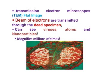
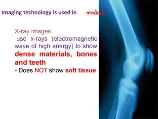
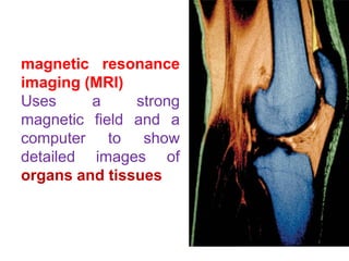
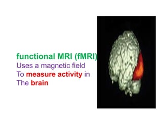
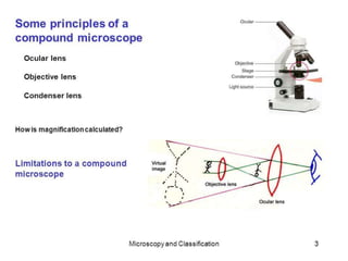
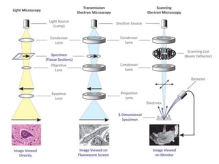
Technology provides biologists with new tools to study living and non-living specimens. Microscopes use light and lenses to provide enlarged images, with scanning electron microscopes using electrons to generate 3D images of non-living surfaces. Transmission electron microscopes also use electrons to create flat, highly magnified images that can see viruses and nanoparticles by transmitting beams through dead specimens. Imaging technology in medicine includes X-rays to view dense bones and teeth, magnetic resonance imaging to show detailed images of organs and tissues, and functional MRI to measure brain activity.


