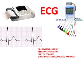
1. ECG
DR. GAYATRI D. HANDE
ASSISTANT PROFESSOR
DEPT OF ZOOLOGY
SHRI SHIVAJI SCIENCE COLLEGE, AMRAVATI
2.
3.
4.
5.
6. Electrocardiographs detect the electrical
signals associated with cardiac activity and
produce an ECG, a graphic record of the voltage
versus time.
They are used to diagnose and assist in
treating some types of heart disease and
arrhythmias, determine a patient’s response to
drug therapy, and reveal trends or changes in
heart function.
7.
8.
9. Multichannel electrocardiographs record
signals from two or more leads
simultaneously and are frequently used in
place of single-channel units.
Some electrocardiographs can perform
automatic measurement and interpretation
of the ECG as a selectable or optional
feature.
29. P Wave
• Atrial activation begins in the SA node
– Spreads in radial fashion to depolarize the right
atrium, interatrial septum, then the left atrium
– Last area of the left atrium to be activated is the
tip of the left atrial appendage
• Normal amplitude
– Seldom exceeds 0.25 mV (2.5 small squares)
normally in limb leads
– In precordial leads, positive component is
normally less than 0.15 mV