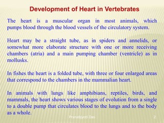
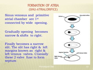
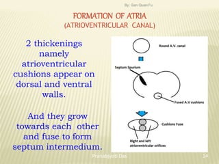
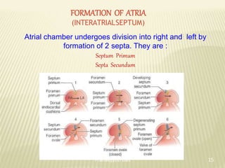
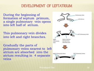
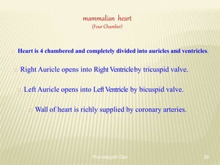
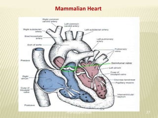
Evolution
OF HEART
Pranabjyoti Das
Arunodoi Jr College
Development of Heart in Vertebrates
The heart is a muscular organ in most animals, which
pumps blood through the blood vessels of the circulatory system.
Heart may be a straight tube, as in spiders and annelids, or
somewhat more elaborate structure with one or more receiving
chambers (atria) and a main pumping chamber (ventricle) as in
mollusks.
In fishes the heart is a folded tube, with three or four enlarged areas
that correspond to the chambers in the mammalian heart.
In animals with lungs like amphibians, reptiles, birds, and
mammals, the heart shows various stages of evolution from a single
to a double pump that circulates blood to the lungs and to the body
as a whole.
2
Pranabjyoti Das
The cardiovascular system is one of the first body systems to
appear within the embryo. The heart is an unpaired organ but its
origin is bilateral. In an embryo the mesenchyme forms a group
of endocardial cells below the pharynx. These cells become
arranged to form a pair of thin endothelial tubes. The two
endothelial tubes soon fuse to form a single endocardial tube
lying longitudinally below the pharynx.
3
Pranabjyoti Das
The splanchnic mesoderm lying below the endoderm gets folded
longitudinally around the endocardial tube. This two-layered tube
will form the heart in which the splanchnic mesoderm thickens to
form a myocardium or muscular wall of the heart and an outer
thin epicardium or visceral pericardium. The endocardial tube
becomes the lining of the heart known as endocardium.
4
Pranabjyoti Das
Folds of splanchnic mesoderm meet above to form a dorsal
mesocardium which suspends the heart in the coelom. Soon a
transverse septum is formed behind the heart which divides the
coelom into two chambers, an anterior pericardial cavity
enclosing the heart and a posterior abdominal cavity.
5
Pranabjyoti Das
Originally the heart is a
straight tube but it
starts increasing in its
length and becomes S-
shaped because its ends
are fixed.
Appearance of valves,
constriction, partitions
in the heart and
differential thickenings
of its walls form three or
four chambers in the
heart
6
Pranabjyoti Das
Primitive Heart Tube
The development of the heart
begins with the formation of
the primitive heart tube by
following the folding of the
embryo.
At first lateral folding creates
the heart tube by bringing
together two precursor regions,
and then cephalocaudal folding
positions the heart tube in the
future thorax.
7
Pranabjyoti Das
Initially, the heart tube is suspended within the pericardial cavity
by a membrane which subsequently degenerates to allow for
further growth. From superior to inferior, the primitive heart tube
is now comprised of six distinct regions.
They are…
1. Aortic roots
2. Truncus arteriosus
3. Bulbus cordis
4. Ventricle
5. Atrium
6. Sinus venosus
8
Pranabjyoti Das
The heart tube continues to elongate, and begins looping during
its development. The bulbus cordis first moves ventrally, then
caudally, and to the right. The caudal portion, i.e. the primitive
ventricle starts moveing dorsally, cranially and finally to the left
This process produces an S-shape that is much closer to the fully
developed heart.
9
Pranabjyoti Das
HEART TUBE
In the 1st diagram the right and left endothelial heart tubes are
seen. And in the 2nd diagram we can see that these two tubes
have fused together form a single tube.
The single tube shows series of dilatation like Bulbus Cordis,
Primitive Ventricle, Primitive Atrium and Sinus Venosus
10
Pranabjyoti Das
BULBUSCORDIS
Lies at arterial end of heart , it is
divisible into:
Proximal Part = Conus
Distal Part = Truncus arteriosus
Truncus arteriosus continuous
distally with aortic sac.
Aortic Sac is continuous distally
with right and left pharyngeal arch
arteries.
Arteries arch backwards to become
continuous with right and left dorsal
aortae.
11
Pranabjyoti Das
SINUS VENOSUS
Lies at venous end of
heart, it has right and
left prolongations
(horns) of sinus
venosus.
Each Horns are joined by:
One vitelline vein
One umbilical vein
One common cardinal vein
12
Pranabjyoti Das
FORMATION OF ATRIA
(SINU-ATRIALORIFICE)
Sinus venosus and primitive
atrial chamber are 1st
connected by wide opening.
Gradually opening becomes
narrow & shifts to right.
Finally becomes a narrow
slit. The slit has right & left
margins known as right &
left venous valves. Cranially
these 2 valve fuse to form
septum
13
Pranabjyoti Das
FORMATION OF ATRIA
(ATRIOVENTRICULAR CANAL)
2 thickenings
namely
atrioventricular
cushions appear on
dorsal and ventral
walls.
And they grow
towards each other
and fuse to form
septum intermedium.
By: Gan Quan Fu
14
Pranabjyoti Das
FORMATION OF ATRIA
(INTERATRIALSEPTUM)
Atrial chamber undergoes division into right and left by
formation of 2 septa. They are :
Septum Primum
Septa Secundum
15
Pranabjyoti Das
DEVELOPMENT OF LEFTATRIUM
During the beginning of
formation of septum primum,
a single pulmonary vein opens
into left half of atrium.
This pulmonary vein divides
into left and right branches.
Gradually the parts of
pulmonary veins nearest to left
atrium are absorbed into the
atrium resulting in 4 separate
veins
16
Pranabjyoti Das
SEPARATION OF AORTA
Spiral Septum appears within truncus arteriosus and
subdivides it into ascending aorta and pulmonary arteries.
They fuse along the axis of the cylinder extending down
towards the ventricles.
Interventricular foramen obliterated by mass of endocardial
tissues from interventricular septum, endocardial cushions &
Spiral Septum.
17
Pranabjyoti Das
DEVELOPMENT OFVENTRICLES
(INTERVENTRICULAR SEPTUM)
Conus of bulbus cordis merge with cavity
of primitive ventricle.
Interventricular septum grows upwards
from the floor of bulbo- ventricular cavity &
divide it into right & left halves.
Two ridges from the wall of the conical part
of bulbar cavity grows towards each other
and fuse form bulbar septum.
The gap between upper edge of
interventricular septum and lower edge
of bulbar septum is filled by cushions.
18
Pranabjyoti Das
Single-Chambered Heart
In Amphioxus (primitive chordate), a true heart is not found. A
part of ventral aorta beneath the pharynx is muscular and
contractile in nature. This muscular body in amphioxus acts as a
heart.
19
Pranabjyoti Das
The heart of fish has only two chambers, namely an auricle and ventricle.
On the dorsal side of the auricle there is a sac-like sinuous which receives
blood from all parts of the body. This sinuous opens into the auricle by an
aperture. Auricle communicate with the ventricle.
Ventricle is continued ventrally and forward by another swollen part called
conus arteriosus. Conus arteriosus extend forward as the ventral aorta
Two Chambered Heart
20
Pranabjyoti Das
In cyclostomes, there are four chambers arranged in a linear order- a
thin-walled sinus venosus, a slightly muscular atrium (auricle), a muscular
ventricle and a muscular conus arteriosus or bulbus cordis. It lies in the body
cavity in which other visceral organs are also present. Out of four chambers,
only atrium and ventricle correspond to the four chambers of the higher
vertebrates. In the evolution of heart many changes have taken place.
Two-Chambered Heart
21
Pranabjyoti Das
The heart of frog (amphibians) has 2 auricles and one ventricle.
2 auricles are seperted by an interauricular septum.
Sinus venosus on the dorsal side, opens into the right auricle
through the sinu- auricular aperature.
Left auricle- contains oxygenated blood.
The 2 auricles open into the only ventricle by a common aperature
which is guarded by the auriculo-ventricular valve
Three Chambered Heart
22
Pranabjyoti Das
Three Chambered Heart
In amphibians, the dorsal atrium shifts anterior to ventricle. The sinus venosus
opens into right atrium dorsally and not posteriorly. The atrium is completely
divided into right and left chambers and has no foramen ovale in the inter-
auricular septum, which remains open in dipnoans. Deep pockets develop in
the ventricular cavity. The conus arteriosus divides into systemic and
pulmonary vessels by a spiral valve. In lung less salamanders, the inter- atrial
septum is incomplete and pulmonary veins are absent.
23
Pranabjyoti Das
Avian heart
(Four chambered)
Birds have a 4-chambered heart with 2 atria and 2 ventricles,
with complete separation of oxygenated and de-oxygenated
blood.
The right ventricle pumps blood to the lungs, while the left
ventricle pumps blood to the rest of the body.
The three vena cavae or two precavals and a post caval empty
the blood directly into the right auricle. The left auricle receives
four pulmonary veins from the lungs.
The right auriculo ventricular aperture is guarded by two large
muscular flap like valve and the left by three valves.
24
Pranabjyoti Das
Heart of a Bird
In birds, the ventricle is completely divided into two, so that the heart is four
chambered. There is complete separation of venous and arterial blood. The
systemic aorta leaves the left ventricle and carries blood to the head and body.
While the pulmonary artery leaves the right ventricle and carries blood to the
lungs for oxygenation.
25
Pranabjyoti Das
mammalian heart
(Four Chamber)
Heart is 4 chambered and completely divided into auricles and ventricles.
Right Auricle opens into RightVentricleby tricuspid valve.
Left Auricle opens into LeftVentricle by bicuspid valve.
Wall of heart is richly supplied by coronary arteries.
26
Pranabjyoti Das
Mammalian Heart
27
Pranabjyoti Das
Convergence is the tendency of independent species to
evolve similarly when yhey are subjected to the same
environmental conditions.
The primitive blueprint for the heart and circulatory system
emerged with the arrival of the third mesodermal germ layer
in bilaterally symetrical animals. Since then, they have
evolved from a single layered tube to a multiple chambered
heart in the course of time.
Although the heart is physiologically specific to the anatomy
of each individual species, it has evolved along the same
directional path.
conclusion
28
Pranabjyoti Das
29
Pranabjyoti Das

Evolution of Heart

  • 1.
  • 2.
    Development of Heartin Vertebrates The heart is a muscular organ in most animals, which pumps blood through the blood vessels of the circulatory system. Heart may be a straight tube, as in spiders and annelids, or somewhat more elaborate structure with one or more receiving chambers (atria) and a main pumping chamber (ventricle) as in mollusks. In fishes the heart is a folded tube, with three or four enlarged areas that correspond to the chambers in the mammalian heart. In animals with lungs like amphibians, reptiles, birds, and mammals, the heart shows various stages of evolution from a single to a double pump that circulates blood to the lungs and to the body as a whole. 2 Pranabjyoti Das
  • 3.
    The cardiovascular systemis one of the first body systems to appear within the embryo. The heart is an unpaired organ but its origin is bilateral. In an embryo the mesenchyme forms a group of endocardial cells below the pharynx. These cells become arranged to form a pair of thin endothelial tubes. The two endothelial tubes soon fuse to form a single endocardial tube lying longitudinally below the pharynx. 3 Pranabjyoti Das
  • 4.
    The splanchnic mesodermlying below the endoderm gets folded longitudinally around the endocardial tube. This two-layered tube will form the heart in which the splanchnic mesoderm thickens to form a myocardium or muscular wall of the heart and an outer thin epicardium or visceral pericardium. The endocardial tube becomes the lining of the heart known as endocardium. 4 Pranabjyoti Das
  • 5.
    Folds of splanchnicmesoderm meet above to form a dorsal mesocardium which suspends the heart in the coelom. Soon a transverse septum is formed behind the heart which divides the coelom into two chambers, an anterior pericardial cavity enclosing the heart and a posterior abdominal cavity. 5 Pranabjyoti Das
  • 6.
    Originally the heartis a straight tube but it starts increasing in its length and becomes S- shaped because its ends are fixed. Appearance of valves, constriction, partitions in the heart and differential thickenings of its walls form three or four chambers in the heart 6 Pranabjyoti Das
  • 7.
    Primitive Heart Tube Thedevelopment of the heart begins with the formation of the primitive heart tube by following the folding of the embryo. At first lateral folding creates the heart tube by bringing together two precursor regions, and then cephalocaudal folding positions the heart tube in the future thorax. 7 Pranabjyoti Das
  • 8.
    Initially, the hearttube is suspended within the pericardial cavity by a membrane which subsequently degenerates to allow for further growth. From superior to inferior, the primitive heart tube is now comprised of six distinct regions. They are… 1. Aortic roots 2. Truncus arteriosus 3. Bulbus cordis 4. Ventricle 5. Atrium 6. Sinus venosus 8 Pranabjyoti Das
  • 9.
    The heart tubecontinues to elongate, and begins looping during its development. The bulbus cordis first moves ventrally, then caudally, and to the right. The caudal portion, i.e. the primitive ventricle starts moveing dorsally, cranially and finally to the left This process produces an S-shape that is much closer to the fully developed heart. 9 Pranabjyoti Das
  • 10.
    HEART TUBE In the1st diagram the right and left endothelial heart tubes are seen. And in the 2nd diagram we can see that these two tubes have fused together form a single tube. The single tube shows series of dilatation like Bulbus Cordis, Primitive Ventricle, Primitive Atrium and Sinus Venosus 10 Pranabjyoti Das
  • 11.
    BULBUSCORDIS Lies at arterialend of heart , it is divisible into: Proximal Part = Conus Distal Part = Truncus arteriosus Truncus arteriosus continuous distally with aortic sac. Aortic Sac is continuous distally with right and left pharyngeal arch arteries. Arteries arch backwards to become continuous with right and left dorsal aortae. 11 Pranabjyoti Das
  • 12.
    SINUS VENOSUS Lies atvenous end of heart, it has right and left prolongations (horns) of sinus venosus. Each Horns are joined by: One vitelline vein One umbilical vein One common cardinal vein 12 Pranabjyoti Das
  • 13.
    FORMATION OF ATRIA (SINU-ATRIALORIFICE) Sinusvenosus and primitive atrial chamber are 1st connected by wide opening. Gradually opening becomes narrow & shifts to right. Finally becomes a narrow slit. The slit has right & left margins known as right & left venous valves. Cranially these 2 valve fuse to form septum 13 Pranabjyoti Das
  • 14.
    FORMATION OF ATRIA (ATRIOVENTRICULARCANAL) 2 thickenings namely atrioventricular cushions appear on dorsal and ventral walls. And they grow towards each other and fuse to form septum intermedium. By: Gan Quan Fu 14 Pranabjyoti Das
  • 15.
    FORMATION OF ATRIA (INTERATRIALSEPTUM) Atrialchamber undergoes division into right and left by formation of 2 septa. They are : Septum Primum Septa Secundum 15 Pranabjyoti Das
  • 16.
    DEVELOPMENT OF LEFTATRIUM Duringthe beginning of formation of septum primum, a single pulmonary vein opens into left half of atrium. This pulmonary vein divides into left and right branches. Gradually the parts of pulmonary veins nearest to left atrium are absorbed into the atrium resulting in 4 separate veins 16 Pranabjyoti Das
  • 17.
    SEPARATION OF AORTA SpiralSeptum appears within truncus arteriosus and subdivides it into ascending aorta and pulmonary arteries. They fuse along the axis of the cylinder extending down towards the ventricles. Interventricular foramen obliterated by mass of endocardial tissues from interventricular septum, endocardial cushions & Spiral Septum. 17 Pranabjyoti Das
  • 18.
    DEVELOPMENT OFVENTRICLES (INTERVENTRICULAR SEPTUM) Conusof bulbus cordis merge with cavity of primitive ventricle. Interventricular septum grows upwards from the floor of bulbo- ventricular cavity & divide it into right & left halves. Two ridges from the wall of the conical part of bulbar cavity grows towards each other and fuse form bulbar septum. The gap between upper edge of interventricular septum and lower edge of bulbar septum is filled by cushions. 18 Pranabjyoti Das
  • 19.
    Single-Chambered Heart In Amphioxus(primitive chordate), a true heart is not found. A part of ventral aorta beneath the pharynx is muscular and contractile in nature. This muscular body in amphioxus acts as a heart. 19 Pranabjyoti Das
  • 20.
    The heart offish has only two chambers, namely an auricle and ventricle. On the dorsal side of the auricle there is a sac-like sinuous which receives blood from all parts of the body. This sinuous opens into the auricle by an aperture. Auricle communicate with the ventricle. Ventricle is continued ventrally and forward by another swollen part called conus arteriosus. Conus arteriosus extend forward as the ventral aorta Two Chambered Heart 20 Pranabjyoti Das
  • 21.
    In cyclostomes, thereare four chambers arranged in a linear order- a thin-walled sinus venosus, a slightly muscular atrium (auricle), a muscular ventricle and a muscular conus arteriosus or bulbus cordis. It lies in the body cavity in which other visceral organs are also present. Out of four chambers, only atrium and ventricle correspond to the four chambers of the higher vertebrates. In the evolution of heart many changes have taken place. Two-Chambered Heart 21 Pranabjyoti Das
  • 22.
    The heart offrog (amphibians) has 2 auricles and one ventricle. 2 auricles are seperted by an interauricular septum. Sinus venosus on the dorsal side, opens into the right auricle through the sinu- auricular aperature. Left auricle- contains oxygenated blood. The 2 auricles open into the only ventricle by a common aperature which is guarded by the auriculo-ventricular valve Three Chambered Heart 22 Pranabjyoti Das
  • 23.
    Three Chambered Heart Inamphibians, the dorsal atrium shifts anterior to ventricle. The sinus venosus opens into right atrium dorsally and not posteriorly. The atrium is completely divided into right and left chambers and has no foramen ovale in the inter- auricular septum, which remains open in dipnoans. Deep pockets develop in the ventricular cavity. The conus arteriosus divides into systemic and pulmonary vessels by a spiral valve. In lung less salamanders, the inter- atrial septum is incomplete and pulmonary veins are absent. 23 Pranabjyoti Das
  • 24.
    Avian heart (Four chambered) Birdshave a 4-chambered heart with 2 atria and 2 ventricles, with complete separation of oxygenated and de-oxygenated blood. The right ventricle pumps blood to the lungs, while the left ventricle pumps blood to the rest of the body. The three vena cavae or two precavals and a post caval empty the blood directly into the right auricle. The left auricle receives four pulmonary veins from the lungs. The right auriculo ventricular aperture is guarded by two large muscular flap like valve and the left by three valves. 24 Pranabjyoti Das
  • 25.
    Heart of aBird In birds, the ventricle is completely divided into two, so that the heart is four chambered. There is complete separation of venous and arterial blood. The systemic aorta leaves the left ventricle and carries blood to the head and body. While the pulmonary artery leaves the right ventricle and carries blood to the lungs for oxygenation. 25 Pranabjyoti Das
  • 26.
    mammalian heart (Four Chamber) Heartis 4 chambered and completely divided into auricles and ventricles. Right Auricle opens into RightVentricleby tricuspid valve. Left Auricle opens into LeftVentricle by bicuspid valve. Wall of heart is richly supplied by coronary arteries. 26 Pranabjyoti Das
  • 27.
  • 28.
    Convergence is thetendency of independent species to evolve similarly when yhey are subjected to the same environmental conditions. The primitive blueprint for the heart and circulatory system emerged with the arrival of the third mesodermal germ layer in bilaterally symetrical animals. Since then, they have evolved from a single layered tube to a multiple chambered heart in the course of time. Although the heart is physiologically specific to the anatomy of each individual species, it has evolved along the same directional path. conclusion 28 Pranabjyoti Das
  • 29.