Downloaded 1,079 times


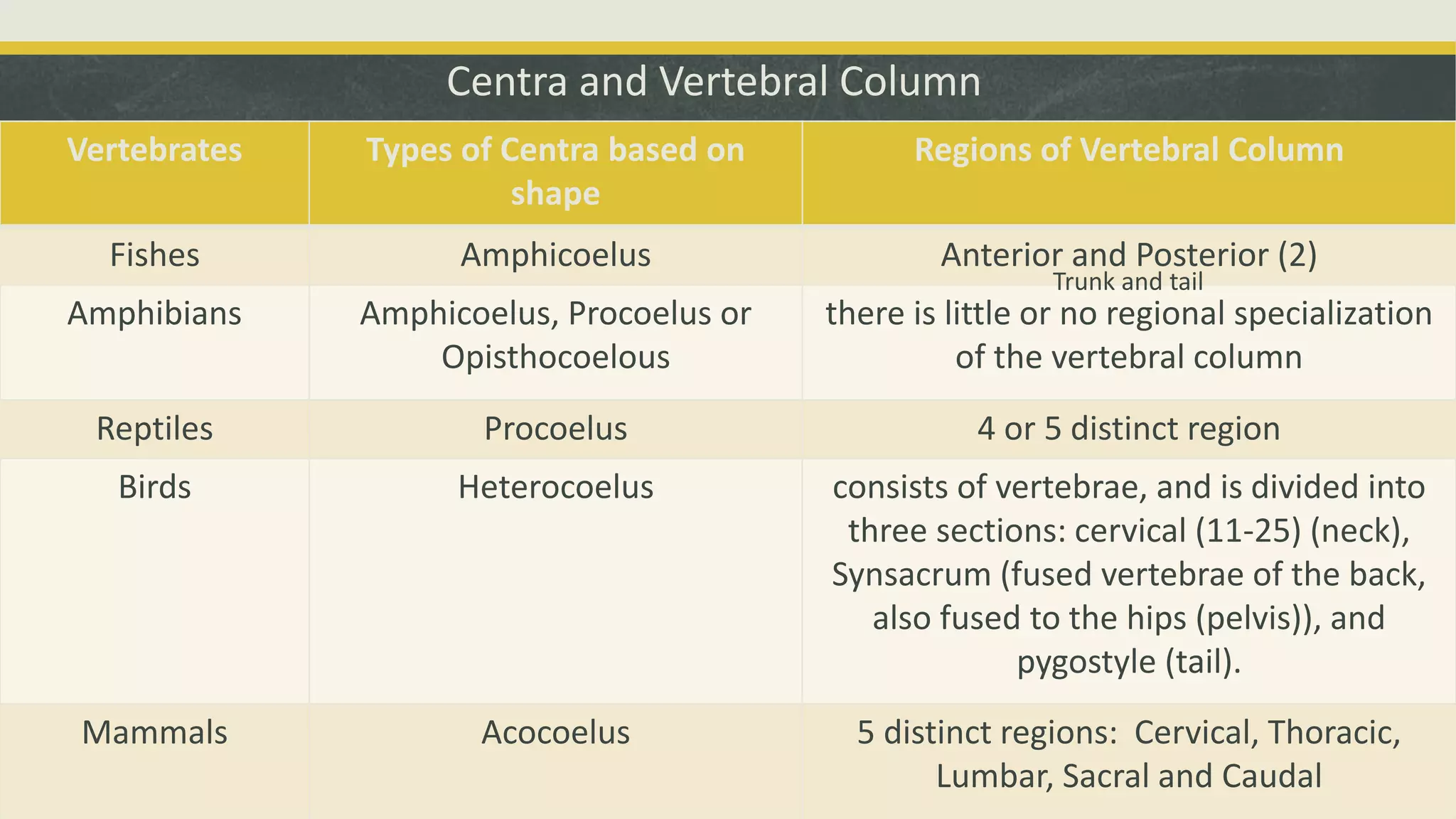
![Figure 2.33. Comparison of vertebrae of primitive
tetrapods and modern amniotes. The rachitomous type
(shown also in cross section, X.S.) occurred in
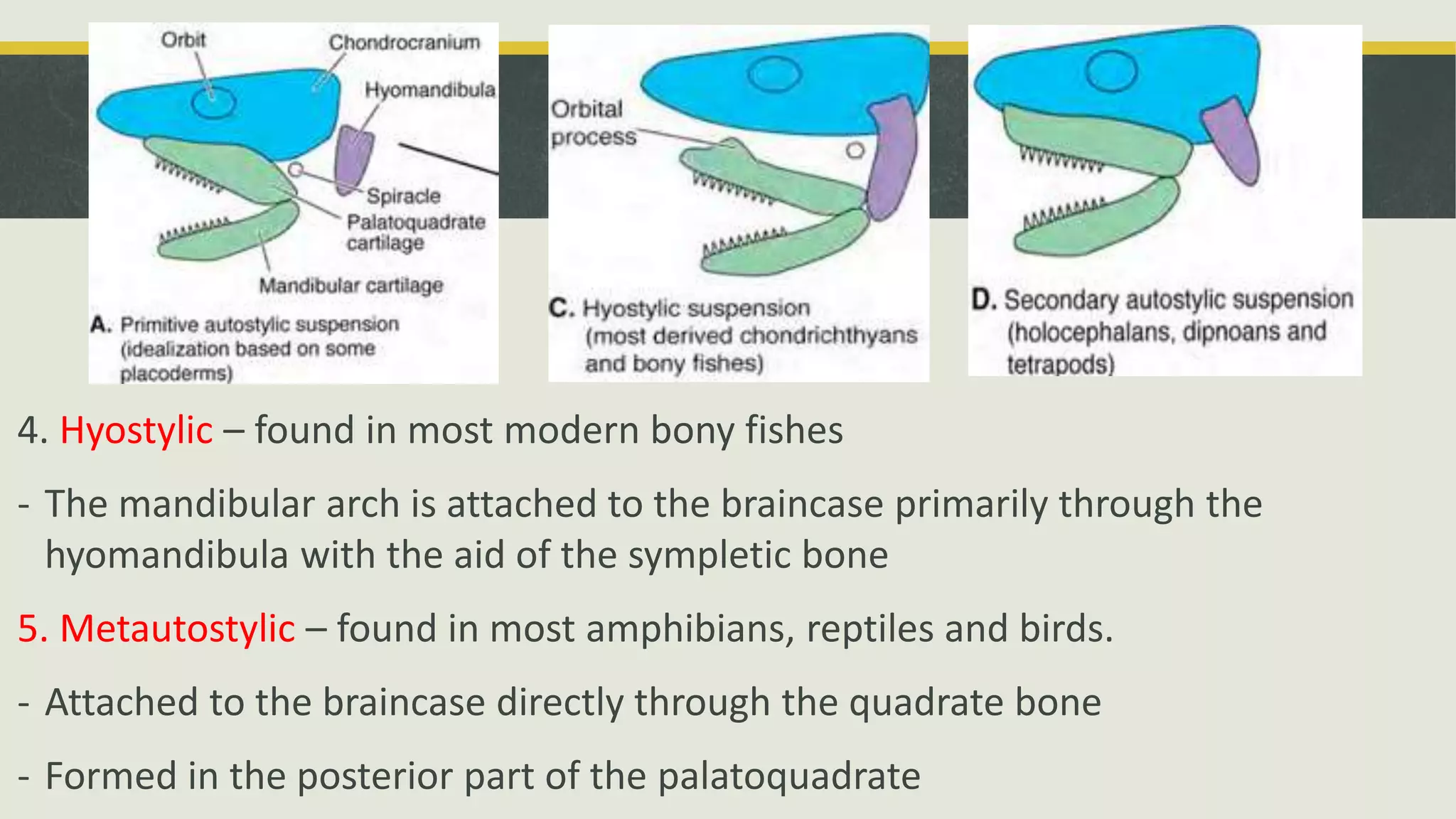
crossopterygians and in the earliest amphibians. B is from
a labyrinthodont in the reptile line. B1 and B2 are from
other labyrinthodonts. Whether the modern amphibian
centrum represents a hypocentrum (diagonal lines) or a
pleurocentrum (stippled) is not certain. The unmarked part
of the vertebra is the neural arch. Adapted, with
permission, from Kent, G. C. 4th ed. Comparative
anatomy of the vertebrates. St. Louis: C. V. Mosby Co.;
1978. [134]](https://image.slidesharecdn.com/group2-skeletalsystem-140802130106-phpapp01/75/Comparative-Anatomy-Skeletal-System-34-2048.jpg)
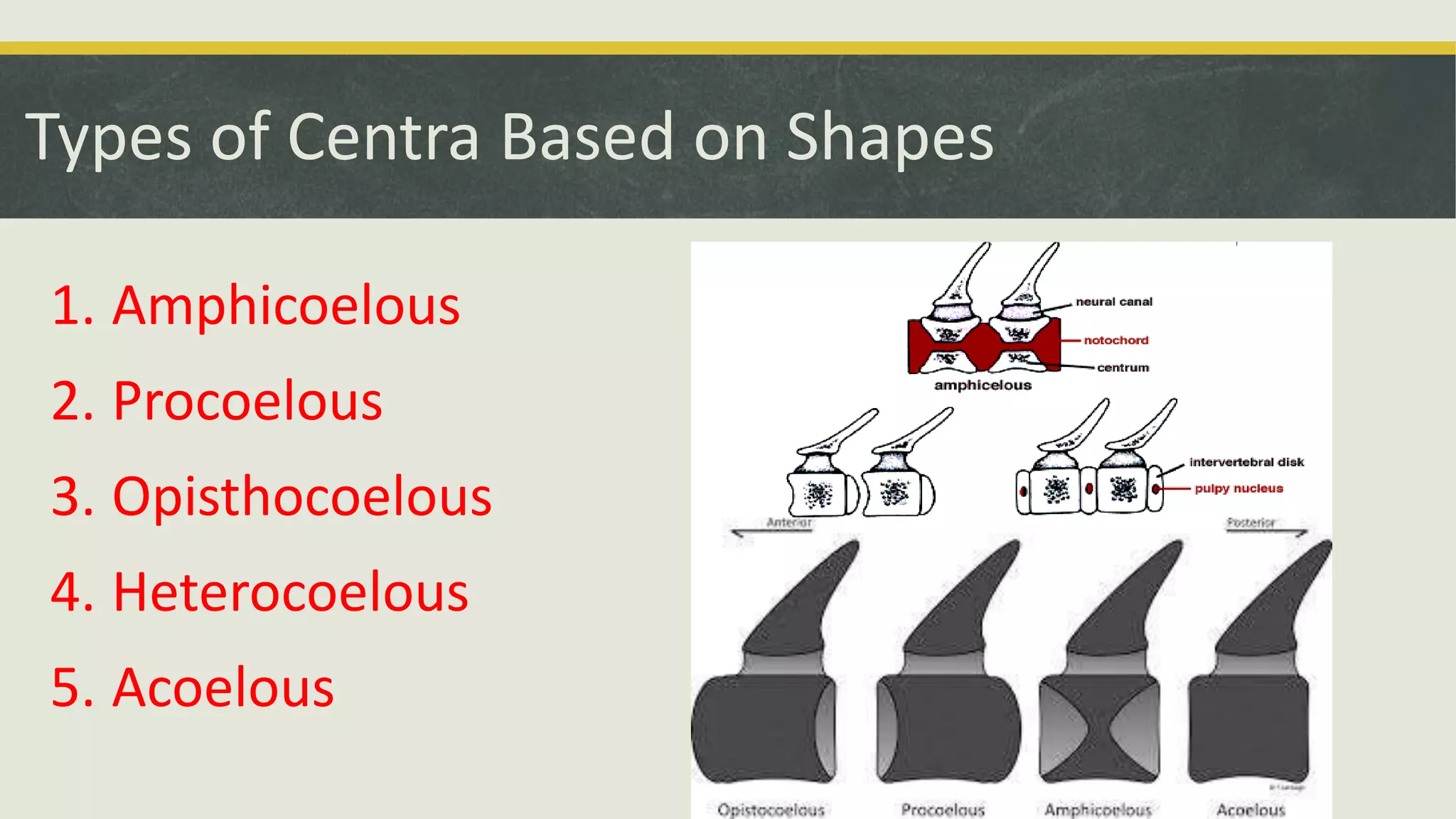
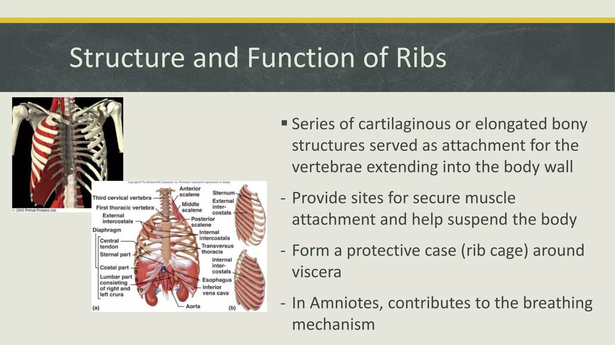
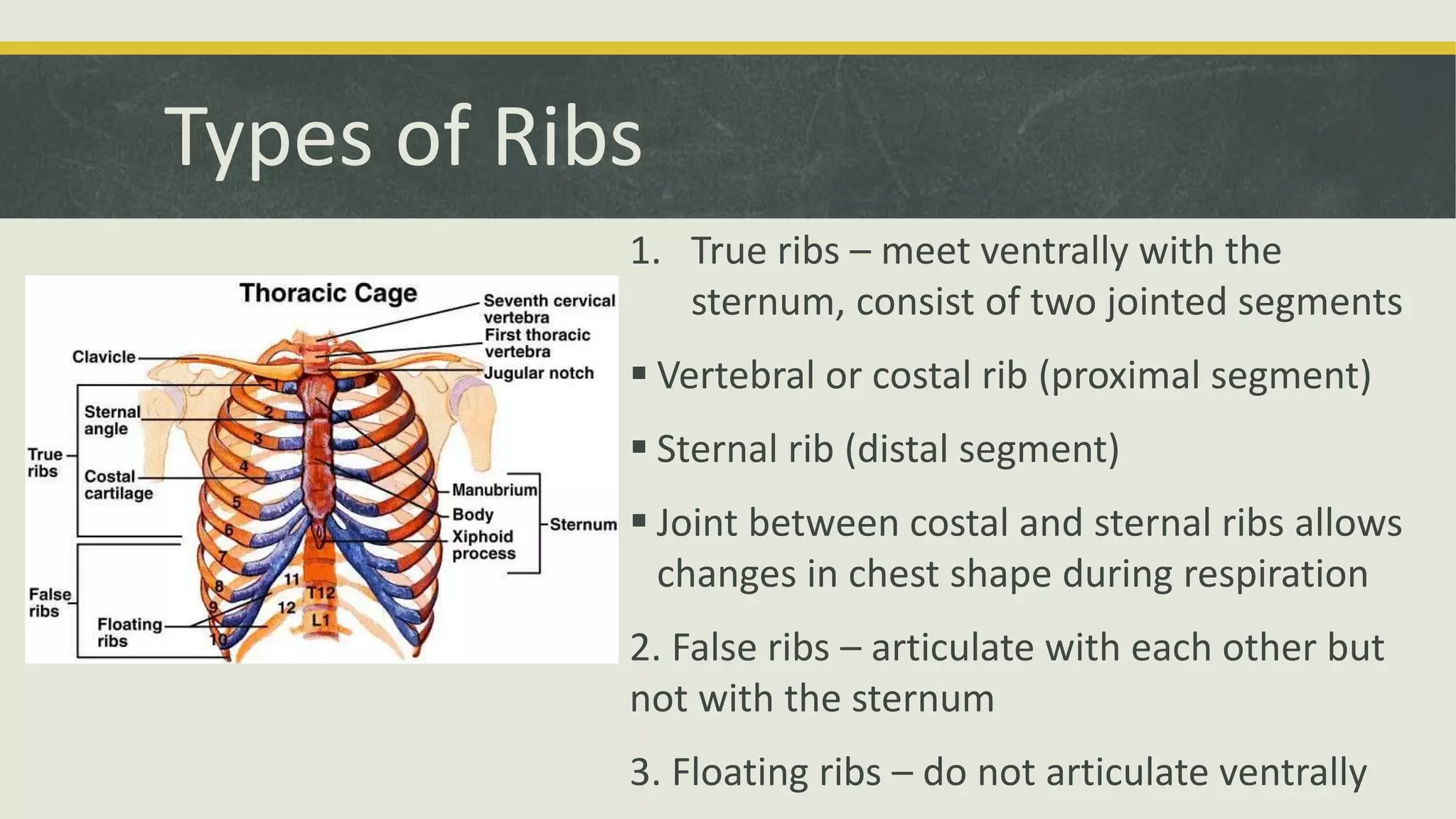

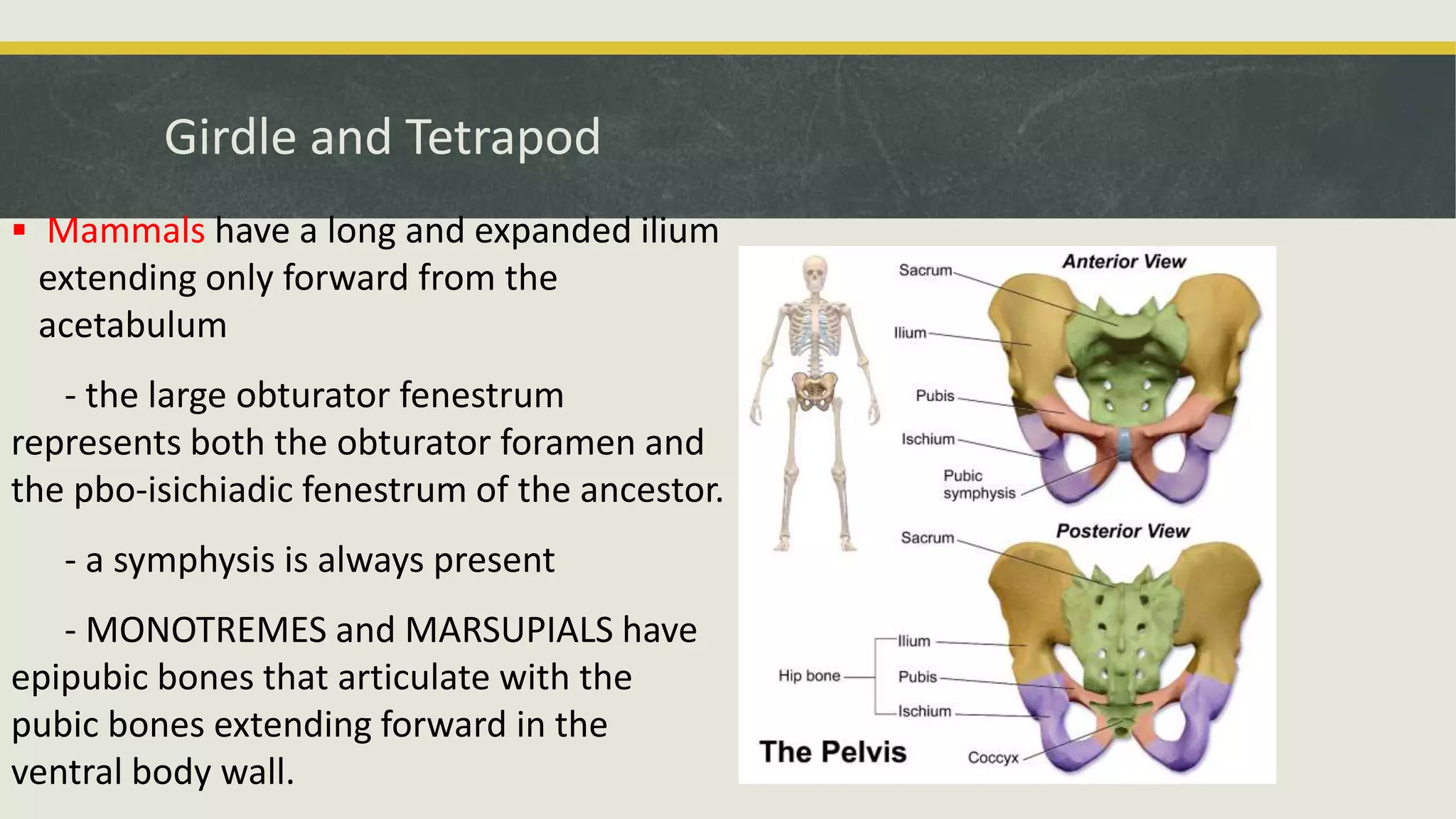
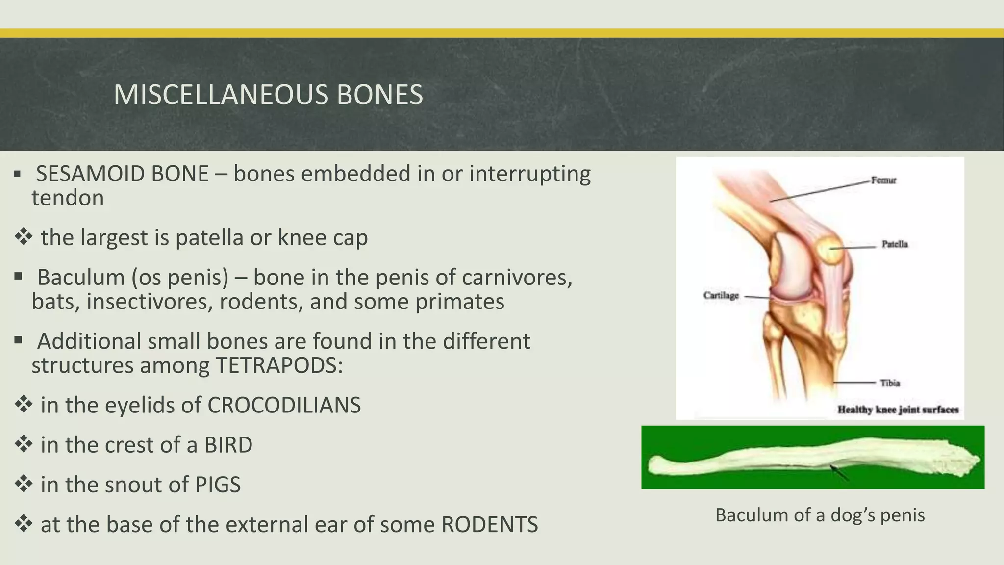


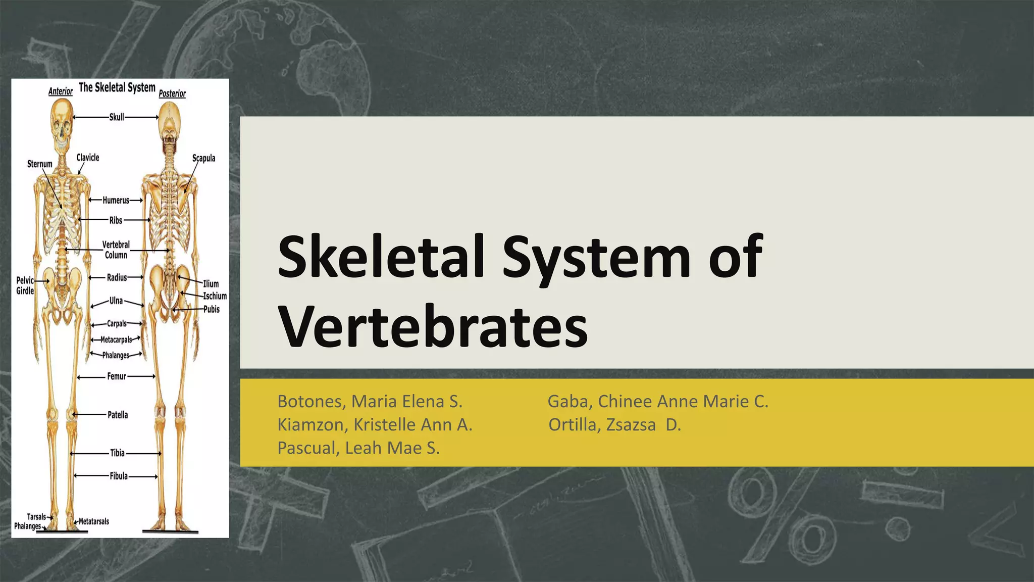
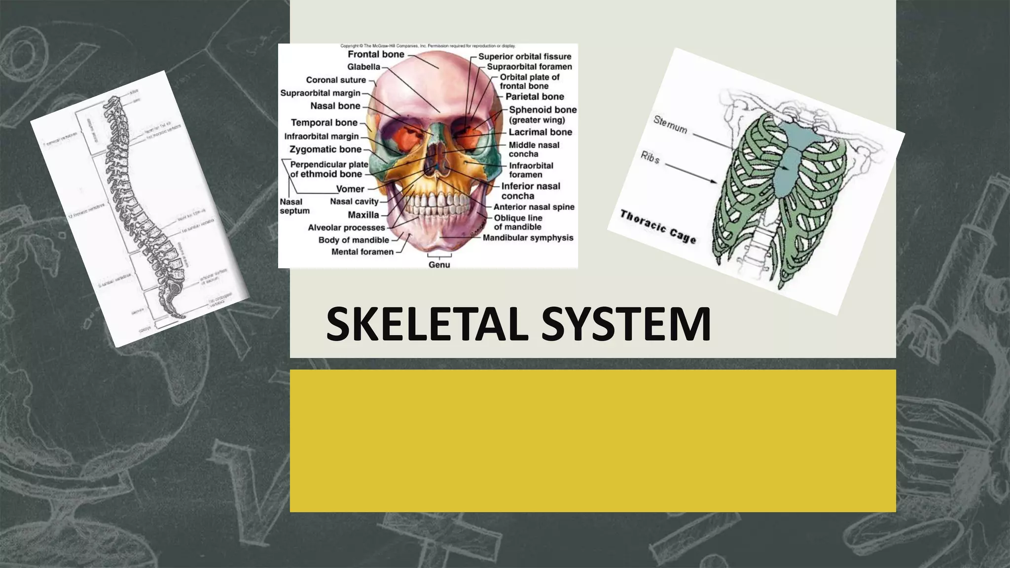
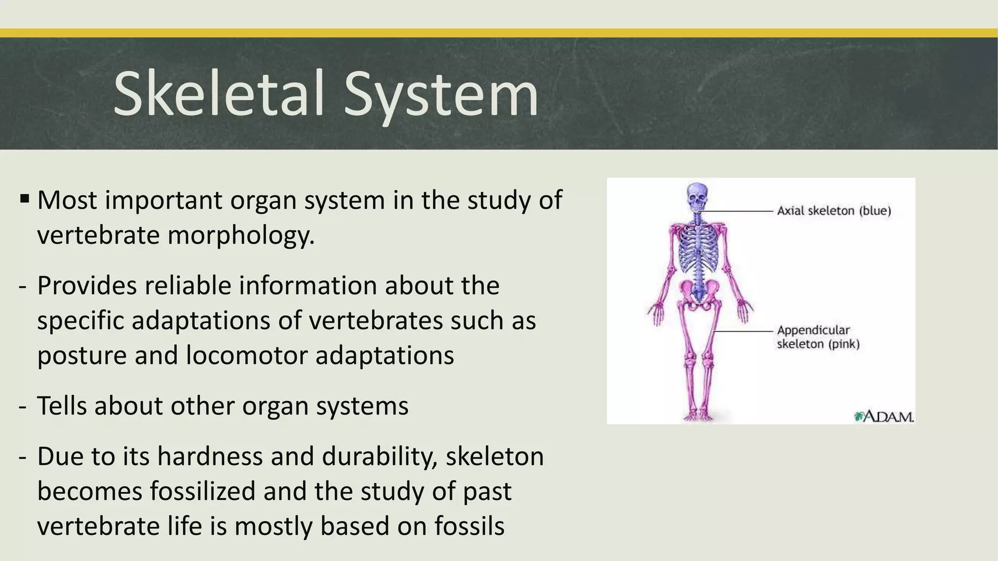
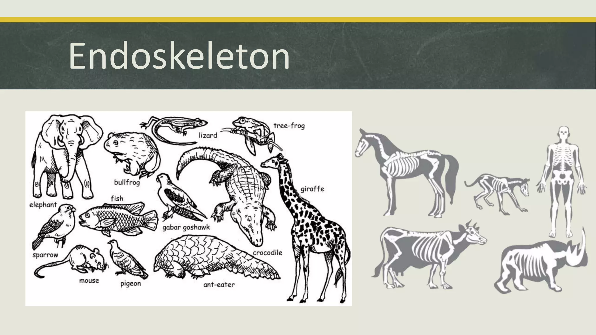
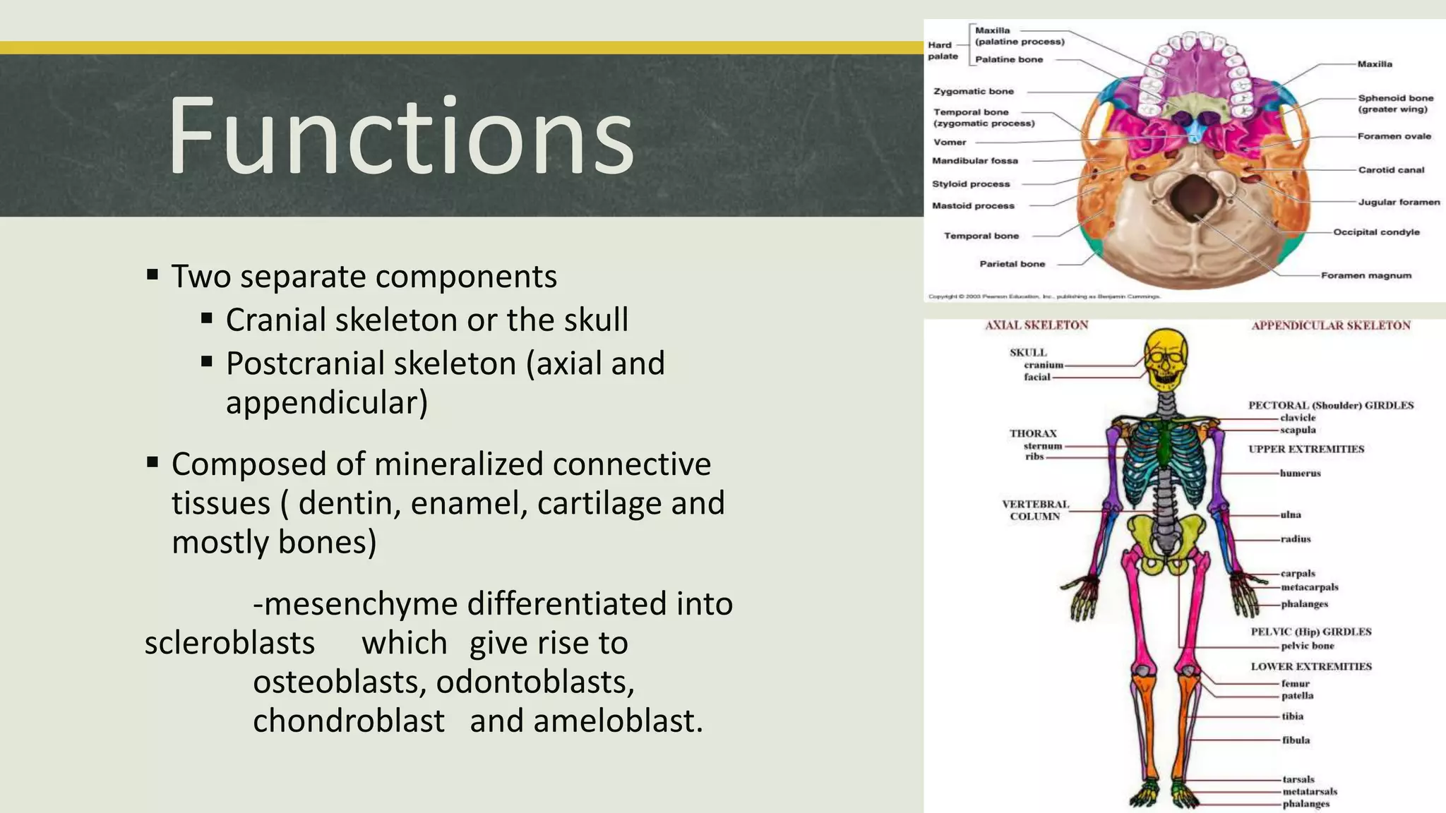
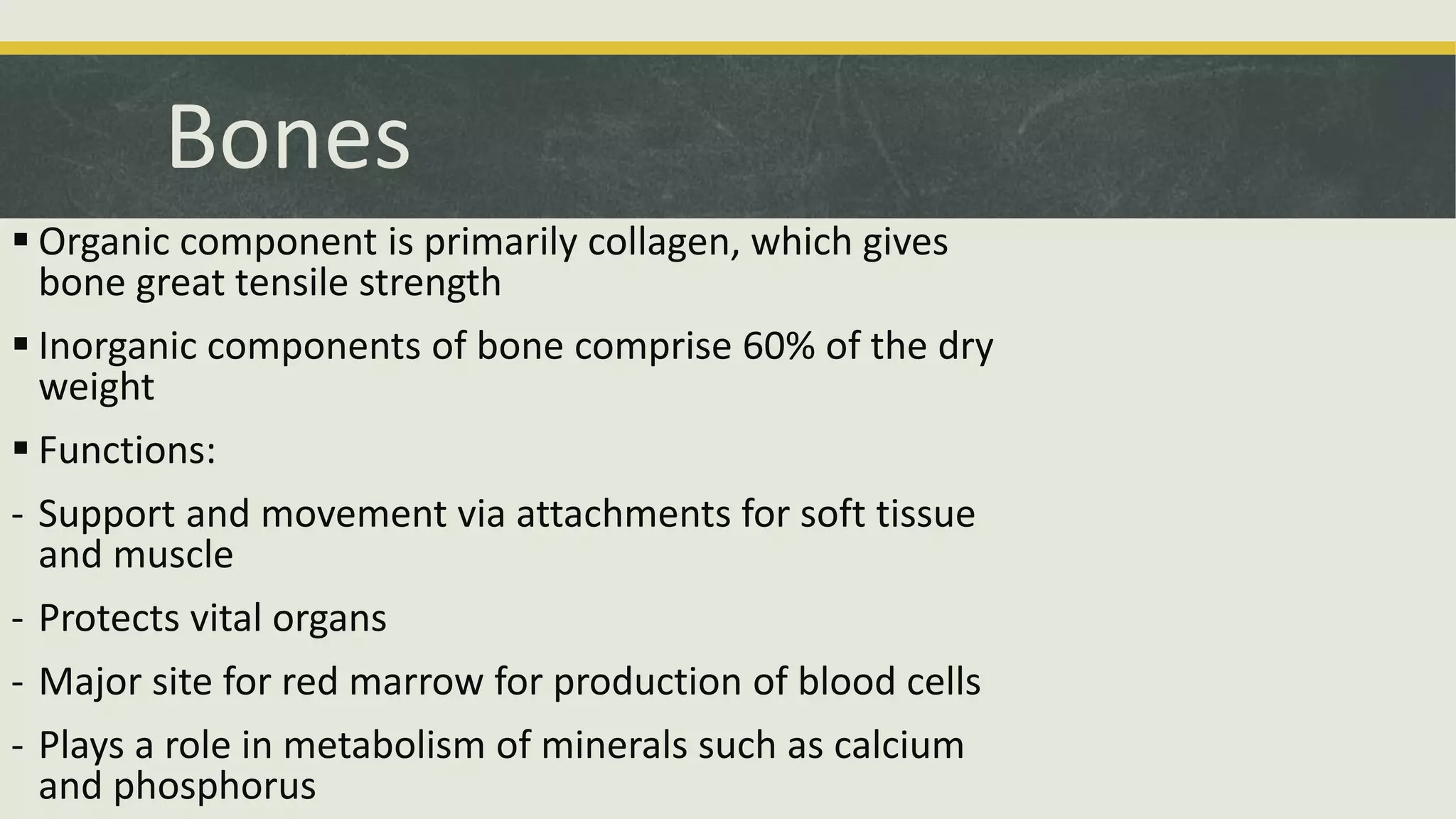
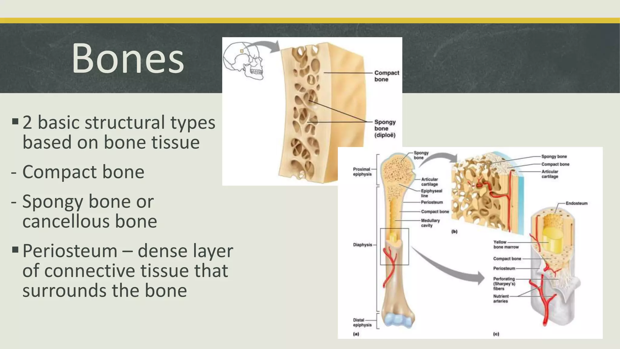
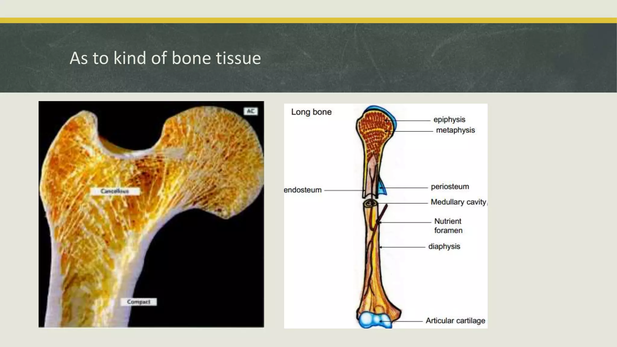
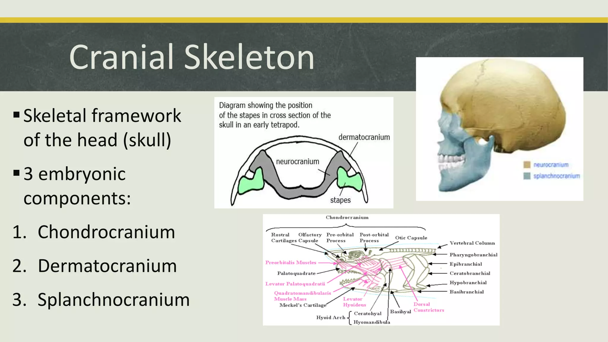
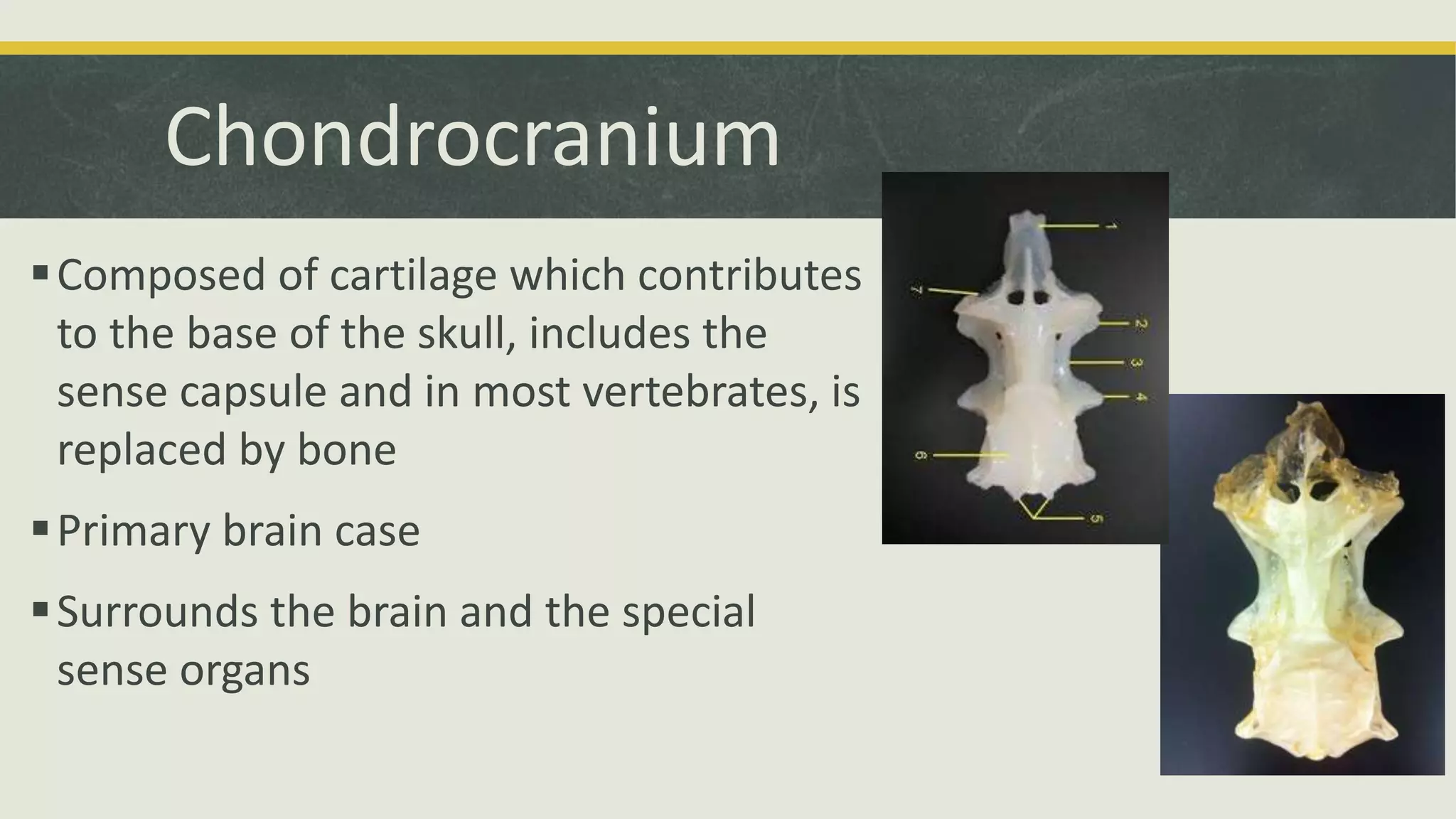
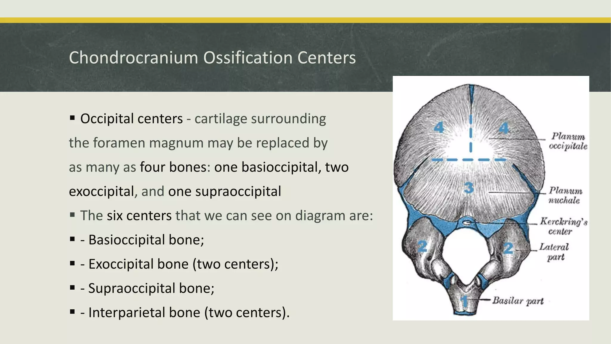
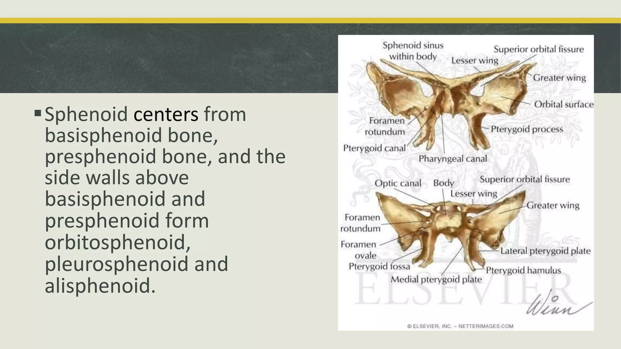
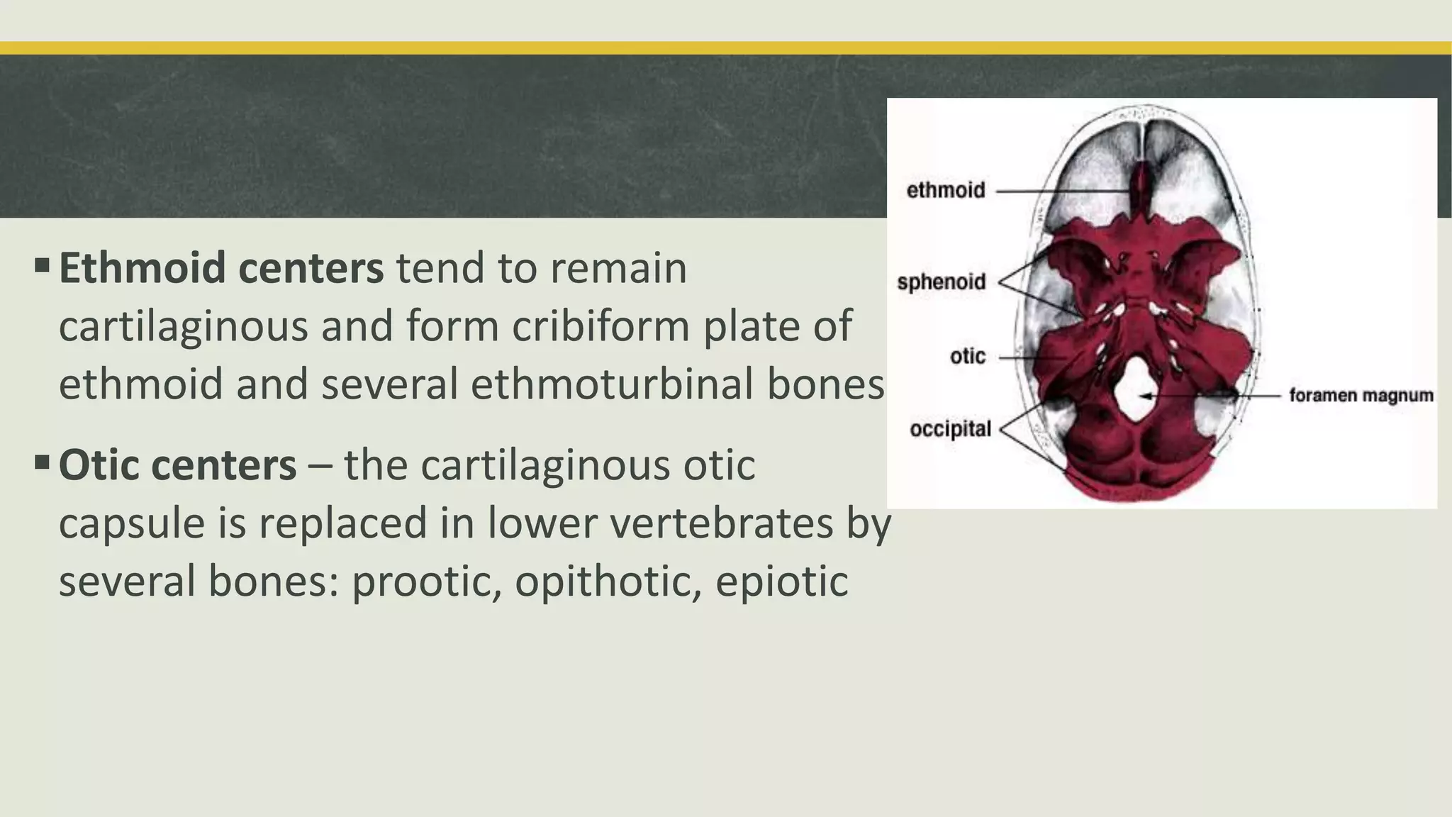
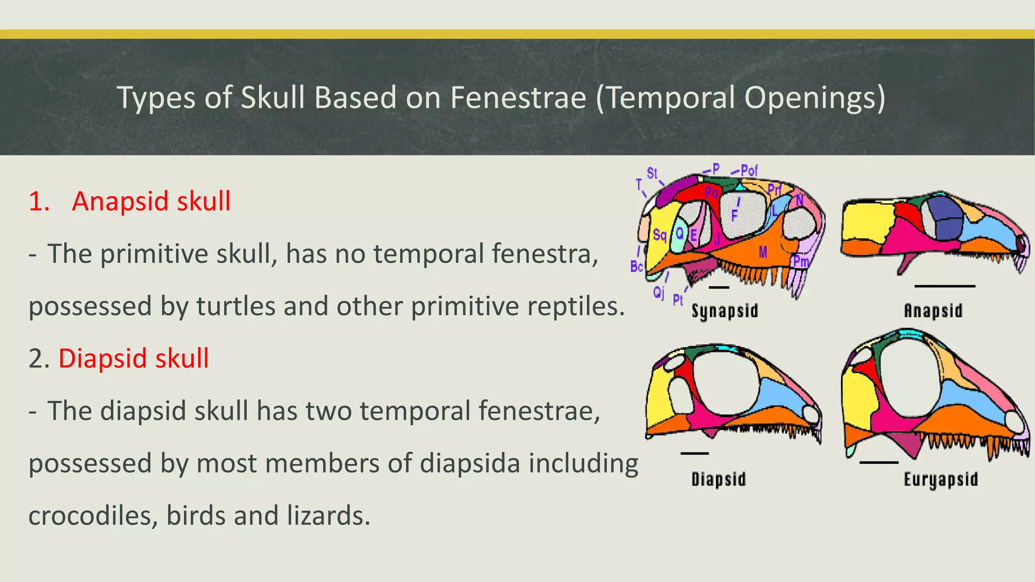
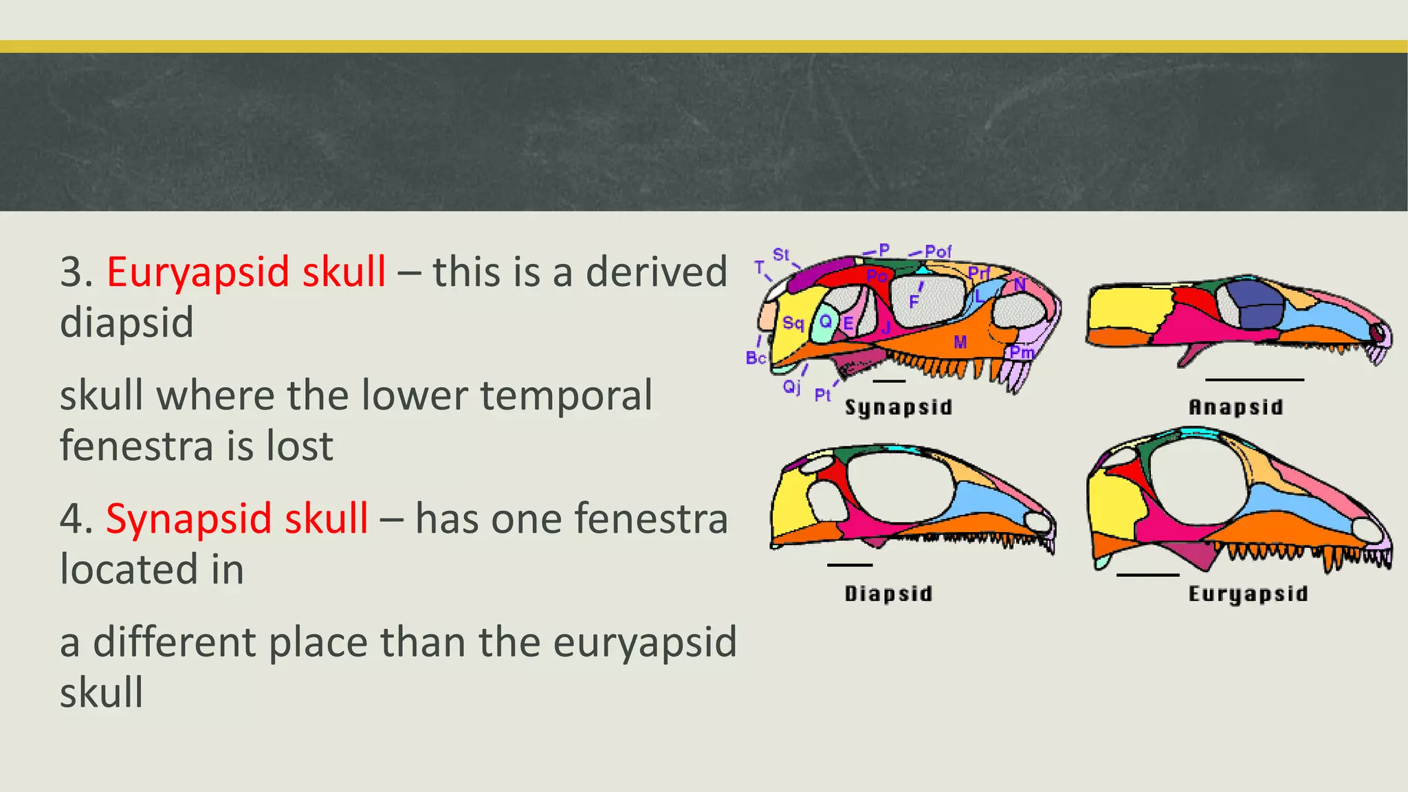
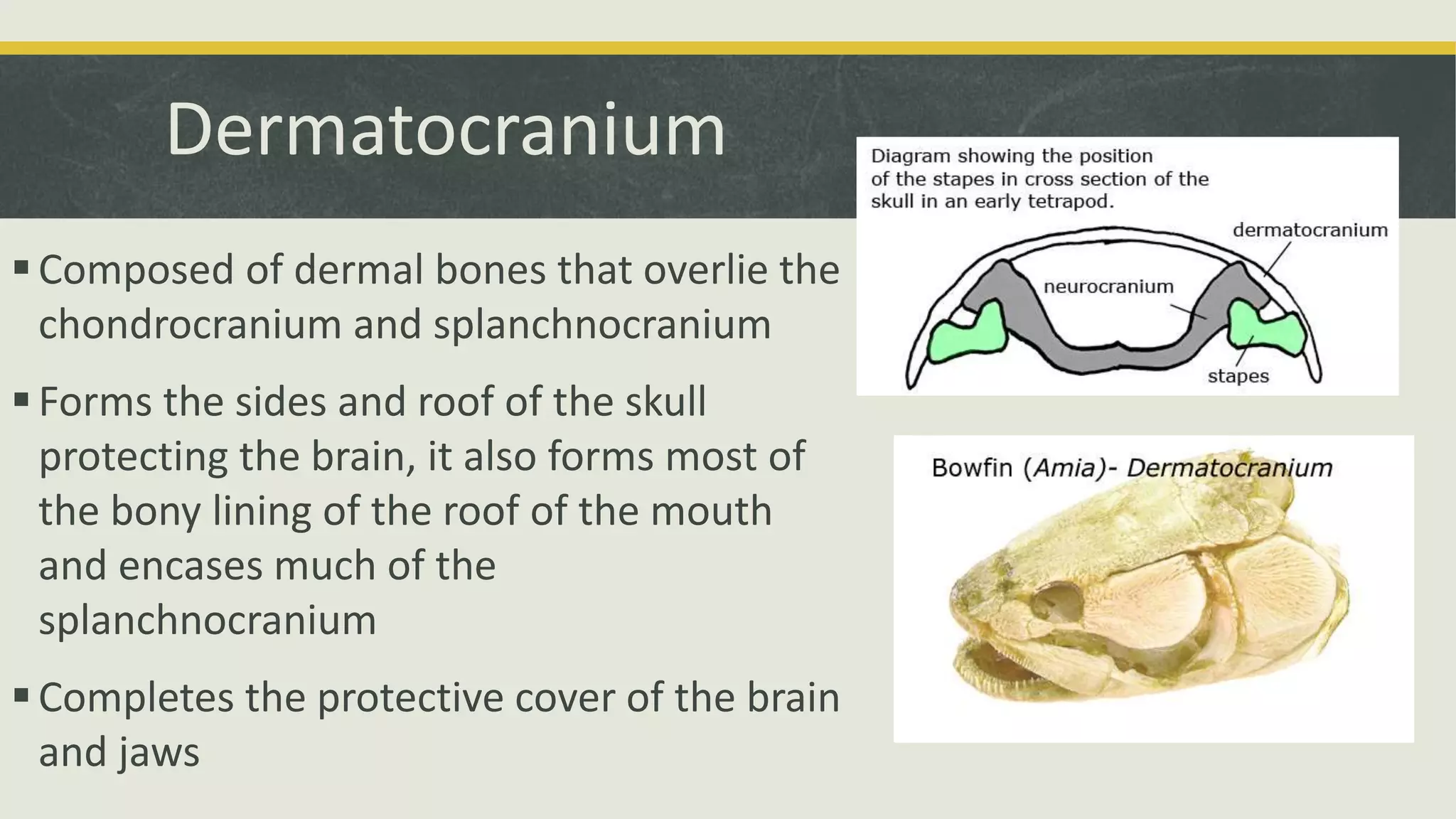
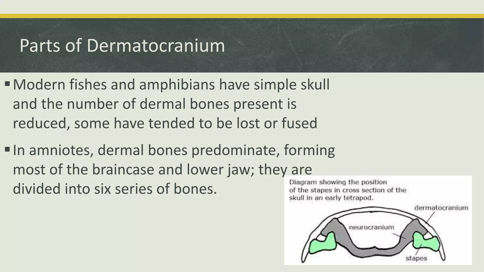
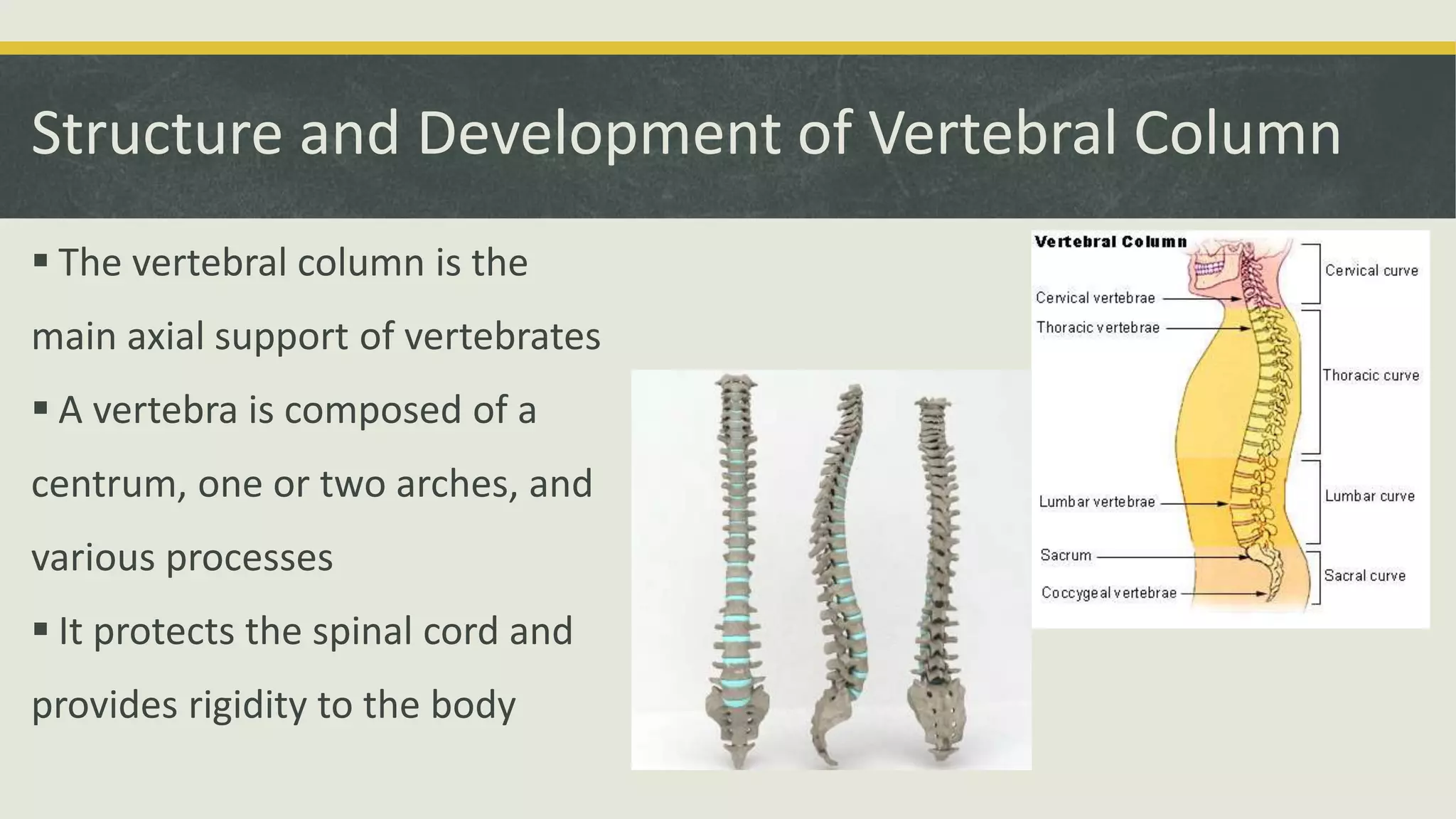
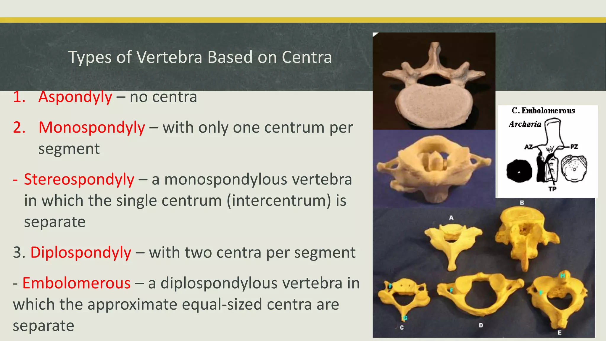
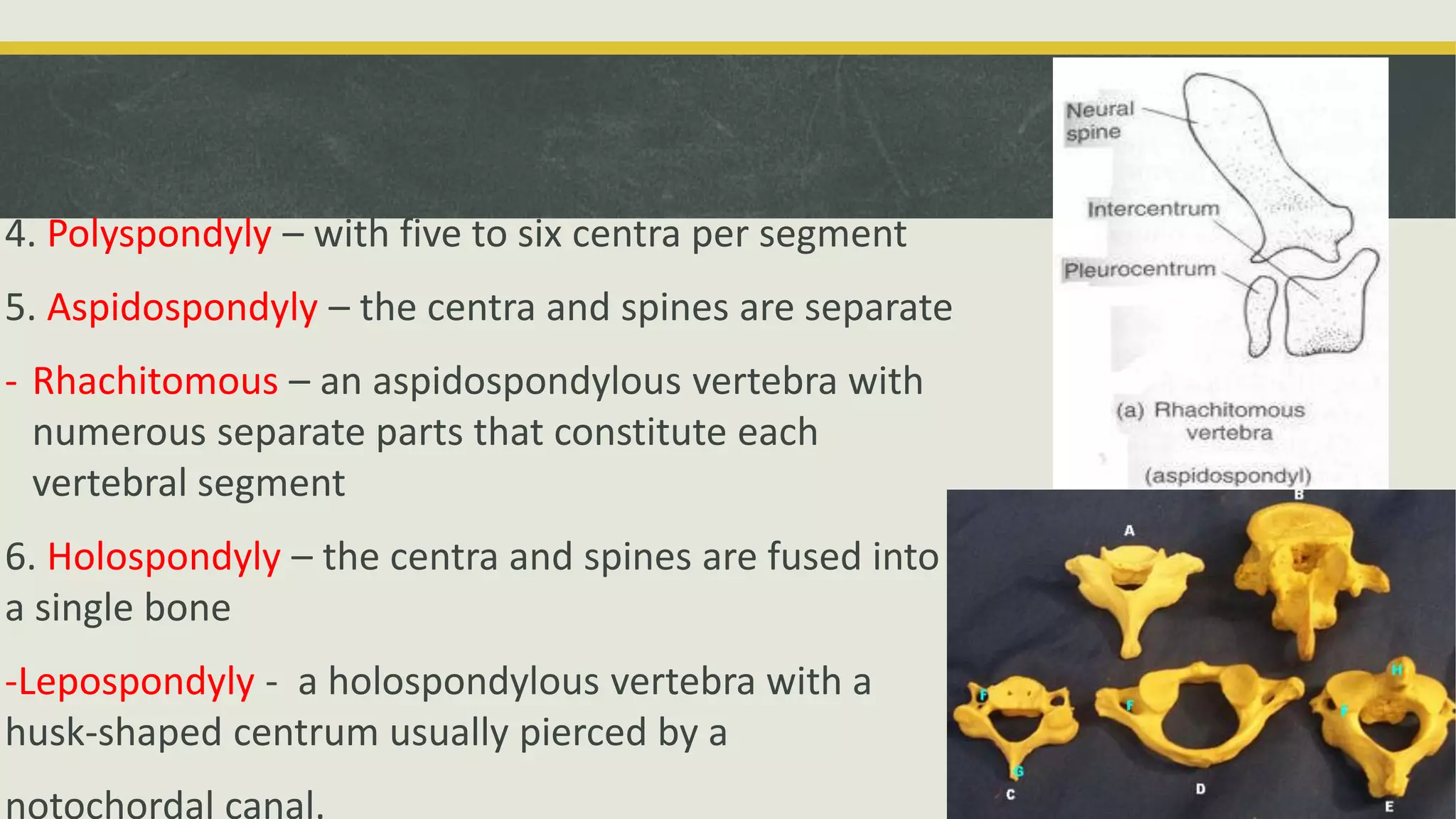
The skeletal system is the most important organ system for studying vertebrate morphology. It provides shape, support, and protection to the body. The skeletal system consists of two main parts - the axial skeleton which includes the skull and vertebral column, and the appendicular skeleton. The axial skeleton gives the body its basic form and protects the spinal cord and vital organs. It develops from cartilage that later ossifies into bone. The skull is made up of the neurocranium, dermatocranium, and splanchnocranium. Vertebrae come in different shapes depending on the number and fusion of centra. Ribs attach to vertebrae and form a protective rib cage in most vertebrates


![Jaw suspension in vertebrates [autosaved]](https://cdn.slidesharecdn.com/ss_thumbnails/jawsuspensioninvertebratesautosaved-201219155254-thumbnail.jpg?width=640&height=640&fit=bounds)


