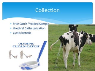
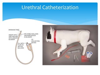
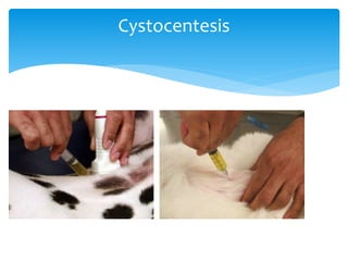
Urine Examination
Muzammal Tariq
 Swine Kidney Worm Infection
 Stephanurus dentatus
 Dioctophyme renale
 Schistosomiasis
 Filariasis
 Capillaria
Parasites
 Free Catch / Voided Sample
 Urethral Catheterization
 Cystocentesis
Collection
Urethral Catheterization
Cystocentesis
 evaluated within 30 min, but if this is not possible, it
may be refrigerated for up to 24 hr
 Color
 Clarity
 Odor
Physical Examination
 Specific gravity
 Semiquantitative, Colorimetric Reagent Strips
 Ph
 Protein
 Glucose
 Ketone
 Bilirubin/ urobilonogen
Other TEST
 Urine SedimentRed Blood Cells:
 White Blood Cells
 Epithelial Cells
 Cylindruria (Casts)
 Infectious Organisms
 Crystals
 Lipids
 Plant Material
 Bladder Tumor Antigen Test
 Specific gravity is defined as the ratio of the weight of
a volume of liquid to the weight of an equal volume of
distilled water
 The urine specific gravity (SG) is determined using a
refractometer designed for veterinary samples
 Cat= 1.015 to 1.050
 Dog= 1.015 to 1.040
 Cattle= 1.030–1.045
Specific gravity
 The saline should be clear and free of any visible
contamination.
 The microscope should have been calibrated within
the last 12 months, and the objectives and oculars
used for the calibration procedure should be used for
all measurements on the microscope. Calibration
factors should be pasted in front of or on the body of
the microscope.
QUALITY CONTROL
 If a 24 hour urine sample was collected, allow the
specimen to sediment for 2 hours, and decant the
major portion of the supernatant. There may be 100
to 200 ml of sediment left. If a first voided urine
specimen is recieved, use the entire specimen.
 Place the remaining urine specimen (sediment) in
centrifuge tubes.
 Centrifuge the specimen at 500 x g for 5 min
PROCEDURE
 Decant the supernatant fluid
 With a pipette, mix and aspirate the sediment
 Place a drop of the sediment on a microscope slide
 Place a cover glass on top of the sediment
 Observe the specimen under the cover glass at
magnifications of x100 and x400. Examine the entire
coverslip at x100 and at least half the coverslip at
x400
 trichomoniasis = motlie flagellates are seen
 schistosoma haematobium = Eggs(eggs for flame
activity (motile cilia) or live miracidia)
 D. renalae= eggs
 Stephanurus dentatus = eggs
Results
 Urine pH is typically acidic in dogs and cats and
alkaline in horses and ruminants, but varies
depending on diet, medications, or presence of
disease.
Urine pH
 http://www.msdvetmanual.com/clinical-pathology-and-
procedures/diagnostic-procedures-for-the-private-practice-
laboratory/urinalysis
 http://parasitologyillustrated.com/procedures/urine_parasi
tes_procedure.html
 http://parasitologyillustrated.com/classes_of_parasites/pro
tozoa/Flagellates/t_vaginalis.html
 http://www.scielo.br/scielo.php?script=sci_arttext&pid=S1
984-29612016005010104&lng=en&nrm=iso&tlng=en
For More Info

Urine and sputum examination

  • 1.
  • 2.
     Swine KidneyWorm Infection  Stephanurus dentatus  Dioctophyme renale  Schistosomiasis  Filariasis  Capillaria Parasites
  • 3.
     Free Catch/ Voided Sample  Urethral Catheterization  Cystocentesis Collection
  • 4.
  • 5.
  • 6.
     evaluated within30 min, but if this is not possible, it may be refrigerated for up to 24 hr  Color  Clarity  Odor Physical Examination
  • 7.
     Specific gravity Semiquantitative, Colorimetric Reagent Strips  Ph  Protein  Glucose  Ketone  Bilirubin/ urobilonogen Other TEST
  • 8.
     Urine SedimentRedBlood Cells:  White Blood Cells  Epithelial Cells  Cylindruria (Casts)  Infectious Organisms  Crystals  Lipids  Plant Material  Bladder Tumor Antigen Test
  • 9.
     Specific gravityis defined as the ratio of the weight of a volume of liquid to the weight of an equal volume of distilled water  The urine specific gravity (SG) is determined using a refractometer designed for veterinary samples  Cat= 1.015 to 1.050  Dog= 1.015 to 1.040  Cattle= 1.030–1.045 Specific gravity
  • 10.
     The salineshould be clear and free of any visible contamination.  The microscope should have been calibrated within the last 12 months, and the objectives and oculars used for the calibration procedure should be used for all measurements on the microscope. Calibration factors should be pasted in front of or on the body of the microscope. QUALITY CONTROL
  • 11.
     If a24 hour urine sample was collected, allow the specimen to sediment for 2 hours, and decant the major portion of the supernatant. There may be 100 to 200 ml of sediment left. If a first voided urine specimen is recieved, use the entire specimen.  Place the remaining urine specimen (sediment) in centrifuge tubes.  Centrifuge the specimen at 500 x g for 5 min PROCEDURE
  • 12.
     Decant thesupernatant fluid  With a pipette, mix and aspirate the sediment  Place a drop of the sediment on a microscope slide  Place a cover glass on top of the sediment  Observe the specimen under the cover glass at magnifications of x100 and x400. Examine the entire coverslip at x100 and at least half the coverslip at x400
  • 13.
     trichomoniasis =motlie flagellates are seen  schistosoma haematobium = Eggs(eggs for flame activity (motile cilia) or live miracidia)  D. renalae= eggs  Stephanurus dentatus = eggs Results
  • 14.
     Urine pHis typically acidic in dogs and cats and alkaline in horses and ruminants, but varies depending on diet, medications, or presence of disease. Urine pH
  • 15.
     http://www.msdvetmanual.com/clinical-pathology-and- procedures/diagnostic-procedures-for-the-private-practice- laboratory/urinalysis  http://parasitologyillustrated.com/procedures/urine_parasi tes_procedure.html http://parasitologyillustrated.com/classes_of_parasites/pro tozoa/Flagellates/t_vaginalis.html  http://www.scielo.br/scielo.php?script=sci_arttext&pid=S1 984-29612016005010104&lng=en&nrm=iso&tlng=en For More Info

Editor's Notes

  • #12 Microscopic examination of urine sediment should be part of a routine urinalysis. For centrifugation, 3–5 mL of urine is transferred to a conical centrifuge tube. Urine is centrifuged at 1,000–1,500 rpm for ~3–5 min. The supernatant is decanted, leaving ~0.5 mL of urine and sediment in the tip of the conical tube. The sediment is resuspended by tapping the tip of the conical tube against the table several times. A few drops of the sediment are transferred to a glass slide, and a cover slip is applied. Examination of unstained urine is recommended for routine samples. Microscopic examination is performed at 100× (for crystals, casts, and cells) and 400× (for cells and bacteria) magnifications. Contrast of the sample is enhanced by closing the iris diaphragm and lowering the condenser of the microscope. Stains such as Sedistain® and new methylene blue can be used to aid in cell identification but may dilute the specimen and introduce artifacts such as stain precipitate and crystals. Use of a modified Wright stain increases the sensitivity, specificity, and positive and negative predictive values for detection of bacteria. For some tests, air dried, stained smears are necessary.