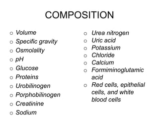
EXAMINATION OF URINE
COMPOSITION
o Volume
o Specific gravity
o Osmolality
o pH
o Glucose
o Proteins
o Urobilinogen
o Porphobilinogen
o Creatinine
o Sodium
o Urea nitrogen
o Uric acid
o Potassium
o Chloride
o Calcium
o Formiminoglutamic
acid
o Red cells, epithelial
cells, and white
blood cells
INDICATIONS FOR
URINALYSIS
1. Suspected renal diseases
2. Detection of urinary tract infection
3. Detection of Metabolic disorders
4. Detection of plasma cell dyscariasis
5. Diagnosis of Pregnancy
Collection of urine sample
• A single specimen: This may be a first
morning voiding, a random specimen, or a
post-prandial specimen.
• The first voided specimen in the morning
1. is the most concentrated and
2. has acidic pH in which formed elements
(cells and casts) are well preserved.
1. This specimen is used for routine
examination,
2. fasting glucose,
3. proteins,
4. nitrite,
5. microscopic analysis for cellular
elements,
6. pregnancy test,
7. orthostatic proteinuria, and
8. bacteriological analysis.
• The random specimen is a single
specimen collected at any time of day. It is
used for routine urine examination.
• Post-prandial specimen (collected 2
hours after a meal in the afternoon) is
sometimes requested for estimation of
glucose (to monitor insulin therapy in
diabetes mellitus) or of urobilinogen.
24-hour specimen: After getting up in the
morning, the first urine is discarded.
All the urine voided subsequently during the
rest of the day and the night is collected in a
large bottle (clean bottle of 2 liter capacity
with a cap).
The first urine after getting up in the morning
on the next day is also collected.
• The urine should be preserved at 4-6°C during
the period of collection.
• The container is then immediately transported
to the laboratory.
• The urine is thoroughly mixed and an aliquot is
used for testing.
• This method is used for quantitative estimation
of proteins and hormones.
COLLECTION METHODS
• Midstream specimen:
• Used for all types of examinations.
• After voiding initial half of urine into the toilet, a
part of urine is collected in the bottle.
• First half of stream serves to flush out
contaminating cells and microbes from urethra
and perineum.
• Subsequent stream is collected which is from
the urinary bladder.
• Clean-catch specimen:
• Recommended for bacteriologic culture.
• In men- glans penis is sufficiently exposed and
cleaned with soap and water.
• In women urethral opening should be exposed,
washed with soapy cotton balls, rinsed with
water-saturated cotton, and holding the labia
apart
• Initial urine allowed to pass and the remaining is
voided into the bottle (amount 20-100 ml).
• This method avoids contamination of urine with
the vaginal fluids.
• Catheter specimen:
• Bacteriological study or culture in
bedridden,
• Ill patients or in patients with obstruction of
urinary tract.
• Usually avoided in ambulatory patients-
bcz it carries the risk of introduction of
infection.
• Infants:
• A clean plastic bag can be attached around the
baby’s genitalia and left in place for some time.
• For bacteriologic examination, urine is aspirated
from bladder by passing a needle just above
symphysis pubis.
Changes -Standing Urine at
Room Temperature
1. Increase in pH
2. Formation of crystals
3. Loss of ketone bodies
4. Decrease in glucose
5. Oxidation of bilirubin to biliverdin
6. Oxidation of urobilinogen to urobilin
7. Bacterial proliferation
8. Disintegration of cellular elements
• Urine sample must be tested in the
laboratory within 2 hours of collection to
get the correct results.
Preservation of Urine Sample
• Can be kept in the refrigerator for a
maximum of 8 hours.
• Refrigeration (4-6°C)
• For routine urinalysis, preservatives
should be avoided, as they interfere with
reagent strip techniques and chemical test
for protein.
• Hydrochloric acid: used for preservation of a
24hour urine sample for adrenaline,
noradrenaline, vanillylmandelic acid, and
steroids.
• Toluene: It forms a thin layer over the surface
and acts as a physical barrier for bacteria and
air. It is used for measurement of chemicals.
• Boric acid: A general preservative.
• Thymol: It inhibits bacteria and fungi.
• Formalin: excellent chemical for preservation of
formed elements.
PHYSICAL EXAMINATION
1. Volume - only the 24-hr specimen of urine
needs to be measured and reported.
Adult average is 600-2000 ml.
2. Polyuria volume > 2000 ml/24 hours.
diabetes mellitus (osmotic diuresis),
diabetes insipidus (failure of secretion of
antidiuretic hormone), chronic renal
failure (loss of concentrating ability of
kidneys) or diuretic therapy.
• Oliguria - volume < 400 ml/24 hours.
Causes include febrile states, acute
glomerulonephritis (decreased glomerular
filtration), congestive cardiac failure or
dehydration (decreased renal blood flow).
• Anuria means urinary output < 100 ml/24
hours or complete cessation of urine
output. It occurs in acute tubular necrosis
(e.g. in shock, hemolytic transfusion
reaction), acute glomerulonephritis, and
complete urinary tract obstruction.
COLOR
• Normal urine color in a fresh state is pale
yellow or amber and is due to the
presence of various pigments collectively
called urochrome.
• Depending on the state of hydration urine
may normally be colorless (over hydration)
or dark yellow (dehydration).
• Colorless
Appearance
• Normal, freshly voided urine is clear in
appearance. Causes of cloudy or turbid
urine are listed in Table 1.3. Foamy urine
occurs in the presence of excess proteins
or bilirubin.
Odor
• Freshly voided urine has a typical aromatic
odor due to volatile organic acids. After
standing, urine develops ammoniacal odor
(formation of ammonia occurs when urea
is decomposed by bacteria).
• Some abnormal odors with associated
conditions are:
• Fruity: Ketoacidosis, starvation
• Mousy or musty: Phenylketonuria •
• Fishy: Urinary tract infection with Proteus,
tyrosinaemia.
• Ammoniacal: Urinary tract infection with
Escherichia coli, old standing urine.
• Foul: Urinary tract infection
• Sulfurous: Cystinuria.
Specific Gravity (SG)
• Normal SG of urine is 1.003 to 1.030
• SG increases as solute concentration
increases and decreases when
temperature rises
Reaction and pH
• The pH is the scale for measuring acidity
or alkalinity (acid if pH is < 7.0; alkaline if
pH is > 7.0; neutral if pH is
CHEMICAL EXAMINATION
• Normally, kidneys excrete scant amount
of protein in urine (up to 150 mg/24 hours).
• Proteinuria refers to protein excretion in
urine greater than 150 mg/24 hours in
adults.
Causes of Proteinuria
Tests for Detection of
Proteinuria
1. Heat and acetic acid test (Boiling test)
Principle: proteins get precipitated when
boiled in an acidic solution.
Reagent strip test:
Priniciple: “protein error of indicators”
• Sulphosalicylic acid test:
Addition of sulphosalicylic acid to the urine
causes formation of a white precipitate if
proteins are present
Quantitative Estimation of
Proteins
• Diagnosis of nephrotic syndrome
• Detection of microalbuminuria or early
diabetic nephropathy
• To follow response to therapy in renal
disease Proteinuria >1500 mg/ 24 hours
indicates glomerular disease;
• proteinuria >3500 mg/24 hours is called
as nephrotic range proteinuria; in tubular,
hemodynamic and post renal diseases,
proteinuria is usually < 1500 mg/ 24 hours.
Bence Jones Proteinuria
• Bence Jones proteins are monoclonal
immunoglobulin light chains (either κ or λ)
that are synthesized by neoplastic plasma
cells.
GLUCOSE
• The main indication for testing for glucose
in urine is detection of unsuspected
diabetes mellitus or follow-up of known
diabetic patients.
• Normally a very small amount of glucose
is excreted in urine (< 500 mg/24 hours or
<15 mg/dl) that cannot be detected by the
routine tests.
• Presence of detectable amounts of
glucose in urine is called as Glucosuria.
Causes of Glycosuria
Tests for Detection of Glucose
in Urine
Copper reduction methods:
Benedict’s qualitative test: When urine is
boiled in Benedict’s qualitative solution, blue
alkaline copper sulphate is reduced to red-
brown cuprous oxide if a reducing agent is
present.
But not specific for Glucose
Method
• Take 5 ml of Benedict’s qualitative reagent
in a test tube
• (composition of Benedict’s qualitative
reagent: copper sulphate 17.3 gram,
sodium carbonate 100 gram, sodium
citrate 173 gram, distilled water 1000 ml).
2. Add 0.5 ml (or 8 drops) of urine. Mix
well.
• 3. Boil over a flame for 2 minutes.
• 4. Allow to cool at room temperature.
• 5. Note the color change, if any.
• The result is reported in grades as follows Nil: no
change from blue color
• Trace: Green without precipitate
• 1+ (approx. 0.5 grams/dl): Green with
precipitate
• 2+ (approx. 1.0 grams/dl): Brown precipitate
• 3+ (approx. 1.5 grams/dl: Yellow-orange
precipitate
• 4+ ( > 2.0 grams/dl): Brick- red precipitate.
• Clinitest tablet method (Copper reduction
tablet test): This is a modified form of
Benedict’s test in which the reagents are
present in a tablet form (copper sulphate,
citric acid, sodium carbonate, and
anhydrous sodium hydroxide). Sensitivity
is 200 mgs/dl of glucose.
2. Reagent strip method
• This test is specific for glucose and is
therefore preferred over Benedict’s test.
Ketones
• Excretion of ketone bodies (acetoacetic
acid, β-hydroxybutyric acid, and acetone)
in urine is called as ketonuria.
• Ketones are breakdown products of fatty
acids and their presence in urine is
indicative of excessive fatty acid
metabolism to provide energy.
Causes of Ketonuria
• Normally ketone bodies are not detectable in the
urine of healthy persons.
• If energy requirements cannot be met by
metabolism of glucose (due to defective
carbohydrate metabolism,
• low carbohydrate intake, or increased metabolic
needs),
• then energy is derived from breakdown of fats.
This leads to the formation of ketone bodies
1. Decreased utilization of carbohydrates
2. Decreased availability of carbohydrates
in the diet.
3. Increased metabolic needs
Tests for Detection of Ketones in
Urine
Urine.pptx

Urine.pptx

  • 1.
  • 2.
    COMPOSITION o Volume o Specificgravity o Osmolality o pH o Glucose o Proteins o Urobilinogen o Porphobilinogen o Creatinine o Sodium o Urea nitrogen o Uric acid o Potassium o Chloride o Calcium o Formiminoglutamic acid o Red cells, epithelial cells, and white blood cells
  • 3.
    INDICATIONS FOR URINALYSIS 1. Suspectedrenal diseases 2. Detection of urinary tract infection 3. Detection of Metabolic disorders 4. Detection of plasma cell dyscariasis 5. Diagnosis of Pregnancy
  • 4.
    Collection of urinesample • A single specimen: This may be a first morning voiding, a random specimen, or a post-prandial specimen. • The first voided specimen in the morning 1. is the most concentrated and 2. has acidic pH in which formed elements (cells and casts) are well preserved.
  • 5.
    1. This specimenis used for routine examination, 2. fasting glucose, 3. proteins, 4. nitrite, 5. microscopic analysis for cellular elements, 6. pregnancy test, 7. orthostatic proteinuria, and 8. bacteriological analysis.
  • 6.
    • The randomspecimen is a single specimen collected at any time of day. It is used for routine urine examination. • Post-prandial specimen (collected 2 hours after a meal in the afternoon) is sometimes requested for estimation of glucose (to monitor insulin therapy in diabetes mellitus) or of urobilinogen.
  • 7.
    24-hour specimen: Aftergetting up in the morning, the first urine is discarded. All the urine voided subsequently during the rest of the day and the night is collected in a large bottle (clean bottle of 2 liter capacity with a cap). The first urine after getting up in the morning on the next day is also collected.
  • 8.
    • The urineshould be preserved at 4-6°C during the period of collection. • The container is then immediately transported to the laboratory. • The urine is thoroughly mixed and an aliquot is used for testing. • This method is used for quantitative estimation of proteins and hormones.
  • 9.
    COLLECTION METHODS • Midstreamspecimen: • Used for all types of examinations. • After voiding initial half of urine into the toilet, a part of urine is collected in the bottle. • First half of stream serves to flush out contaminating cells and microbes from urethra and perineum. • Subsequent stream is collected which is from the urinary bladder.
  • 10.
    • Clean-catch specimen: •Recommended for bacteriologic culture. • In men- glans penis is sufficiently exposed and cleaned with soap and water. • In women urethral opening should be exposed, washed with soapy cotton balls, rinsed with water-saturated cotton, and holding the labia apart • Initial urine allowed to pass and the remaining is voided into the bottle (amount 20-100 ml). • This method avoids contamination of urine with the vaginal fluids.
  • 11.
    • Catheter specimen: •Bacteriological study or culture in bedridden, • Ill patients or in patients with obstruction of urinary tract. • Usually avoided in ambulatory patients- bcz it carries the risk of introduction of infection.
  • 12.
    • Infants: • Aclean plastic bag can be attached around the baby’s genitalia and left in place for some time. • For bacteriologic examination, urine is aspirated from bladder by passing a needle just above symphysis pubis.
  • 13.
    Changes -Standing Urineat Room Temperature 1. Increase in pH 2. Formation of crystals 3. Loss of ketone bodies 4. Decrease in glucose 5. Oxidation of bilirubin to biliverdin 6. Oxidation of urobilinogen to urobilin 7. Bacterial proliferation 8. Disintegration of cellular elements
  • 14.
    • Urine samplemust be tested in the laboratory within 2 hours of collection to get the correct results.
  • 15.
    Preservation of UrineSample • Can be kept in the refrigerator for a maximum of 8 hours. • Refrigeration (4-6°C) • For routine urinalysis, preservatives should be avoided, as they interfere with reagent strip techniques and chemical test for protein.
  • 16.
    • Hydrochloric acid:used for preservation of a 24hour urine sample for adrenaline, noradrenaline, vanillylmandelic acid, and steroids. • Toluene: It forms a thin layer over the surface and acts as a physical barrier for bacteria and air. It is used for measurement of chemicals. • Boric acid: A general preservative. • Thymol: It inhibits bacteria and fungi. • Formalin: excellent chemical for preservation of formed elements.
  • 17.
    PHYSICAL EXAMINATION 1. Volume- only the 24-hr specimen of urine needs to be measured and reported. Adult average is 600-2000 ml. 2. Polyuria volume > 2000 ml/24 hours. diabetes mellitus (osmotic diuresis), diabetes insipidus (failure of secretion of antidiuretic hormone), chronic renal failure (loss of concentrating ability of kidneys) or diuretic therapy.
  • 18.
    • Oliguria -volume < 400 ml/24 hours. Causes include febrile states, acute glomerulonephritis (decreased glomerular filtration), congestive cardiac failure or dehydration (decreased renal blood flow).
  • 19.
    • Anuria meansurinary output < 100 ml/24 hours or complete cessation of urine output. It occurs in acute tubular necrosis (e.g. in shock, hemolytic transfusion reaction), acute glomerulonephritis, and complete urinary tract obstruction.
  • 20.
    COLOR • Normal urinecolor in a fresh state is pale yellow or amber and is due to the presence of various pigments collectively called urochrome. • Depending on the state of hydration urine may normally be colorless (over hydration) or dark yellow (dehydration).
  • 21.
  • 22.
    Appearance • Normal, freshlyvoided urine is clear in appearance. Causes of cloudy or turbid urine are listed in Table 1.3. Foamy urine occurs in the presence of excess proteins or bilirubin.
  • 23.
    Odor • Freshly voidedurine has a typical aromatic odor due to volatile organic acids. After standing, urine develops ammoniacal odor (formation of ammonia occurs when urea is decomposed by bacteria).
  • 24.
    • Some abnormalodors with associated conditions are: • Fruity: Ketoacidosis, starvation • Mousy or musty: Phenylketonuria • • Fishy: Urinary tract infection with Proteus, tyrosinaemia. • Ammoniacal: Urinary tract infection with Escherichia coli, old standing urine. • Foul: Urinary tract infection • Sulfurous: Cystinuria.
  • 25.
    Specific Gravity (SG) •Normal SG of urine is 1.003 to 1.030 • SG increases as solute concentration increases and decreases when temperature rises
  • 26.
    Reaction and pH •The pH is the scale for measuring acidity or alkalinity (acid if pH is < 7.0; alkaline if pH is > 7.0; neutral if pH is
  • 27.
  • 28.
    • Normally, kidneysexcrete scant amount of protein in urine (up to 150 mg/24 hours). • Proteinuria refers to protein excretion in urine greater than 150 mg/24 hours in adults.
  • 29.
  • 30.
    Tests for Detectionof Proteinuria 1. Heat and acetic acid test (Boiling test) Principle: proteins get precipitated when boiled in an acidic solution.
  • 31.
    Reagent strip test: Priniciple:“protein error of indicators”
  • 32.
    • Sulphosalicylic acidtest: Addition of sulphosalicylic acid to the urine causes formation of a white precipitate if proteins are present
  • 33.
    Quantitative Estimation of Proteins •Diagnosis of nephrotic syndrome • Detection of microalbuminuria or early diabetic nephropathy • To follow response to therapy in renal disease Proteinuria >1500 mg/ 24 hours indicates glomerular disease; • proteinuria >3500 mg/24 hours is called as nephrotic range proteinuria; in tubular, hemodynamic and post renal diseases, proteinuria is usually < 1500 mg/ 24 hours.
  • 34.
    Bence Jones Proteinuria •Bence Jones proteins are monoclonal immunoglobulin light chains (either κ or λ) that are synthesized by neoplastic plasma cells.
  • 35.
    GLUCOSE • The mainindication for testing for glucose in urine is detection of unsuspected diabetes mellitus or follow-up of known diabetic patients. • Normally a very small amount of glucose is excreted in urine (< 500 mg/24 hours or <15 mg/dl) that cannot be detected by the routine tests.
  • 36.
    • Presence ofdetectable amounts of glucose in urine is called as Glucosuria.
  • 37.
  • 38.
    Tests for Detectionof Glucose in Urine Copper reduction methods: Benedict’s qualitative test: When urine is boiled in Benedict’s qualitative solution, blue alkaline copper sulphate is reduced to red- brown cuprous oxide if a reducing agent is present. But not specific for Glucose
  • 39.
    Method • Take 5ml of Benedict’s qualitative reagent in a test tube • (composition of Benedict’s qualitative reagent: copper sulphate 17.3 gram, sodium carbonate 100 gram, sodium citrate 173 gram, distilled water 1000 ml). 2. Add 0.5 ml (or 8 drops) of urine. Mix well. • 3. Boil over a flame for 2 minutes. • 4. Allow to cool at room temperature. • 5. Note the color change, if any.
  • 40.
    • The resultis reported in grades as follows Nil: no change from blue color • Trace: Green without precipitate • 1+ (approx. 0.5 grams/dl): Green with precipitate • 2+ (approx. 1.0 grams/dl): Brown precipitate • 3+ (approx. 1.5 grams/dl: Yellow-orange precipitate • 4+ ( > 2.0 grams/dl): Brick- red precipitate.
  • 41.
    • Clinitest tabletmethod (Copper reduction tablet test): This is a modified form of Benedict’s test in which the reagents are present in a tablet form (copper sulphate, citric acid, sodium carbonate, and anhydrous sodium hydroxide). Sensitivity is 200 mgs/dl of glucose.
  • 42.
    2. Reagent stripmethod • This test is specific for glucose and is therefore preferred over Benedict’s test.
  • 43.
    Ketones • Excretion ofketone bodies (acetoacetic acid, β-hydroxybutyric acid, and acetone) in urine is called as ketonuria. • Ketones are breakdown products of fatty acids and their presence in urine is indicative of excessive fatty acid metabolism to provide energy.
  • 44.
    Causes of Ketonuria •Normally ketone bodies are not detectable in the urine of healthy persons. • If energy requirements cannot be met by metabolism of glucose (due to defective carbohydrate metabolism, • low carbohydrate intake, or increased metabolic needs), • then energy is derived from breakdown of fats. This leads to the formation of ketone bodies
  • 45.
    1. Decreased utilizationof carbohydrates 2. Decreased availability of carbohydrates in the diet. 3. Increased metabolic needs
  • 46.
    Tests for Detectionof Ketones in Urine