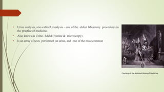
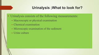
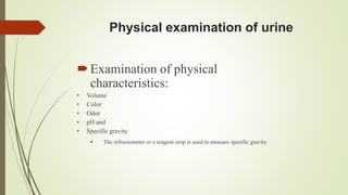
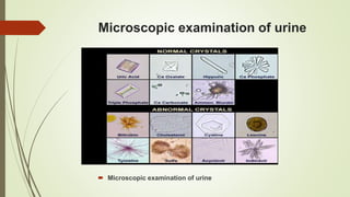
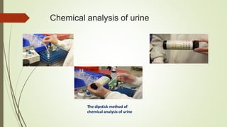
• Urine analysis, also called Urinalysis – one of the oldest laboratory procedures in
the practice of medicine.
• Also knows as Urine- R&M (routine & microscopy)
• Is an array of tests performed on urine, and one of the most common
Courtesy of the National Library of Medicine
Why urinalysis?
• General evaluation of health
• Diagnosis of disease or disorders of the kidneys or urinary tract
• Diagnosis of other systemic disease that affect kidney function
• Monitoring of patients with diabetes
• Screening for drug abuse (eg. Sulfonamide or aminoglycosides)
Collection of urine specimens
• Improper collection---- may invalidate the results
• Containers for collection of urine should be wide mouthed, clean
and dry.
• Analyzed within 2 hours of collection else requires
refrigeration.
Types of urine sample
Sample type Sampling Purpose
Random specimen No specific time
most common, taken
anytime of day
Routine screening, chemical
& FEME(Full examination &
microscopic examination)
Morning sample First urine in the morning,
most concentrated
Pregnancy test, microscopic
test
Clean catch midstream Discard first few ml, collect
the rest
Culture
24 hours All the urine passed during
the day and night and next
day Ist sample is collected.
used for quantitative and
qualitative analysis of
substances
Postprandial 2 hours after meal Determine glucose in
diabetic monitoring
Supra-pubic aspired Needle aspiration Obtaining sterile urine
Urinalysis ;What to look for?
• Urinalysis consists of the following measurements:
– Macroscopic or physical examination
– Chemical examination
– Microscopic examination of the sediment
– Urine culture
Physical examination of urine
Examination of physical
characteristics:
• Volume
• Color
• Odor
• pH and
• Specific gravity
 The refractometer or a reagent strip is used to measure specific gravity
Physical examination continued…
 Volume
 Normal- 1-2.5 L/day
 Oliguria- Urine Output < 400ml/day Seen in
– Dehydration
– Shock
– Acute glomerulonephritis
– Renal Failure
 Polyuria- Urine Output > 2.5 L/day Seen in
– Increased water ingestion
– Diabetes mellitus and insipidus.
 Anuria- Urine output < 100ml/day Seen in renal shut down
Physical examination continued…
 Color
 Normal- pale yellow in color due to pigments urochrome, urobilin and uroerythrin.
 Cloudiness may be caused by excessive cellular material or protein, crystallization or
precipitation of non pathological salts upon standing at room temperature or in the
refrigerator.
 Colour of urine depending upon it’s constituents.
Physical examination continued…
Color • Abnormal colors:
• Colorless – diabetes, diuretics.
• DeepYellow – concentrated urine,
 excess bile pigments, jaundice
Physical examination continued…
Odour• Normal - aromatic due to the volatile fatty acids
• On long standing – ammonical (decomposition of urea forming ammonia
which gives a strong ammonical smell)
• Foul, offensive - pus or inflammation
• Sweet - Diabetes
• Fruity - Ketonuria
• Maple syrup-like - Maple Syrup Urine Disease
• Rancid - Tyrosinaemia
• Characteristic "rotten egg" odor -
Physical examination continued…
pH
• Reflects ability of kidney to maintain normal hydrogen ion
concentration in plasma & ECF
• Urine pH ranges from 4.5 to 8
• Normally it is slightly acidic lying between 6 – 6.5.
• Tested by:
– litmus paper
– pH paper
– dipsticks
• Acidic Urine –Ketosis (diabetes, starvation, fever),systemic acidosis,
UTI- E.coli, acidification therapy
Physical examination continued…
Specific gravity
It is measurement of urine density which reflects the
ability of the kidney to concentrate or dilute the urine
relative to the plasma from which it is filtered.
• Measured by:
– urinometer
– refractometer
– dipsticks
Physical examination continued…
 Specific gravity : Normal :- 1.001- 1.040.
 Increase in Specific Gravity - Low water intake, Diabetes mellitus,
Albuminuruia,Acute nephritis.
 Decrease in Specific Gravity -Absence ofADH, Renal Tubular
damage.
 Fixed specific gravity (isosthenuria)=1.010
Microscopic examination of urine
• A sample of well-mixed urine (usually 10-15 ml) is centrifuged in a test tube at relatively
low speed (about 2000-3,000 rpm) for 5-10 minutes which produces a concentration of
sediment (cellular matter) at the bottom of the tube.
• A drop of sediment is poured onto a glass slide, a thin slice of glass (a coverslip) is place
over it ond observed under microscope
Microscopic examination of urine
• A variety of normal and abnormal cellular
elements may be seen in urine sediment such as:
• Red blood cells
• White blood cells
• Mucus
• Various epithelial cells
• Various crystals
• Bacteria
• Casts
Microscopic examination of urine
• Hematuria is the presence of abnormal numbers of red cells in urine due to any of
several possible causes.
– glomerular damage,
– tumors which erode the urinary tract anywhere along its length,
– kidney trauma,
– urinary tract stones,
– acute tubular necrosis,
– upper and lower urinary tract infections,
– nephrotoxins
• WBC in high numbers indicate inflammation or infection somewhere along
the urinary or
Microscopic examination of urine
Red blood cells in urine appear as refractile
disks
White blood cells in urine
Microscopic examination of urine
Casts
• Urinary casts are cylindrical aggregations of particles that form in the distal nephron,
dislodge, and pass into the urine. In urinalysis they indicate kidney disease.
• They form via precipitation of Tamm- Horsfall mucoprotein which is secreted by renal
tubule cells.
Microscopic examination of urine
Hyaline Cast Granular Cast
Microscopic examination of urine
 Microscopic examination of urine
Chemical analysis of urine
• The chemical analysis of urine us undertaken to
evaluate the levels of the following component:
– Protein
– Glucose
– Ketones
– Occult blood
– Bilirubin
– Urobilinogen
– Bile salts
Chemical analysis of urine
• The presence of normal and abnormal chemical elements in the
urine are detected using dry reagent strips called dipsticks.
• When the test strip is dipped in urine the reagents are activated
and a chemical reaction occurs.
• The chemical reaction results in a specific color change.
• After a specific amount of time has elapse, this color change is
compared against a reference color chart provided.
Chemical analysis of urine
The dipstick method of
chemical analysis of urine
Chemical analysis of urine
 Proteins in urine:
• Detected by heat coagulation or dipstick method
• Urine proteins come from plasma protein and Tomm-Horsfall (T-H) glycoprotein
• healthy individuals excrete <150 mg/d of total protein and <30 mg/d of albumin.
Chemical analysis of urine

urine analysis.pptx

  • 2.
    • Urine analysis,also called Urinalysis – one of the oldest laboratory procedures in the practice of medicine. • Also knows as Urine- R&M (routine & microscopy) • Is an array of tests performed on urine, and one of the most common Courtesy of the National Library of Medicine
  • 3.
    Why urinalysis? • Generalevaluation of health • Diagnosis of disease or disorders of the kidneys or urinary tract • Diagnosis of other systemic disease that affect kidney function • Monitoring of patients with diabetes • Screening for drug abuse (eg. Sulfonamide or aminoglycosides)
  • 4.
    Collection of urinespecimens • Improper collection---- may invalidate the results • Containers for collection of urine should be wide mouthed, clean and dry. • Analyzed within 2 hours of collection else requires refrigeration.
  • 5.
    Types of urinesample Sample type Sampling Purpose Random specimen No specific time most common, taken anytime of day Routine screening, chemical & FEME(Full examination & microscopic examination) Morning sample First urine in the morning, most concentrated Pregnancy test, microscopic test Clean catch midstream Discard first few ml, collect the rest Culture 24 hours All the urine passed during the day and night and next day Ist sample is collected. used for quantitative and qualitative analysis of substances Postprandial 2 hours after meal Determine glucose in diabetic monitoring Supra-pubic aspired Needle aspiration Obtaining sterile urine
  • 6.
    Urinalysis ;What tolook for? • Urinalysis consists of the following measurements: – Macroscopic or physical examination – Chemical examination – Microscopic examination of the sediment – Urine culture
  • 7.
    Physical examination ofurine Examination of physical characteristics: • Volume • Color • Odor • pH and • Specific gravity  The refractometer or a reagent strip is used to measure specific gravity
  • 8.
    Physical examination continued… Volume  Normal- 1-2.5 L/day  Oliguria- Urine Output < 400ml/day Seen in – Dehydration – Shock – Acute glomerulonephritis – Renal Failure  Polyuria- Urine Output > 2.5 L/day Seen in – Increased water ingestion – Diabetes mellitus and insipidus.  Anuria- Urine output < 100ml/day Seen in renal shut down
  • 9.
    Physical examination continued… Color  Normal- pale yellow in color due to pigments urochrome, urobilin and uroerythrin.  Cloudiness may be caused by excessive cellular material or protein, crystallization or precipitation of non pathological salts upon standing at room temperature or in the refrigerator.  Colour of urine depending upon it’s constituents.
  • 10.
    Physical examination continued… Color• Abnormal colors: • Colorless – diabetes, diuretics. • DeepYellow – concentrated urine,  excess bile pigments, jaundice
  • 11.
    Physical examination continued… Odour•Normal - aromatic due to the volatile fatty acids • On long standing – ammonical (decomposition of urea forming ammonia which gives a strong ammonical smell) • Foul, offensive - pus or inflammation • Sweet - Diabetes • Fruity - Ketonuria • Maple syrup-like - Maple Syrup Urine Disease • Rancid - Tyrosinaemia • Characteristic "rotten egg" odor -
  • 12.
    Physical examination continued… pH •Reflects ability of kidney to maintain normal hydrogen ion concentration in plasma & ECF • Urine pH ranges from 4.5 to 8 • Normally it is slightly acidic lying between 6 – 6.5. • Tested by: – litmus paper – pH paper – dipsticks • Acidic Urine –Ketosis (diabetes, starvation, fever),systemic acidosis, UTI- E.coli, acidification therapy
  • 13.
    Physical examination continued… Specificgravity It is measurement of urine density which reflects the ability of the kidney to concentrate or dilute the urine relative to the plasma from which it is filtered. • Measured by: – urinometer – refractometer – dipsticks
  • 14.
    Physical examination continued… Specific gravity : Normal :- 1.001- 1.040.  Increase in Specific Gravity - Low water intake, Diabetes mellitus, Albuminuruia,Acute nephritis.  Decrease in Specific Gravity -Absence ofADH, Renal Tubular damage.  Fixed specific gravity (isosthenuria)=1.010
  • 15.
    Microscopic examination ofurine • A sample of well-mixed urine (usually 10-15 ml) is centrifuged in a test tube at relatively low speed (about 2000-3,000 rpm) for 5-10 minutes which produces a concentration of sediment (cellular matter) at the bottom of the tube. • A drop of sediment is poured onto a glass slide, a thin slice of glass (a coverslip) is place over it ond observed under microscope
  • 16.
    Microscopic examination ofurine • A variety of normal and abnormal cellular elements may be seen in urine sediment such as: • Red blood cells • White blood cells • Mucus • Various epithelial cells • Various crystals • Bacteria • Casts
  • 17.
    Microscopic examination ofurine • Hematuria is the presence of abnormal numbers of red cells in urine due to any of several possible causes. – glomerular damage, – tumors which erode the urinary tract anywhere along its length, – kidney trauma, – urinary tract stones, – acute tubular necrosis, – upper and lower urinary tract infections, – nephrotoxins • WBC in high numbers indicate inflammation or infection somewhere along the urinary or
  • 18.
    Microscopic examination ofurine Red blood cells in urine appear as refractile disks White blood cells in urine
  • 19.
    Microscopic examination ofurine Casts • Urinary casts are cylindrical aggregations of particles that form in the distal nephron, dislodge, and pass into the urine. In urinalysis they indicate kidney disease. • They form via precipitation of Tamm- Horsfall mucoprotein which is secreted by renal tubule cells.
  • 20.
    Microscopic examination ofurine Hyaline Cast Granular Cast
  • 21.
    Microscopic examination ofurine  Microscopic examination of urine
  • 22.
    Chemical analysis ofurine • The chemical analysis of urine us undertaken to evaluate the levels of the following component: – Protein – Glucose – Ketones – Occult blood – Bilirubin – Urobilinogen – Bile salts
  • 23.
    Chemical analysis ofurine • The presence of normal and abnormal chemical elements in the urine are detected using dry reagent strips called dipsticks. • When the test strip is dipped in urine the reagents are activated and a chemical reaction occurs. • The chemical reaction results in a specific color change. • After a specific amount of time has elapse, this color change is compared against a reference color chart provided.
  • 24.
    Chemical analysis ofurine The dipstick method of chemical analysis of urine
  • 25.
    Chemical analysis ofurine  Proteins in urine: • Detected by heat coagulation or dipstick method • Urine proteins come from plasma protein and Tomm-Horsfall (T-H) glycoprotein • healthy individuals excrete <150 mg/d of total protein and <30 mg/d of albumin.
  • 26.