Stool/feces is the end product of digestive system of the body. Following digestion and absorption of the essential food ingredients in the stomach and intestine, the undigested food and unabsorbed secretions of stomach, liver, pancreas and intestine appear in stool.
KFT are used for evaluating kidney functions. there are several routine tests such as urea, creatinine and uric acid. Calculation of eGFR is recommended by national kidney organization whenever creatinine serum is measured.
An illustrative presentation on Microscopic examination of Urine for Medical, Dental, Pharmacology and Biotechnology students to facilitate easy- learning and self-study..
It is fluid which is present in
the abdominal cavity.
The peritoneal cavity is a potential
space lined by mesothelium of the
visceral n parietal peritoneum.
Urine examination how to approach final.ppt1Sachin Verma
Dr. Sachin Verma is a young, diligent and dynamic physician. He did his graduation from IGMC Shimla and MD in Internal Medicine from GSVM Medical College Kanpur. Then he did his Fellowship in Intensive Care Medicine (FICM) from Apollo Hospital Delhi. He has done fellowship in infectious diseases by Infectious Disease Society of America (IDSA). He has also done FCCS course and is certified Advance Cardiac Life support (ACLS) and Basic Life Support (BLS) provider by American Heart Association. He has also done a course in Cardiology by American College of Cardiology and a course in Diabetology by International Diabetes Centre. He specializes in the management of Infections, Multiorgan Dysfunctions and Critically ill patients and has many publications and presentations in various national conferences under his belt. He is currently working in NABH Approved Ivy super-specialty Hospital Mohali as Consultant Intensivists and Physician.
Laboratory and diagnostic examination(urine analysis)anjalatchi
laboratory investigation like urine and stool test like meaning, type of test, interpretation nurses role in laboratory investigation collectin and transportation etc.
Urine is a waste product that is produced by the kidneys in their process of cleaning the blood and is made up of water and dissolved waste products.
The waste products are substances that the body does not need and that can be harmful to our organs if accumulated in the body.
The test measures the amount of sugar in a urine sample. Normal urine does not contain glucose. Microscopic Examination. A variety of normal and abnormal.
Microscopic examination of urine Casts • Urinary casts are cylindrical aggregations of particles that form in the distal nephron, dislodge
Recomendações da OMS sobre cuidados maternos e neonatais para uma experiência pós-natal positiva.
Em consonância com os ODS – Objetivos do Desenvolvimento Sustentável e a Estratégia Global para a Saúde das Mulheres, Crianças e Adolescentes, e aplicando uma abordagem baseada nos direitos humanos, os esforços de cuidados pós-natais devem expandir-se para além da cobertura e da simples sobrevivência, de modo a incluir cuidados de qualidade.
Estas diretrizes visam melhorar a qualidade dos cuidados pós-natais essenciais e de rotina prestados às mulheres e aos recém-nascidos, com o objetivo final de melhorar a saúde e o bem-estar materno e neonatal.
Uma “experiência pós-natal positiva” é um resultado importante para todas as mulheres que dão à luz e para os seus recém-nascidos, estabelecendo as bases para a melhoria da saúde e do bem-estar a curto e longo prazo. Uma experiência pós-natal positiva é definida como aquela em que as mulheres, pessoas que gestam, os recém-nascidos, os casais, os pais, os cuidadores e as famílias recebem informação consistente, garantia e apoio de profissionais de saúde motivados; e onde um sistema de saúde flexível e com recursos reconheça as necessidades das mulheres e dos bebês e respeite o seu contexto cultural.
Estas diretrizes consolidadas apresentam algumas recomendações novas e já bem fundamentadas sobre cuidados pós-natais de rotina para mulheres e neonatos que recebem cuidados no pós-parto em unidades de saúde ou na comunidade, independentemente dos recursos disponíveis.
É fornecido um conjunto abrangente de recomendações para cuidados durante o período puerperal, com ênfase nos cuidados essenciais que todas as mulheres e recém-nascidos devem receber, e com a devida atenção à qualidade dos cuidados; isto é, a entrega e a experiência do cuidado recebido. Estas diretrizes atualizam e ampliam as recomendações da OMS de 2014 sobre cuidados pós-natais da mãe e do recém-nascido e complementam as atuais diretrizes da OMS sobre a gestão de complicações pós-natais.
O estabelecimento da amamentação e o manejo das principais intercorrências é contemplada.
Recomendamos muito.
Vamos discutir essas recomendações no nosso curso de pós-graduação em Aleitamento no Instituto Ciclos.
Esta publicação só está disponível em inglês até o momento.
Prof. Marcus Renato de Carvalho
www.agostodourado.com
Lung Cancer: Artificial Intelligence, Synergetics, Complex System Analysis, S...Oleg Kshivets
RESULTS: Overall life span (LS) was 2252.1±1742.5 days and cumulative 5-year survival (5YS) reached 73.2%, 10 years – 64.8%, 20 years – 42.5%. 513 LCP lived more than 5 years (LS=3124.6±1525.6 days), 148 LCP – more than 10 years (LS=5054.4±1504.1 days).199 LCP died because of LC (LS=562.7±374.5 days). 5YS of LCP after bi/lobectomies was significantly superior in comparison with LCP after pneumonectomies (78.1% vs.63.7%, P=0.00001 by log-rank test). AT significantly improved 5YS (66.3% vs. 34.8%) (P=0.00000 by log-rank test) only for LCP with N1-2. Cox modeling displayed that 5YS of LCP significantly depended on: phase transition (PT) early-invasive LC in terms of synergetics, PT N0—N12, cell ratio factors (ratio between cancer cells- CC and blood cells subpopulations), G1-3, histology, glucose, AT, blood cell circuit, prothrombin index, heparin tolerance, recalcification time (P=0.000-0.038). Neural networks, genetic algorithm selection and bootstrap simulation revealed relationships between 5YS and PT early-invasive LC (rank=1), PT N0—N12 (rank=2), thrombocytes/CC (3), erythrocytes/CC (4), eosinophils/CC (5), healthy cells/CC (6), lymphocytes/CC (7), segmented neutrophils/CC (8), stick neutrophils/CC (9), monocytes/CC (10); leucocytes/CC (11). Correct prediction of 5YS was 100% by neural networks computing (area under ROC curve=1.0; error=0.0).
CONCLUSIONS: 5YS of LCP after radical procedures significantly depended on: 1) PT early-invasive cancer; 2) PT N0--N12; 3) cell ratio factors; 4) blood cell circuit; 5) biochemical factors; 6) hemostasis system; 7) AT; 8) LC characteristics; 9) LC cell dynamics; 10) surgery type: lobectomy/pneumonectomy; 11) anthropometric data. Optimal diagnosis and treatment strategies for LC are: 1) screening and early detection of LC; 2) availability of experienced thoracic surgeons because of complexity of radical procedures; 3) aggressive en block surgery and adequate lymph node dissection for completeness; 4) precise prediction; 5) adjuvant chemoimmunoradiotherapy for LCP with unfavorable prognosis.
- Video recording of this lecture in English language: https://youtu.be/lK81BzxMqdo
- Video recording of this lecture in Arabic language: https://youtu.be/Ve4P0COk9OI
- Link to download the book free: https://nephrotube.blogspot.com/p/nephrotube-nephrology-books.html
- Link to NephroTube website: www.NephroTube.com
- Link to NephroTube social media accounts: https://nephrotube.blogspot.com/p/join-nephrotube-on-social-media.html
263778731218 Abortion Clinic /Pills In Harare ,sisternakatoto
263778731218 Abortion Clinic /Pills In Harare ,ABORTION WOMEN’S CLINIC +27730423979 IN women clinic we believe that every woman should be able to make choices in her pregnancy. Our job is to provide compassionate care, safety,affordable and confidential services. That’s why we have won the trust from all generations of women all over the world. we use non surgical method(Abortion pills) to terminate…Dr.LISA +27730423979women Clinic is committed to providing the highest quality of obstetrical and gynecological care to women of all ages. Our dedicated staff aim to treat each patient and her health concerns with compassion and respect.Our dedicated group ABORTION WOMEN’S CLINIC +27730423979 IN women clinic we believe that every woman should be able to make choices in her pregnancy. Our job is to provide compassionate care, safety,affordable and confidential services. That’s why we have won the trust from all generations of women all over the world. we use non surgical method(Abortion pills) to terminate…Dr.LISA +27730423979women Clinic is committed to providing the highest quality of obstetrical and gynecological care to women of all ages. Our dedicated staff aim to treat each patient and her health concerns with compassion and respect.Our dedicated group of receptionists, nurses, and physicians have worked together as a teamof receptionists, nurses, and physicians have worked together as a team wwww.lisywomensclinic.co.za/
Ethanol (CH3CH2OH), or beverage alcohol, is a two-carbon alcohol
that is rapidly distributed in the body and brain. Ethanol alters many
neurochemical systems and has rewarding and addictive properties. It
is the oldest recreational drug and likely contributes to more morbidity,
mortality, and public health costs than all illicit drugs combined. The
5th edition of the Diagnostic and Statistical Manual of Mental Disorders
(DSM-5) integrates alcohol abuse and alcohol dependence into a single
disorder called alcohol use disorder (AUD), with mild, moderate,
and severe subclassifications (American Psychiatric Association, 2013).
In the DSM-5, all types of substance abuse and dependence have been
combined into a single substance use disorder (SUD) on a continuum
from mild to severe. A diagnosis of AUD requires that at least two of
the 11 DSM-5 behaviors be present within a 12-month period (mild
AUD: 2–3 criteria; moderate AUD: 4–5 criteria; severe AUD: 6–11 criteria).
The four main behavioral effects of AUD are impaired control over
drinking, negative social consequences, risky use, and altered physiological
effects (tolerance, withdrawal). This chapter presents an overview
of the prevalence and harmful consequences of AUD in the U.S.,
the systemic nature of the disease, neurocircuitry and stages of AUD,
comorbidities, fetal alcohol spectrum disorders, genetic risk factors, and
pharmacotherapies for AUD.
New Directions in Targeted Therapeutic Approaches for Older Adults With Mantl...i3 Health
i3 Health is pleased to make the speaker slides from this activity available for use as a non-accredited self-study or teaching resource.
This slide deck presented by Dr. Kami Maddocks, Professor-Clinical in the Division of Hematology and
Associate Division Director for Ambulatory Operations
The Ohio State University Comprehensive Cancer Center, will provide insight into new directions in targeted therapeutic approaches for older adults with mantle cell lymphoma.
STATEMENT OF NEED
Mantle cell lymphoma (MCL) is a rare, aggressive B-cell non-Hodgkin lymphoma (NHL) accounting for 5% to 7% of all lymphomas. Its prognosis ranges from indolent disease that does not require treatment for years to very aggressive disease, which is associated with poor survival (Silkenstedt et al, 2021). Typically, MCL is diagnosed at advanced stage and in older patients who cannot tolerate intensive therapy (NCCN, 2022). Although recent advances have slightly increased remission rates, recurrence and relapse remain very common, leading to a median overall survival between 3 and 6 years (LLS, 2021). Though there are several effective options, progress is still needed towards establishing an accepted frontline approach for MCL (Castellino et al, 2022). Treatment selection and management of MCL are complicated by the heterogeneity of prognosis, advanced age and comorbidities of patients, and lack of an established standard approach for treatment, making it vital that clinicians be familiar with the latest research and advances in this area. In this activity chaired by Michael Wang, MD, Professor in the Department of Lymphoma & Myeloma at MD Anderson Cancer Center, expert faculty will discuss prognostic factors informing treatment, the promising results of recent trials in new therapeutic approaches, and the implications of treatment resistance in therapeutic selection for MCL.
Target Audience
Hematology/oncology fellows, attending faculty, and other health care professionals involved in the treatment of patients with mantle cell lymphoma (MCL).
Learning Objectives
1.) Identify clinical and biological prognostic factors that can guide treatment decision making for older adults with MCL
2.) Evaluate emerging data on targeted therapeutic approaches for treatment-naive and relapsed/refractory MCL and their applicability to older adults
3.) Assess mechanisms of resistance to targeted therapies for MCL and their implications for treatment selection
Pulmonary Thromboembolism - etilogy, types, medical- Surgical and nursing man...VarunMahajani
Disruption of blood supply to lung alveoli due to blockage of one or more pulmonary blood vessels is called as Pulmonary thromboembolism. In this presentation we will discuss its causes, types and its management in depth.
These simplified slides by Dr. Sidra Arshad present an overview of the non-respiratory functions of the respiratory tract.
Learning objectives:
1. Enlist the non-respiratory functions of the respiratory tract
2. Briefly explain how these functions are carried out
3. Discuss the significance of dead space
4. Differentiate between minute ventilation and alveolar ventilation
5. Describe the cough and sneeze reflexes
Study Resources:
1. Chapter 39, Guyton and Hall Textbook of Medical Physiology, 14th edition
2. Chapter 34, Ganong’s Review of Medical Physiology, 26th edition
3. Chapter 17, Human Physiology by Lauralee Sherwood, 9th edition
4. Non-respiratory functions of the lungs https://academic.oup.com/bjaed/article/13/3/98/278874
micro teaching on communication m.sc nursing.pdfAnurag Sharma
Microteaching is a unique model of practice teaching. It is a viable instrument for the. desired change in the teaching behavior or the behavior potential which, in specified types of real. classroom situations, tends to facilitate the achievement of specified types of objectives.
TEST BANK for Operations Management, 14th Edition by William J. Stevenson, Ve...kevinkariuki227
TEST BANK for Operations Management, 14th Edition by William J. Stevenson, Verified Chapters 1 - 19, Complete Newest Version.pdf
TEST BANK for Operations Management, 14th Edition by William J. Stevenson, Verified Chapters 1 - 19, Complete Newest Version.pdf
TEST BANK for Operations Management, 14th Edition by William J. Stevenson, Ve...
Urine examination
1. 1
Urine examination
Introduction
Urine is the waste product in ourbody.The kidneys formurine which passes through the ureters to the bladderforstorage
prior to excretion. Waste productsofprotein metabolismare excreted,electrolyte balance is maintained and the pH (acid-
base balance) is maintained by the excretion of hydrogen ions. Nephron is the functional unit of kidney.
Examination of urine is a fundamentalinvestigation in patients in whomkidney disorders orinfections ofthe urinary tract
are suspected.There are alsomany patientswhoexhibit no clinicalsymptoms,butin whompreviously unrecognized urinary
tract infections can be diagnosed by urine examination.
There are three processes involved in the formation of urine:
1. Simple filtration
2. Selective re-absorptions
3. Secretion.
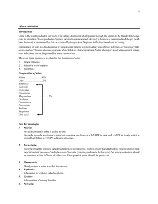
Composition of urine:
Water …………………96%
Urea ……………………2%
Ammonia
Calcium
Chlorides
Creatinine
Magnesium ……….. 2%
Oxalates
Phosphates
Potassium
Sodium
Sulphates
Uric acid
Few Terminologies
1 Pyuria:
Pus cells present in urine is called pyuria.
Normally pus cells are absent in urine but some time may be seen in 1-3/HPF in male and 1-5/HPF in female which in
normal but if there is >5/HPF indicates abnormal.
2. Bacteriuria:
Bacteria present in urine are called bacteriuria.In normal urine, there is absent bacteria but long time incubationthere
may be bacteria because ofmultiplication ofbacteria.(Urine is good media forbacteria). So,urine examination should
be examined within 1-2 hour of collection. If not possible urine should be preserved.
3. Haematuria
Blood present in urine is called haematuria.
4. Nephritis:
Inflamation of nephron called nephritis.
5. Cystitis:
Inflammation of urinary bladder.
6. Polyuria:
2. 2
Excessive output of urine. (>2000 ml).
Conditions- Diabetes Mellitus, Diabetes insipidus, Recovery fromacute renal failure (ARF), Diuretic therapy
7. Oligouria:
Less than 500 ml of urine per day in adults,
Severe oliguria (<100 ml of urine per day in adults)
Conditions- ARF, Vomiting, Fever, Burns.
8. Nocturia:
Excessive urination at night is called Nocturia.
9. Dysuria:
Painful urination is called dysuria.
10. Anuria:
Absence of urination or < 50 ml per 12 hours.
1. Renal threshold: The maximum capacity of kidney to absorbe glucose i.e 160-180mg/dL is called renal threshold.
Glucose appears in the urine when the blood glucose level is more than 10 mmol/l (180 mg%).
12.Renal glycosuria:It is a conditionin which the renalthresholdforglucoseis decreases.Glomerularfiltration rate (GFR)
will be normalbut defect on the renalabsorption.It is unrelated todiabetes mellitus.It does not showany symptoms of
diabetes.
Volume of urine: 0.7 to 2 litre/ day (24 hrs).
Collection of urine specimens
Containers forthe collection ofurine should be wide-mouthed,clean and dry.Labelpatient name,lab no,age/sex,date etc.
If the urine specimen has to be transported for any length of time it should contain an appropriate preservative to prevent
bacterial overgrowth or hatching of viable ova.
Types of urine specimen
1. Early morning urine specimen
Early morning urine provides the most concentrated sample. It is better for urine pregnancy test.
2. Random urine specimen
A randomurine sample, taken at any time of the day, will enable the laboratory to screen for substances which are
indicators of kidney infection.
3. 24-Hour urine specimen
The 24-hour urine specimen is collected in a clean gallon with a stopper.On the first morning the patient getsup and
urinates; this urine is not collected.Allthe urine passed during the rest ofthe day and night is collected in the bottle.
The next morning the patient getsup and collects thefirst urine ofthe morning in the bottle (8 amto 8am). The bottle
should then betaken immediately to the laboratory.Measure the volume ofurine with a measuring cylinderand record
it.
4. Midstream urine specimen
While passingurine,the patient placesan opencontainerin the streamofurine and collects about 20to 30 ml of urine.
The container should be covered immediately. This sample is suitable of urine culture.
5. Catheter urine sample
Collection of urine using a catheter must be carried out by a qualified physician or nurse. The procedure is used for
certain bacteriological tests, mainly in women. Usually, however, a specimen collected in the normal way following
thorough cleansing is acceptable for this purpose.
3. 3
6. Suprapubic aspiration
This method is suitable for infants. Sample is collected by physician or experience nurse or other medical person.
Preservation of urine specimens
Urine passed at a clinic and examined immediately, does not require preservation.
Preservative methods for urine sample.
1. Refrigerator method
2. Chemical method
1. Refrigerator method:
In refrigerator 2-8°C is better.
2. Chemical methods
Mainly acids are usedas a chemical preservative.
i. Concentrate HCl: 10-30 ml HCL is used to 24hrs urine sample. This is suitable for calcium, phosphate, urea,
creatinine urine test.
ii. Toluene: 5-10 ml is used to 24hrs urine sample. This is suitable for protein, uric acid etc.
iii. Chloroform: Same as toluene.
iv. Acetic acid: 10 ml is used to 24hrs urine sample. This is suitable for vitamin test.
v. Boric acid: This is most common preservative used in lab. 1-2 grams boric acid is used in 120 ml of urine. It is
also used in urine culture. 10 gmfor 24hrs urine.
vi. Formalin: 1-2 drops formalin is used in 30ml of urine.
vii. Thymol: 0.1gm per 100ml of urine sample.
Urine Routine Examination
Collection of urine specimens
Containers for the collection of urine should be wide-mouthed, clean and dry with screw capped. Label patient name, lab
no, age/sex, date etc. If the urine specimen has to be transported for any length of time it should contain an appropriate
preservative to prevent bacterial overgrowth.
Urine routine examination:
1. Physical examination (Macroscopic examination)
2. Chemical examination
3. Microscopic examination test
1. Physical examination:
Physical examination is done by naked eye.
a. Colour
b. Transparency
c. pH
4. 4
d. Specific gravity (Sp. G)
Colour:
Urine is normally clear straw-yellow or light yellow in colour. More concentrated urine may appear dark yellow.
The colour of urine is light yellow or straw yellow because present of urobilin.
The presence of blood cells or excess salts may make the urine appear cloudy.
Pigments frombile substances may make the urine appear deep yellow or brown.
Appearance Possible Cause
Cloudy ● Bacterial urinary infection
Red and cloudy
(Due to red cells)
● Urinary schistosomiasis
● Bacterial infection
Brown and cloudy
(Due to haemoglobin)
● Malaria haemoglobinuria
● Other conditions that cause
intravascular haemolysis.
Yellow-brown, orgreen-brown
(Due to bilirubin)
● Acute viral hepatitis
● Obstructive jaundice
Yellow-orange
Due to urobilin i.e. oxidized
urobilinogen
● Haemolysis
● Hepatocellular jaundice
Milky-white ● Filariasis, Pus
Colourless Very dilute urine, polyuria
Transparency
Normally fresh voided urine is clear. If urine is not clear we report as turbid. The following condition urine may turbid,
1. UTI
2. Filariasis
3. High protein present in urine.
Specific gravity
Measured by urinometer - implies the capacity of kidney to concentrate urine.
Normal value: 1.015-1.030. Depends upon - State of hydration & solute load
Increased specific gravity is called hypersthenuria.
Decreased specific gravity is called hyposthenuria.
Values more than 1.030 indicates
Severe dehydration
DM (diabetes mellitus)
Adrenal insufficiency
Values less than 1.015 indicates
Increase water intake
Diabetes insipidus
Chronic Nephritis
5. 5
Specific gravity of urine can be measuredwith
1. Urinometer 2. Refractometer
3. Dip Sticks 4. Osmometer
5. Specific gravity bottle 6. Specific gravity beads
Fig: Urinometer
Fig: Urinometers
pH
Normal freshly passedurine is slightly acid (around6.0).In certain conditions,the pHofthe urine may increaseordecrease.
Normal range is 4.5 to 8.5.
Average is 6.0
Determination of urine pH
1. By using pH meter
2. By using pH strip
Increasedcondition: After heavy meals, Proteus infection, Dietary products.
Decreasedcondition: Afterheavy exercise,Metabolic Acidosis,Chronic respiratory acidosis,vegetarian diet,high protein
intake.
6. 6
2. Chemical examination:
a. Urine sugar test
b. Urine protein test or urine albumin test
URINE SUGAR TEST:
Normally sugar is absent in urine. If glucose is present in urine is called glycosuria. Glycosuria is condition of diabetes
mellitus. We can performurine sugar by following methods.
i. Benedict's is method
ii. Urine strip method
iii. Fehling test
Urine sugar test by Benedict's method:
Benedict’s test is used for testing reducing sugars. This test is also called ad reducing sugar test. A reducing sugar is a
carbohydratepossessing eithera free aldehydeorfree ketone functionalgroupas partofits molecularstructure.This includes
all monosaccharides(eg.glucose,fructose,galactose)andmany disaccharides,including lactoseandmaltosebut not sucrose
(sucrose is non-reducing sugar).
It can be used as a semi-quantitative test.
Benedict’s test is most commonly used to test forthe presence ofglucose in urine. Glucose found to be present in urine is
an indication ofDiabetes mellitus. Glucose is a reducing substance.It reduces the blue coppersulfate in benedict solution
to red copper oxide, which is insoluble.
Principle:
Reducing sugars under alkaline and heat conditions, copper sulphate (Cu2+
) reduced to cuprous oxide (Cu2O) and gives
green, yellow, orange or brick red precipitate.
Cupric ions (Cu2+
) + Urine glucose
𝐴𝑙𝑘𝑎𝑙𝑖𝑛𝑒 𝑐𝑜𝑛𝑑𝑖𝑡𝑖𝑜𝑛
𝐻𝑒𝑎𝑡
> Cuprous oxide (Cu2O)
(Blue colour)
Precipitate to green, yellow, orange or brick red.
Materials and reagents
1. Test-tubes
2. Wooden test-tube holder
3. Test-tube rack
4. Beaker
5. Bunsen burner or spirit lamp
6. Dropper pipette
7. Graduated pipette, 5ml
8. Benedict solution.
7. 7
Procedure:
1. Pipette 5 ml of Benedict solution into a test-tube.
2. Add 8 drops (0.5ml) of urine and mixwell.
3. Boil overa Bunsen burnerorspirit lamp for 2 minutes orplace the test-tube in a beakerat boiling waterfor5 minutes.
4. Place the test-tube in the test-tube rack and allow to cool to roomtemperature.
5. Examine the colour change of the solution and any precipitate, report the result.
Reporting
1. No colour change (blue) Nil
2. Slightly colour change Trace
3. Green colour + (0.5%)
4. Yellow colour ++ (0.5-1.5%)
5. Orange colour +++ (1.5 - 2%)
6. Red colour (brick colour) ++++ (>2%)
Precautions:
1. Used clean and dry test tube.
2. Benedict’s solution should be clean and dry.
3. Test tube mouth should be away fromface.
Composition of Benedict's solution:
1. Solution A:
i. Copper sulphate 17.3 gram
ii. Distilled water 300 ml
2. Solution B:
i. Sodiumcarbonate 100 gram
ii. Sodiumcitrate 175 gram
iii. Distilled water 700 ml
Mix Solution A and B then pour on bottle and label it and store at roomtemperature.
Copper sulphate provides cupric ion (Cu++ ion) in solution.
Sodium citrate prevents precipitation of cupric ion as cuprous hydroxide by forming a loose cupric sodium citrate
complex.
8. 8
Sodiumcarbonate provides the alkaline pH to the solution
False positive results
1. Chloral hydrate
2. Isoniazid
3. Aminosalicylic acid
4. Nalidixic acid
5. Nitrofurantoin
6. Vitamin C.
7. Glucuronides.
8. Patient under dextrose infusion.
9. Thymol
10. Chloroform.
ii. Urine sugar detection by stripmethod
Principle:
Glucose is oxidized to gluconolactone by the enzyme glucose oxidase (specific for glucose) with the release of hydrogen
peroxide.A chromogen isthen oxidized by the hydrogenperoxide and a peroxidase enzyme is usedto convert the chromogen
from a reduced colourless state to a coloured oxidized state.
Advantages of strip method:
1. Quick
2. Cheap
3. Easy to perform
4. More specific
5. More sensitive
URINE PROTEIN TEST
Normally healthy people’surine contains only a smallamount ofurinary proteins (<150 mg perday)but not detectprotein
in urine by quantitative method.Normally we cannot detectprotein in urine.If protein detect in urine is called proteinuria.
Albumin is the commonest protein excreted in urine due to its relatively small size (molecular weight 66,000) and high
concentrationin plasma.Proteinuria is an importantpathologicalfinding with severalimportant causes,mostly renalorigin.
Methods of urine protein detection:
i. Heat and acetic acid method
ii. Sulphosalicylic acid method
iii. Urine strip method
Heat and acetic acid method:
Principle:
Heat causesthe proteinsin urine to coagulate.Proteins are more
susceptible to precipitation whenthey are at the pHoftheir isoelectric point,
which is usually low.
Requirements
1. Test tubes and test tube holder
9. 9
2. Spirit lamp
3. Dropper
4. Timer
5. Reagent 5% acetic acid
6. Sample urine
Procedure:
1. Take 3-5 ml of urine in a test tube.
2. Heat the upper portion of the urine.
3. Add 2-3 drops of 5% acetic acid.
4. Observe the white coagulumor turbid against a dark background.
Result:
1. No change (Clear) Nil
2. Slightly cloudy Trace
3. Cloudy + (Average 30 mg/dl)
4. Cloudy with small precipitate ++ (Average 100 mg/dl)
5. Cloudy with moderate precipitate +++ (Average 300 mg/dl)
6. Cloudy with large precipitate ++++ (Average 1000 mg/dl)
ii. Urine protein test by Sulphosalicylic acidmethod
This method is sensitive and commonly uses method for urine protein test.
Principle
Protein in the urine is precipitatedbysulphosalicylic acid.The degree ofturbidity is relatedto the quantityofprotein present
in urine.
Requirements
1. Test tube with test rack
2. Test tube holder
3. Timer
4. Pipette
5. Sample urine
6. Reagent 3% Sulphosalicylic acid
Procedure
1. Take 3ml of urine taken in a test tube,
2. Add 3ml of 3% Sulphosalicylic acid and mixwell.
3. Observe the result against a dark background.
Note: Control (C) also required.
Result
1. No change (Clear) Nil
2. Slightly cloudy Trace
3. Cloudy +
4. Cloudy with small precipitate ++
10. 10
5. Cloudy with moderate precipitate +++
6. Cloudy with large precipitate ++++
Protein test by stripmethod:
Urine protein strip tests detect mainly albumin. The test area is impregnated with the indicator tetrabromophenol blue. In
the presence of protein there is a change in the colour of the indicator fromlight yellow to green-blue depending on the
amount of protein present in urine.
Clinical significance
Proteinuria is seen in following conditions.
1. Urinary tract infection (UTI)
2. Glomerulonephritis
3. Diabetic nephropathy
4. Pyelonephritis
5. Nephrotic syndrome
6. Sever illness
7. Last stage of pregnancy
8. Fever
9. High protein diet
10. Infection
11. Toxiemia of pregnancy
12. Systemic Lupus Erythematosus (SLE)
13. Exposure to severe cold
14. Multiple myeloma
Microscopic Examination
Microscopic examination of urine is done form the deposit of urine.
1. Take clean and dry test tube and label it.
2. Pour urine around 3-5 ml in test tube.
3. Centrifuge at 3000 rpmfor 3-5 minutes.
4. Then discard supernatant part and pour deposit part in center of slide.
5. Cover with cover slip then observe on microscope at 10X the 40x.
Microscopic findings of urine:
i. RBCs
ii. Pus cells
iii. Epithelial cells
iv. Yeast cells
v. Bacteria
vi. Spermcells
vii. Casts
11. 11
viii. Crystal
ix. Parasites
X. Mucous threads
1. RBCs:
Normally RBC is not presentin urine.More than one RBC perhigh powerfield is abnormal.The presence ofRBC in
urine is called haematuria.
The red blood cells swell up and broken down in diluted urine. The red cells may show crenated margins.
Clinical Significance: Red blood cells are found in
a) Pyelonephritis b) Renal stones c) Cystitis d) Polycythemia vera
Normal RBC
Crenated RBC
Swelling RBC
Figure of RBCs
2. Pus cells:
Pus cells present in urine is called pyuria.Normally pus cells are absent in urine butsome time may be seen in 1-3/HPF
in male and 1-5/HPF in female which in normal but if there is >5/HPF indicates abnormal.
Large number of pus cells indicates bacterial infection of urinary tract.
Figure: Pus cells
3. Epithelial cells:
These cells have a single rounded nucleus. Squamous epithelial cells present in urine in moderate numbers have no
pathological importance.
12. 12
Clinical Significance: Presence of all other epithelial cells indicates pathological condition.
Renal tubular epithelial cells: Unstained cells have almost the same size as that of a neutrophil but contain a large
round nucleus. Oval fat bodies are the cells containing fat globules. The nucleus, is not visible.
Bladder epithelial cells: Unstained cells are larger than renal tubular cells having a round nucleus and vary in size
depending on depth of origin.
Squamous epithelial cells: These are unstained large flattened cells with abundant cytoplasmand a small round
nucleus. The cell may be folded or rolled.
Figure: Epithelial cells
4. Bacteria:
Bacteria are not normally present in urine. However, unless specimens are collected under sterile conditions,a few
bacteria are usually present as a result of vaginal, urethral, external genitalia, or collection-container contamination.
These contaminant bacteria multiply rapidly in specimens that remain at room temperature for extended periods, but
are of no clinical significance. They may produce a positive nitrite test result and also frequently result in a pH
above 8, indicating an unacceptable specimen.
Bacteria may be present in the form of cocci (spherical) or bacilli (rods). Owing to their small size, they must be
observed and reported using high-power magnification.
They are reported as few, moderate, or many per high-power field. To be considered significant for UTI, bacteria
should be accompanied by WBCs.
Figure: Bacteria
13. 13
5. Yeast cells:
Yeast cells appear in the urine as small, refractile oval structures that may or may not contain a bud. In severe infections,
they may appear as branched, mycelial forms. Yeast cells are reported as rare, few, moderate, or many per hpf.
Differentiation betweenyeastcells andRBCs can sometimes bedifficult.Carefulobservationforbuddingyeast cells should
be helpful.
In normal urine yeast cells are absent whereas,in case ofdiabetic,immunocompromised patients andwomen with vaginal
moniliasis may present yeast cells.
Fig: Yeast cells
6. Sperm cells
Spermatozoa are easily identified in the urine sediment by theiroval,slightly tapered heads and long tail.Spermatozoa are
occasionally found in the urine in male. In some cases of both men and women contain spermcells in urine in sexual
intercourse,masturbation,ornocturnalemission.They are rarely of clinical significance except in cases ofmale infertility
or retrograde ejaculation in which spermis expelled into the bladder instead of the urethra.
Figure: Sperm cells
7. Casts:
Urinary casts are formed in the lumen of the tubules of the nephrons and are originate from Tamm-Horsfall- protein
(molecular weight 18,000) secreted by renal tubules.
Casts have paralleledges,round or broken.They may be curved orstraight,short orlong.A significant numberofurinary
casts usually indicates the presence of renal disease.
Urinary casts can be divided into two main group: Acellular and Cellular Casts.
Acellular Casts Cellular Casts
14. 14
Hyaline Casts Red Blood Cell Casts
Granular Casts White Blood Cell Casts
Waxy Casts Bacterial Casts
Fatty Casts Epithelial Cell Casts
Casts Composition AssociatedConditions
Hyaline casts
Solidified Tamm-Horsfall
mucoprotein
Normal individuals
Dehydration
Heavy exercise
Granular casts
Various celltypes (Degenerationof
cellular casts, Aggregates of
plasma proteins or
immunoglobulin light chains)
After sternous exercise
Chronic renal diseases
Acute tubular necrosis
Waxy casts (renal failure
casts)
Various cell types (Final stage of
degeneration of cellular cast)
Severe chronic renal disease
renal amyloidosis
Fatty casts
Lipid droplets within the protein
matrix of the cast
Tubular degeneration
Nephrotic syndrome
Hypothyroidism
RBC Casts Red Blood Cells
Pyelonephritis
Glomerulonephritis
Acute interstitial nephritis
Lupus nephritis
WBC Casts White Blood Cells Glomerulonephritis
Epithelial Cell Casts Renal Tubular Epithelial Cells
Renal tubular necrosis
Viral Diseases
Kidney transplant rejection
15. 15
Figure: Casts
8. Crystals:
Crystals are not usually found in freshly voided urine but appearafterthe urine stands fora while. Crystals are considered
"normal" if they are fromsolutes that are typically found in the urine. Some examples of crystals that can be found in
the urine of healthy individuals includes:amorphous urates,crystalline uric acid, calcium oxalates,amorphous phosphates
and calciumcarbonate.
If the crystals are from solutes that are not normally in the urine,they are considered “abnormal." Abnormalcrystals may
indicate an abnormal metabolic process. Some of these include: cystine, tyrosine and leucine. When crystals
form as urine is being made in the kidney, they may group together to form kidney "stones" or calculi. These stones can
become lodged in the kidney itself or in the ureters, tubes that pass the urine fromkidney to the bladder, causing
extreme pain. Medications,drugs,and x-ray dyes can also crystallize in urine. Associated with renalcalculi, commonly is
oxalate in acidic urine and phosphate in alkaline urine.
21. 21
9. Mucous threads
These are long thin waxy threadsofribbon like structures.These may be present in normalurine but found in inflammation
or irritation of the urinary tract.
10. Urine parasites:
a. Trophozoite of Trichomonas vaginalis:
b. Ova of Schistosoma haematobium
Schistosoma haematobium is established by demonstration of eggs in urine. The specimen should be immediately
centrifuged and the sediment examined by wet mount.
Trichomonasvaginalis motile trophozoitesmay also be foundin the urine,especially in infected male patients.To lookfor
the presence of trophozoites, the urine specimen should be centrifuged and, the sediment mixed with a drop examined by
wet mount. Temporary stains, such as methylene blue or malachite green, are also helpful.
Fig: Trophozoite of Trichomonas vaginalis Fig: Ova of Schistosoma haematobium
23. 23
Special Test
1. Ketone Bodies Test or (Acetone Test)
2. Chyluria Test (Urine chyle Test)
3. Urine Bile Salt Test
4. Urine Bile Pigment Test
5. Urobilinogen Test
1. Ketone bodies test or Acetone test in urine
The present of ketone body in the urine is called ketonuria. Ketonuria seen in mainly uncontrolled diabetic mellitus (type
1), Starvation, Severe vomiting, Glycogen storage disorders, High fat diet.
Acetoacetate, beta-hydroxybutyrate and acetone are collectively referred to as ketone bodies or ketones.
Formation of ketones:
The metabolismof glucose normally provides the body with its energy requirements.If, however,the intake of glucoseis
insufficient as in starvation,orglucosemetabolismis defectivedueto a lackofinsulin as occurs in untreatedoruncontrolled
diabetes,thebody obtainsits energyby breaking down fats.It is this increase in fat metabolismwhich leads to a buildup of
ketones in the body. An accumulation of ketones in the body is called ketosis.
Ketones are toxic to the brain and if present in sufficiently high concentrationin the blood they can contribute to the coma
found in diabetic ketoacidosis.Metabolic acidosis is causedbya variety ofnon-volatile acids accumulatingin the plasma of
patients with diabetic coma.Urine ketone tests detect acetoacetate and acetone. Beta hydroxybutyrate is not detected.
Methods of Ketone bodies test
a. Rothera's test
b. Gerhardt's test
c. Strip test
a) Rothera’s Test
Principle
Ketone bodies react with sodiumnitroprusside at alkaline condition to give purple colour complex.
Requirements:
i. Test tubes
ii. Dropper
iii. Marker
iv. Sample - urine
v. Reagent - Rothera's powder
Rothera's powder
Composition
1. Ammoniumsulphate crystals……….100gm
2. Sodiumnitroprusside crystal.………..3gm
3. Sodiumcarbonate.………………….50 gm
24. 24
Procedure
1. Take pea sized Rothera’s powder and pour in test tube.
2. Add few drops of urine in same tube.
3. Observe the result.
Result
No colour change = Negative
Purple colour = Positive
Note: Excess of sodium nitroprusside in reagent may give false positive result.
Ketone striptests
Severalketone strip testsare available commercially.The reagentstrip is impregnatedwith sodiumnitroprusside buffer.
Interpretations
1. Untreated diabetes mellitus
2. Starvation
3. Prolonged vomiting
4. Glycogen storage disease.
Ketone testing is most often done if have type 1 diabetes and:
Blood sugar is higher than 240mg /dL
Have an illness such as pneumonia, heart attack, or stroke
Nausea or vomiting occur in pregnant
25. 25
2. Urine Chyle test
Introduction:
Present ofchyle in urine is called chyluria.It is complicationof bancroftian filariasis.It occurs whentheurogenitallymphatic
vessels whichare linked to thosethat transportchyle fromthe intestine become blocked andrupture.Milky white appearance
seen when chyle presents in urine.
Principle:
When urine is mixed with organic solvents like ether, chloroform, if urine contain chyle then urine become clear.
Requirements
a. Test tubes
b. Pipette
c. Sample - urine
d. Reagent - organic solvent e.g: chloroform, benzene or diethyl-ether
Procedure:
1. Take one clean and dry test tube and label it.
2. Add 5 ml urine in the test tube.
3. Add 1 to 2 ml reagent.
4. Observe the result.
Result:
If white part disappears or urine is clear: Positive
If no change: Negative
Clinical significance:
This test is used to diagnosis for filariasis.
Bile Salts Test In Urine
Bile salts are normally not excretedin urine but in obstructive jaundice,theyappearin urine.These are sodiumandpotassium
salt of taurocholic acid, glycocholic acid which assists in the emulsion and absorption of dietary lipid by reduce surface
tension.Bile salts are formed in the liverfrom cholesterol.Ifthere is obstructionin bile flow orleakage ofbile in blood then
this salt will appear in urine.
Method:
Hay’s test
Principle
Bile salts reduce the surface tension of urine, which causes sulfur powder to sink to the bottom.
Requirements
26. 26
1. Test tubes
2. Pipette
3. Sample - urine
4. Reagent - Sulphur powder
Procedure
1. Take 2 ml of urine in a test tube.
2. Sprinkle or spray sulfur powder in upper part of urine sample. Do not shake or mix.
3. If the sulfur powder sinks to the bottom, it indicates the presence of bile salts. Run a control using distilled water
instead of urine.
Result
Sulphur powder sink = Positive
Sulphur powder float = Negative
Interpretation
Positive in obstructive jaundice
Bile Pigment test (Urine bilirubin test)
Normal urine does not contain bile pigment.In certain liver disease (jaundice),anemia and infectious disease bile pigments
may pass into blood streamand excreted in urine.
Urine bile pigment test
1. Fouchet's test
2. Foamtest
3. Lugol’s Iodine method
4. Urine strip test
1. Fouchet's test:
Principle
Wet bariumsulphate hasthe capacity to absorbyellowbilirubin. Fouchet’s reagent oxidizes the absorbedbilirubin to green
color biliverdin & blue color bilicyanin.
Requirements:
1. Filter paper
2. Test tubes
3. Funnel
4. Sample (urine)
5. Reagent (i) Barium chloride
(ii) Fouchet's reagent
Composition of fouchet's reagent:
1. Trichloroacetic acid = 25 gram
2. 10% ferric chloride = 10 ml
3. Distilled water = 90 ml
Specimen:
27. 27
Freshly passed urine is required. It should be protected from daylight and fluorescent light because bilirubin is rapidly
oxidized by ultraviolet light to biliverdin which is not detected by the reagents used in the tube or strip tests.
Procedure
1. Take 10 ml of urine in a test tube.
2. Add 5ml of bariumchloride solution followed by pinch of magnesiumsulphate powder.
3. Mix well and keep the solution for 5 minutes.
4. Filter the solution and fold the filter paper and dry it.
5. Add few drops of fouchet’s reagent to the dry unfiltrate precipitate on the filter paper.
6. Observe the colour.
Result
1. Blue green colour = Positive
2. No colour = Negative
Interpretation
Bile pigments are found in urine along with bile salts in severe hepatitis & obstructive jaundice.
Urobilinogen test in Urine
Urobilinogen is normally presentin urine in low concentrations (<4mg/ 24 hrs).It is formed in the intestine frombilirubin,
and a portion of it is absorbed back into the bloodstream.
Increased amount ofurobilinogen in urine indicate liverdisorders in which liver function is impaired. Urobilinogen is high
in haemolytic jaundice but normal in obstructive jaundice.
Method:
1. Ehrlich’s test
2. Strip test
Specimen:
Freshly passed urine is requiredbecause urobilinogenwhich is colourless is rapidly oxidized to the orange pigmenturobilin
which is not detected by Ehrlich’s test or by urobilinogen strip tests.
Ehrlich’s test
Principle:
Urobilinogen reacts with p-dimethylaminobenzaldehyde to form a red condensation product. The intensity of colour
produced corresponds to the concentration of urobilinogen present.
Requirements:
1. Test tubes with test tube rack
2. Pipette
3. Sample = urine
4. Reagent; Ehrlich's reagent
Composition of Ehrlich's reagent:
1. P- dimethylaminobenzaldehyde: 2 gm
2. HCl = 20 ml
28. 28
3. Distilled water: 80 ml
Procedure:
Step1
1. Take 5 ml urine in test tube and add 5 ml Ehrlich’s reagent and mixwell.
2. Wait for 10 minutes.
Pink or red: Positive may be present and process further for confirmation.
Negative: No further confirmation
Step2
3. Mix 5 ml above mixture with 5 ml of saturated sodiumacetate solution.
Colour get intensity of excess of urobilinogen is present and process is further or confirmation.
Step3
4. Mix 5 ml above mixture with 5 ml chloroform, mix thoroughly and allow to separate layer.
Observation:
Lower chloroform= Pink or red = Positive for urobilinogen.
Control
Sometimes colouris faint or doubtfulin such case put a negative control(urine 6N HCL and process in similar as for
urobilinogen).
Interpretation
Increases
1. Hemolytic disease
2. Hepatocellulardamage orhepatic congestion,resultingin less ofthe absorbedurobilinogen being excretedby the liver.
3. Cirrhosis of the liver.