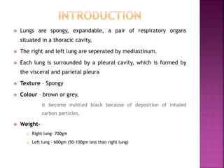
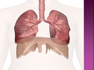
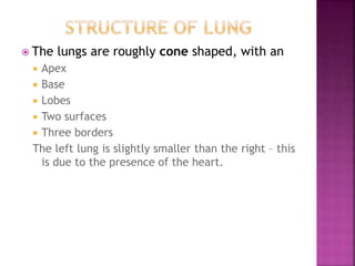
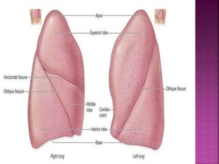
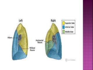
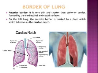
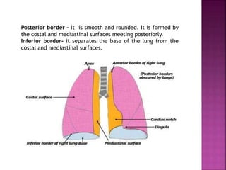
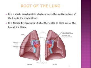
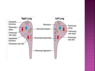
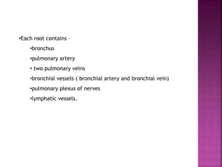
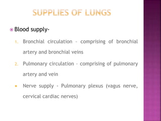
By- Chirag Ajmera
Assistant professor, MJF Group of
Colleges and Hospitals
 Lungs are spongy, expandable, a pair of respiratory organs
situated in a thoracic cavity,
 The right and left lung are seperated by mediastinum.
 Each lung is surrounded by a pleural cavity, which is formed by
the visceral and parietal pleura
 Texture – Spongy
 Colour – brown or grey,
it become mottled black because of deposition of inhaled
carbon particles.
 Weight-
 Right lung- 700gm
 Left lung – 600gm (50-100gm less than right lung)
 The lungs are roughly cone shaped, with an
 Apex
 Base
 Lobes
 Two surfaces
 Three borders
The left lung is slightly smaller than the right – this
is due to the presence of the heart.
Apex
 The apex is blunt and lies above the level of the
anterior end of the first rib.
 It reaches nearly 2.5 cm above the medial one-
third of the clavicle.
Base
 It is semi lunar and concave
 The inferior surface of the lung, which sits on
the diaphragm.
Lobes
 The right lung is divided into 3 lobes (upper, middle and lower)by
two fissures, oblique and horizontal fissure.
 The left lung is divided into two lobes by the oblique fissure.
Fissure
 Oblique fissure- The oblique fissure cuts into the whole thickness
of the lung, except at the hilum. It passes obliquely downwards
and forwards.
 Horizontal fissure- Runs horizontally from the sternum, at the
level of the 4th rib, to meet the oblique fissure in the right lung.
 There are two lung surfaces
 Costal surface
 Medial surface
 The costal surface is smooth and convex. It faces the internal
surface of the chest wall. It is related to the costal pleura, which
separates it from the ribs and innermost intercostal muscles.
 The medial surface is divided into a Posterior or vertebral part,
and an anterior or mediastinal part.
 Mediastinal surface- anterior 2/3rd of the medical surface. It is
related to the mediastinal structures.
 Vertebral surface- posterior 1/3rd of medical surface. It is related to
the vertebral bodies and intervertebral discs.
 Anterior border- it is very thin and shorter than posterior border,
formed by the mediastinal and costal surfaces.
 On the left lung, the anterior border is marked by a deep notch
which is known as the cardiac notch.
Posterior border – it is smooth and rounded. It is formed by
the costal and mediastinal surfaces meeting posteriorly.
Inferior border- it separates the base of the lung from the
costal and mediastinal surfaces.
 It is a short, broad pedicle which connects the medial surface of
the lung to the mediastinum.
 It is formed by structures which either enter or come out of the
lung at the hilum.
•Each root contains –
•bronchus
•pulmonary artery
• two pulmonary veins
•bronchial vessels ( bronchial artery and bronchial vein)
•pulmonary plexus of nerves
•lymphatic vessels.
 Blood supply-
1. Bronchial circulation – comprising of bronchial
artery and bronchial veins
2. Pulmonary circulation – comprising of pulmonary
artery and vein
 Nerve supply - Pulmonary plexus (vagus nerve,
cervical cardiac nerves)
This slides covers introduction, structure, supplies of the lungs

lungs anatomy

  • 1.
    By- Chirag Ajmera Assistantprofessor, MJF Group of Colleges and Hospitals
  • 2.
     Lungs arespongy, expandable, a pair of respiratory organs situated in a thoracic cavity,  The right and left lung are seperated by mediastinum.  Each lung is surrounded by a pleural cavity, which is formed by the visceral and parietal pleura  Texture – Spongy  Colour – brown or grey, it become mottled black because of deposition of inhaled carbon particles.  Weight-  Right lung- 700gm  Left lung – 600gm (50-100gm less than right lung)
  • 4.
     The lungsare roughly cone shaped, with an  Apex  Base  Lobes  Two surfaces  Three borders The left lung is slightly smaller than the right – this is due to the presence of the heart.
  • 6.
    Apex  The apexis blunt and lies above the level of the anterior end of the first rib.  It reaches nearly 2.5 cm above the medial one- third of the clavicle. Base  It is semi lunar and concave  The inferior surface of the lung, which sits on the diaphragm.
  • 7.
    Lobes  The rightlung is divided into 3 lobes (upper, middle and lower)by two fissures, oblique and horizontal fissure.  The left lung is divided into two lobes by the oblique fissure. Fissure  Oblique fissure- The oblique fissure cuts into the whole thickness of the lung, except at the hilum. It passes obliquely downwards and forwards.  Horizontal fissure- Runs horizontally from the sternum, at the level of the 4th rib, to meet the oblique fissure in the right lung.
  • 9.
     There aretwo lung surfaces  Costal surface  Medial surface  The costal surface is smooth and convex. It faces the internal surface of the chest wall. It is related to the costal pleura, which separates it from the ribs and innermost intercostal muscles.  The medial surface is divided into a Posterior or vertebral part, and an anterior or mediastinal part.  Mediastinal surface- anterior 2/3rd of the medical surface. It is related to the mediastinal structures.  Vertebral surface- posterior 1/3rd of medical surface. It is related to the vertebral bodies and intervertebral discs.
  • 10.
     Anterior border-it is very thin and shorter than posterior border, formed by the mediastinal and costal surfaces.  On the left lung, the anterior border is marked by a deep notch which is known as the cardiac notch.
  • 11.
    Posterior border –it is smooth and rounded. It is formed by the costal and mediastinal surfaces meeting posteriorly. Inferior border- it separates the base of the lung from the costal and mediastinal surfaces.
  • 12.
     It isa short, broad pedicle which connects the medial surface of the lung to the mediastinum.  It is formed by structures which either enter or come out of the lung at the hilum.
  • 14.
    •Each root contains– •bronchus •pulmonary artery • two pulmonary veins •bronchial vessels ( bronchial artery and bronchial vein) •pulmonary plexus of nerves •lymphatic vessels.
  • 15.
     Blood supply- 1.Bronchial circulation – comprising of bronchial artery and bronchial veins 2. Pulmonary circulation – comprising of pulmonary artery and vein  Nerve supply - Pulmonary plexus (vagus nerve, cervical cardiac nerves)
  • 16.
    This slides coversintroduction, structure, supplies of the lungs