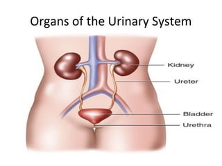
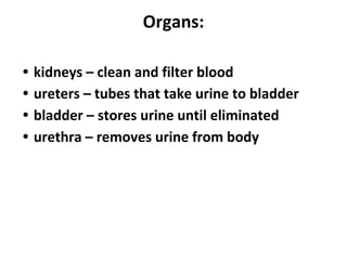
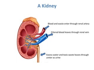
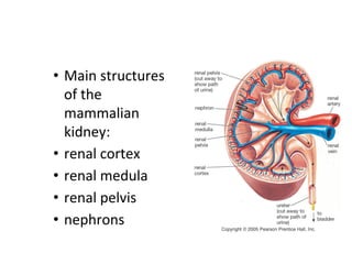
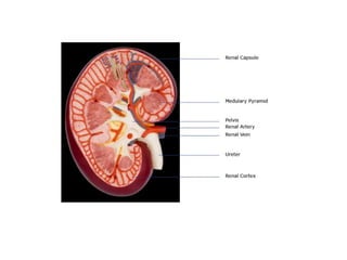
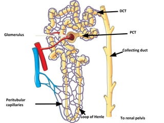
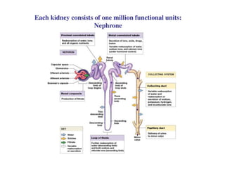
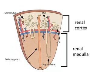
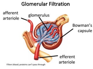
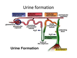
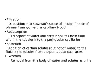
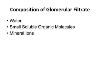
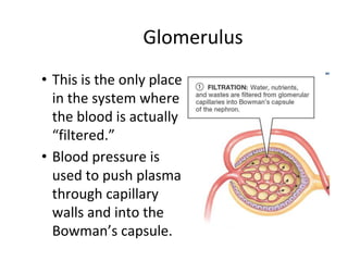
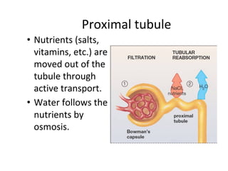
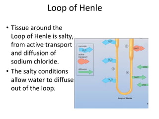
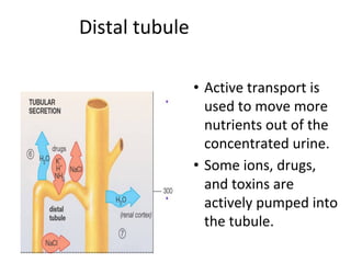
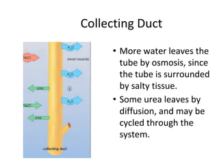
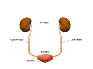
The Urinary System
Ligula Regional Referral Hospital
Samson B.Mkawe
BMedSc,DPT,CSPMedicine
• The Urinary System is a group of organs in the body
concerned with filtering out excess fluid and other
substances from the bloodstream. The substances
are filtered out from the body in the form of urine
• Urine is a liquid produced by the kidneys, collected in
the bladder and excreted through the urethra. Urine
is used to extract excess minerals or vitamins as well
as blood from the body.
FUNCTIONS OF URINARY SYSTEM
1. removal of metabolic wastes & toxins
but we have several organs that serve an excretory
function other than kidneys:
a. kidneys
b. skin-sweat glands rid body of water, minerals,some
nitrogenous wastes (ammonia)
c. Lungs rid body of CO2 from energy metabolism of
cells
d. Liver liver excretes bile pigments, salts, calcium,
some toxins
2. helps to regulate blood volume & pressure
• blood pressure is directly affected by the
volume of fluids retained or removed from
body:
• eg. excessive salts promote water retention
greater volume ----- increases BP
• eg. Dehydration lower volume --- decreases
BP
3. regulation of electrolytes & body Ph
4. regulates erythropoiesis
kidneys produce hormone = erythropoietin that
regulates erythropoiesis
Erythropoiesis is the process which produces red blood cells (erythrocytes). It is stimulated by
decreased O2 in circulation, which is detected by the kidney.
5.Homeostatic organ
6.Acts as blood filter
Organs of the Urinary System
Why is the left kidney slightly higher
than the right?
• The left kidney is located slightly more
superior than the right kidney due to
the larger size of the liver on the rightside of
the body
Organs:
• kidneys – clean and filter blood
• ureters – tubes that take urine to bladder
• bladder – stores urine until eliminated
• urethra – removes urine from body
A Kidney
Blood and waste enter through renal artery
Filtered blood leaves through renal vein
Excess water and toxic waste leaves through
ureter as urine
Kidneys located dorsal body wall, bean-shaped organs Each
kidney is about 4 or 5 inches long, roughly the size of a large
fist.
retroperitoneal - behind parietal peritoneum
• just above waist
• surrounded by renal capsule - barrier against trauma and
spread of infections
• hilum = indentation where vessels and ureter attach
• Frontal Section of Kidney
Cortex-outer zone of kidney
Medulla-interior of kidney
• Main structures
of the
mammalian
kidney:
• renal cortex
• renal medula
• renal pelvis
• nephrons
• Renal Capsule – outer membrane that
surrounds the kidney; it is thin but tough and
fibrous
• Renal Pelvis – basin-like area that collects
urine from the nephrons, it narrows into the
upper end of the ureter
• Cortex – the outer region of the kidney;
extensions of the cortical tissue, contains
about one million blood filtering nephrons
• Nephron – these are the filtration units in the
kidneys
• Medulla – inner region of the kidney contains
8-12 renal pyramids.
• Medullary pyramids – formed by the collecting
ducts, inner part of the kidney
• Renal Artery – branches off of the aorta
bringing waste-filled blood into the kidney for
filtering in the nephrons; Each minute, the
kidneys receive 20% of the blood pumped by
the heart.
• Renal Vein – removes the filtered blood from
the kidneys to the inferior
• Every minute 1300 mL of blood enter the
kidneys, 1299 mL leave the kidney. and 1 mL
leaves as urine.
Nephrone
• Each kidney contains over 1 million nephrons
and thousands of collecting ducts
• is the microscopic structural and functional
unit of the kidney. It is composed of a renal
corpuscle and a renal tubule.
• A nephron is the basic unit of structure in the
kidney
• A nephron is used separate to water, ions and
small molecules from the blood,
Blood Supply of a Nephron
The glomerulus receives blood from a fairly large
afferent arteriole and passes it to a smaller efferent
arteriole.
The efferent arteriole gives rise to the peritubular
capillary system, which surrounds the renal tubule.
Anatomy of the Nephron
• Glomerulus
• Proximal tubule
• Loop of Henle
• Distal tubule
Glomerulus
To renal pelvis
Peritubular
capillaries
Collecting duct
PCT
Loop of Henle
DCT
renal
cortex
renal
medulla
Collecting duct
Loop of Henle
PCT
DCT
Glomerulus
Glomerular Filtration
afferent
arteriole
glomerulus
efferent
arteriole
Bowman’s
capsule
Filters blood; proteins can’t pass through
Urine formation
▪ Filtration
Deposition into Bowman’s space of an ultrafiltrate of
plasma from glomerular capillary blood
▪ Reabsorption
Transport of water and certain solutes from fluid
within the tubules into the peritubular capillaries
▪ Secretion
Addition of certain solutes (but not of water) to the
fluid in the tubules from the peritubular capillaries
▪ Excretion
Removal from the body of water and solutes as urine
Composition of Glomerular Filtrate
• Water
• Small Soluble Organic Molecules
• Mineral Ions
Glomerulus
• This is the only place
in the system where
the blood is actually
“filtered.”
• Blood pressure is
used to push plasma
through capillary
walls and into the
Bowman’s capsule.
Proximal tubule
• Nutrients (salts,
vitamins, etc.) are
moved out of the
tubule through
active transport.
• Water follows the
nutrients by
osmosis.
Loop of Henle
• Tissue around the
Loop of Henle is salty,
from active transport
and diffusion of
sodium chloride.
• The salty conditions
allow water to diffuse
out of the loop.
Distal tubule
• Active transport is
used to move more
nutrients out of the
concentrated urine.
• Some ions, drugs,
and toxins are
actively pumped into
the tubule.
Collecting Duct
• More water leaves the
tube by osmosis, since
the tube is surrounded
by salty tissue.
• Some urea leaves by
diffusion, and may be
cycled through the
system.
Ureter
• ureters are tubes made of smooth muscle
fibers that propel urine from the kidneys to
the urinary bladder.
• There are two ureters, one attached to each
kidney. The upper half of the ureter is located
in the abdomen and the lower half is located
in the pelvic area.
• In the adult, the ureters are usually 25–30 cm
(10–12 in) long and around 3–4 mm
(0.12–0.16 in) in diameter.
Urinary Bladder
ureters
internal sphincters
external sphincters
urethra
• small, size when empty
• can hold up to 800 ml voluntarily
• up to 2000 ml when obstructed
• wall consists of 4 layers (same as GI tract)
Layers
• mucosa -innermost layer
secretes mucous for protection from
corrosive effects of urine
• submucosa –fibrous connective tissue
• muscularis -several smooth muscle layers
• serosa -visceral peritoneum
• involuntary internal & voluntary external
urethral sphincters as bladder expands to hold
urine
• They activate stretch receptors in wall that
monitor volume
• when volume exceeds 200 ml the receptor
signals enter our conscious perception= desire
to urinate
urethra
male:
-dual function,
-rid body of urine,
- release of seminal fluid during orgasm
female:
-single function: rids body of urine
-shorter
-more prone to UTI’s
– Female: 4cm long
– Male:14cm long
Diuresis
• When bladder fills with 200 ml of urine,
stretch receptors transmit impulses to the
CNS and produce a reflex contraction of the
bladder (PNS)
What is urine?
• Urine is a liquid waste product of the body
secreted by the kidneys by a process of blood
filtration and excretion through the urethra.
Composition
• Urine is a transparent solution that can range
from colorless to amber but is usually a pale
yellow. It is normally odorless but can acquire
strong odors due to bacterial action.
• Urine is an aqueous solution of metabolic
wastes such as urea, dissolved salts, and
organic compounds.
• Most noticeably, ammonia is produced by
breakdown of urea, a major component of
urine.
Hazards
• Urea is toxic and can be irritating to skin and
eyes. High concentrations in the blood can
cause damage to organs of the body. Low
concentrations of urea such as in urine are not
dangerous.
volume
The amount of urine produced depends on numerous factors
including :
• state of hydration,
• activities,
• environmental factors,
• age, and health.
• In adult human, the average production is about 1 - 2 L per
day.
• Polyuria is a condition of excessive production of urine (> 2.5
L/day), in contrast to oliguria where < 400 mL are produced
per day, or anuria with a production of < 100 mL per day.
Abnormal urine can have
• Carbohydrates
• Proteins
• Blood
• Bile salts
• Fats
Urinalysis
characteristics:
• smell- ammonia-like
• pH- 4.5-8, ave 6.0
• color- affected by what we eat: salty foods,
vitamins
Guten morgan

URINARY%20SYSTEM.pdf.pdf

  • 1.
    The Urinary System LigulaRegional Referral Hospital Samson B.Mkawe BMedSc,DPT,CSPMedicine
  • 3.
    • The UrinarySystem is a group of organs in the body concerned with filtering out excess fluid and other substances from the bloodstream. The substances are filtered out from the body in the form of urine • Urine is a liquid produced by the kidneys, collected in the bladder and excreted through the urethra. Urine is used to extract excess minerals or vitamins as well as blood from the body.
  • 4.
    FUNCTIONS OF URINARYSYSTEM 1. removal of metabolic wastes & toxins but we have several organs that serve an excretory function other than kidneys: a. kidneys b. skin-sweat glands rid body of water, minerals,some nitrogenous wastes (ammonia) c. Lungs rid body of CO2 from energy metabolism of cells d. Liver liver excretes bile pigments, salts, calcium, some toxins
  • 5.
    2. helps toregulate blood volume & pressure • blood pressure is directly affected by the volume of fluids retained or removed from body: • eg. excessive salts promote water retention greater volume ----- increases BP • eg. Dehydration lower volume --- decreases BP
  • 6.
    3. regulation ofelectrolytes & body Ph 4. regulates erythropoiesis kidneys produce hormone = erythropoietin that regulates erythropoiesis Erythropoiesis is the process which produces red blood cells (erythrocytes). It is stimulated by decreased O2 in circulation, which is detected by the kidney. 5.Homeostatic organ 6.Acts as blood filter
  • 7.
    Organs of theUrinary System
  • 8.
    Why is theleft kidney slightly higher than the right? • The left kidney is located slightly more superior than the right kidney due to the larger size of the liver on the rightside of the body
  • 9.
    Organs: • kidneys –clean and filter blood • ureters – tubes that take urine to bladder • bladder – stores urine until eliminated • urethra – removes urine from body
  • 10.
    A Kidney Blood andwaste enter through renal artery Filtered blood leaves through renal vein Excess water and toxic waste leaves through ureter as urine
  • 11.
    Kidneys located dorsalbody wall, bean-shaped organs Each kidney is about 4 or 5 inches long, roughly the size of a large fist. retroperitoneal - behind parietal peritoneum • just above waist • surrounded by renal capsule - barrier against trauma and spread of infections • hilum = indentation where vessels and ureter attach • Frontal Section of Kidney Cortex-outer zone of kidney Medulla-interior of kidney
  • 12.
    • Main structures ofthe mammalian kidney: • renal cortex • renal medula • renal pelvis • nephrons
  • 14.
    • Renal Capsule– outer membrane that surrounds the kidney; it is thin but tough and fibrous • Renal Pelvis – basin-like area that collects urine from the nephrons, it narrows into the upper end of the ureter
  • 15.
    • Cortex –the outer region of the kidney; extensions of the cortical tissue, contains about one million blood filtering nephrons • Nephron – these are the filtration units in the kidneys • Medulla – inner region of the kidney contains 8-12 renal pyramids. • Medullary pyramids – formed by the collecting ducts, inner part of the kidney
  • 16.
    • Renal Artery– branches off of the aorta bringing waste-filled blood into the kidney for filtering in the nephrons; Each minute, the kidneys receive 20% of the blood pumped by the heart. • Renal Vein – removes the filtered blood from the kidneys to the inferior • Every minute 1300 mL of blood enter the kidneys, 1299 mL leave the kidney. and 1 mL leaves as urine.
  • 17.
    Nephrone • Each kidneycontains over 1 million nephrons and thousands of collecting ducts • is the microscopic structural and functional unit of the kidney. It is composed of a renal corpuscle and a renal tubule. • A nephron is the basic unit of structure in the kidney • A nephron is used separate to water, ions and small molecules from the blood,
  • 18.
    Blood Supply ofa Nephron The glomerulus receives blood from a fairly large afferent arteriole and passes it to a smaller efferent arteriole. The efferent arteriole gives rise to the peritubular capillary system, which surrounds the renal tubule.
  • 19.
    Anatomy of theNephron • Glomerulus • Proximal tubule • Loop of Henle • Distal tubule
  • 20.
  • 22.
  • 23.
  • 24.
  • 25.
    ▪ Filtration Deposition intoBowman’s space of an ultrafiltrate of plasma from glomerular capillary blood ▪ Reabsorption Transport of water and certain solutes from fluid within the tubules into the peritubular capillaries ▪ Secretion Addition of certain solutes (but not of water) to the fluid in the tubules from the peritubular capillaries ▪ Excretion Removal from the body of water and solutes as urine
  • 26.
    Composition of GlomerularFiltrate • Water • Small Soluble Organic Molecules • Mineral Ions
  • 27.
    Glomerulus • This isthe only place in the system where the blood is actually “filtered.” • Blood pressure is used to push plasma through capillary walls and into the Bowman’s capsule.
  • 28.
    Proximal tubule • Nutrients(salts, vitamins, etc.) are moved out of the tubule through active transport. • Water follows the nutrients by osmosis.
  • 29.
    Loop of Henle •Tissue around the Loop of Henle is salty, from active transport and diffusion of sodium chloride. • The salty conditions allow water to diffuse out of the loop.
  • 30.
    Distal tubule • Activetransport is used to move more nutrients out of the concentrated urine. • Some ions, drugs, and toxins are actively pumped into the tubule.
  • 31.
    Collecting Duct • Morewater leaves the tube by osmosis, since the tube is surrounded by salty tissue. • Some urea leaves by diffusion, and may be cycled through the system.
  • 32.
    Ureter • ureters aretubes made of smooth muscle fibers that propel urine from the kidneys to the urinary bladder. • There are two ureters, one attached to each kidney. The upper half of the ureter is located in the abdomen and the lower half is located in the pelvic area. • In the adult, the ureters are usually 25–30 cm (10–12 in) long and around 3–4 mm (0.12–0.16 in) in diameter.
  • 34.
  • 35.
    • small, sizewhen empty • can hold up to 800 ml voluntarily • up to 2000 ml when obstructed • wall consists of 4 layers (same as GI tract)
  • 36.
    Layers • mucosa -innermostlayer secretes mucous for protection from corrosive effects of urine • submucosa –fibrous connective tissue • muscularis -several smooth muscle layers • serosa -visceral peritoneum
  • 37.
    • involuntary internal& voluntary external urethral sphincters as bladder expands to hold urine • They activate stretch receptors in wall that monitor volume • when volume exceeds 200 ml the receptor signals enter our conscious perception= desire to urinate
  • 38.
    urethra male: -dual function, -rid bodyof urine, - release of seminal fluid during orgasm female: -single function: rids body of urine -shorter -more prone to UTI’s – Female: 4cm long – Male:14cm long
  • 40.
    Diuresis • When bladderfills with 200 ml of urine, stretch receptors transmit impulses to the CNS and produce a reflex contraction of the bladder (PNS)
  • 41.
    What is urine? •Urine is a liquid waste product of the body secreted by the kidneys by a process of blood filtration and excretion through the urethra.
  • 42.
    Composition • Urine isa transparent solution that can range from colorless to amber but is usually a pale yellow. It is normally odorless but can acquire strong odors due to bacterial action. • Urine is an aqueous solution of metabolic wastes such as urea, dissolved salts, and organic compounds. • Most noticeably, ammonia is produced by breakdown of urea, a major component of urine.
  • 43.
    Hazards • Urea istoxic and can be irritating to skin and eyes. High concentrations in the blood can cause damage to organs of the body. Low concentrations of urea such as in urine are not dangerous.
  • 44.
    volume The amount ofurine produced depends on numerous factors including : • state of hydration, • activities, • environmental factors, • age, and health. • In adult human, the average production is about 1 - 2 L per day. • Polyuria is a condition of excessive production of urine (> 2.5 L/day), in contrast to oliguria where < 400 mL are produced per day, or anuria with a production of < 100 mL per day.
  • 45.
    Abnormal urine canhave • Carbohydrates • Proteins • Blood • Bile salts • Fats
  • 46.
    Urinalysis characteristics: • smell- ammonia-like •pH- 4.5-8, ave 6.0 • color- affected by what we eat: salty foods, vitamins
  • 49.