Download to read offline


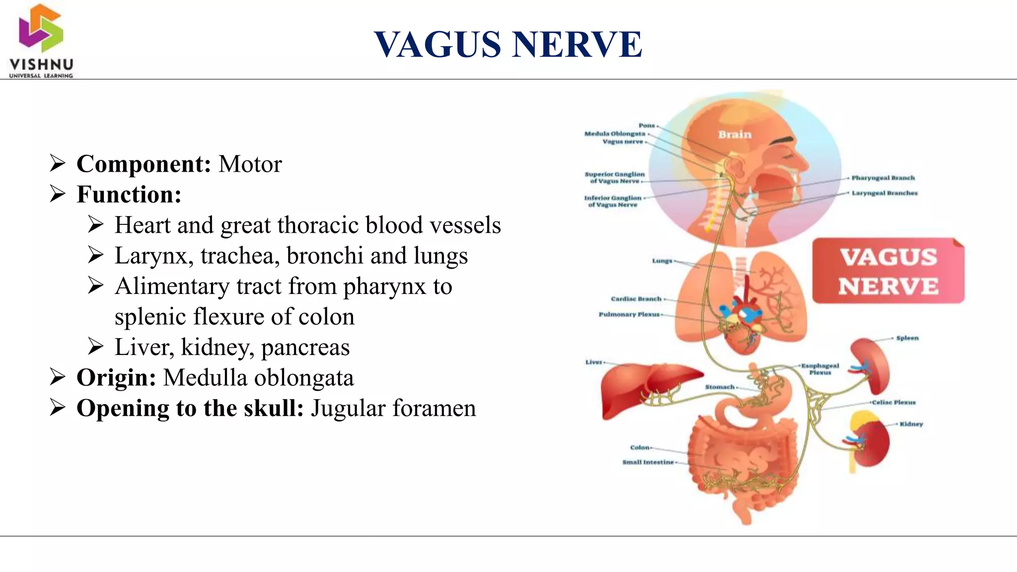


Salivary glands Origin: Medulla oblongata Opening to the skull: Jugular foramen GLOSSOPHARYNGEAL NERVE Component: Mixed Function: Motor: Muscles of larynx Sensory: Larynx, trachea, pharynx, tongue Parasympathetic: Bronchial tree, heart Origin: Medulla oblongata Opening to the skull: Jugular foramen VAGUS NERVE Component: Motor Function:


