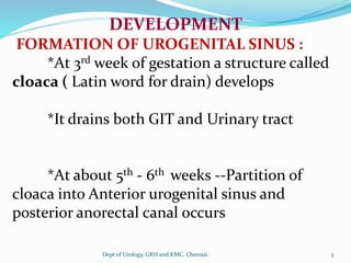
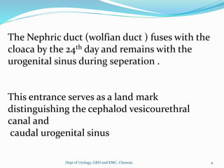
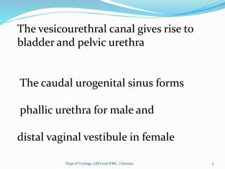
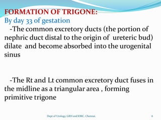
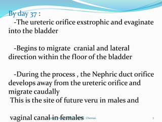
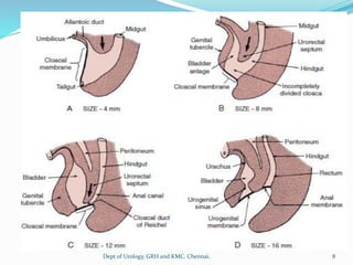
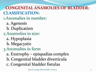
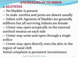
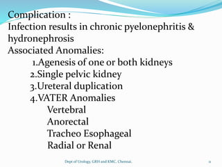
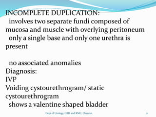
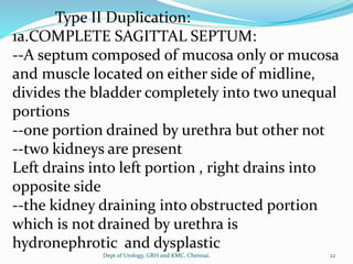
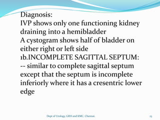
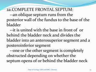
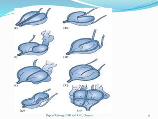
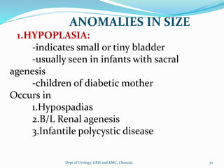
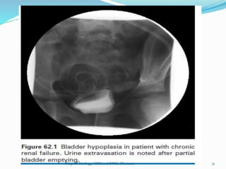
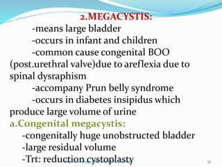
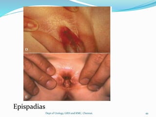
Dept of Urology
Govt Royapettah Hospital and Kilpauk Medical College
Chennai
Moderators:
Professors:
 Prof. Dr. G. Sivasankar, M.S., M.Ch.,
 Prof. Dr. A. Senthilvel, M.S., M.Ch.,
Asst Professors:
 Dr. J. Sivabalan, M.S., M.Ch.,
 Dr. R. Bhargavi, M.S., M.Ch.,
 Dr. S. Raju, M.S., M.Ch.,
 Dr. K. Muthurathinam, M.S., M.Ch.,
 Dr. D. Tamilselvan, M.S., M.Ch.,
 Dr. K. Senthilkumar, M.S., M.Ch.
Dept of Urology, GRH and KMC, Chennai. 2
DEVELOPMENT
FORMATION OF UROGENITAL SINUS :
*At 3rd week of gestation a structure called
cloaca ( Latin word for drain) develops
*It drains both GIT and Urinary tract
*At about 5th - 6th weeks --Partition of
cloaca into Anterior urogenital sinus and
posterior anorectal canal occurs
3
Dept of Urology, GRH and KMC, Chennai.
The Nephric duct (wolfian duct ) fuses with the
cloaca by the 24th day and remains with the
urogenital sinus during seperation .
This entrance serves as a land mark
distinguishing the cephalod vesicourethral
canal and
caudal urogenital sinus
4
Dept of Urology, GRH and KMC, Chennai.
The vesicourethral canal gives rise to
bladder and pelvic urethra
The caudal urogenital sinus forms
phallic urethra for male and
distal vaginal vestibule in female
5
Dept of Urology, GRH and KMC, Chennai.
FORMATION OF TRIGONE:
By day 33 of gestation
-The common excretory ducts (the portion of
nephric duct distal to the origin of ureteric bud)
dilate and become absorbed into the urogenital
sinus
-The Rt and Lt common excretory duct fuses in
the midline as a triangular area , forming
primitive trigone
6
Dept of Urology, GRH and KMC, Chennai.
By day 37 :
-The ureteric orifice exstrophic and evaginate
into the bladder
-Begins to migrate cranial and lateral
direction within the floor of the bladder
-During the process , the Nephric duct orifice
develops away from the ureteric orifice and
migrate caudally
This is the site of future veru in males and
vaginal canal in females 7
Dept of Urology, GRH and KMC, Chennai.
8
Dept of Urology, GRH and KMC, Chennai.
CONGENITAL ANAMOLIES OF BLADDER:
CLASSIFICATION:
1.Anomalies in number:
a. Agenesis
b. Duplication
2.Anamolies in size:
a. Hypoplasia
b. Megacystic
3.Anomalies in form
a. Exstrophy – epispadias complex
b. Congenital bladder diverticula
c. Congenital bladder fistulas
9
Dept of Urology, GRH and KMC, Chennai.
ANOMALIES IN NUMBER
a.AGENESIS
-- No bladder is present
-- In male :urethra and penis are absent usually
-- Infant with Agenesis of bladder are generally
stillborn but all surviving Infants are female
-- Ureter may open ectopically in the external
urethral meatus on each side
-- Ureter may unite and open through a single
orifice
-- Ureter may open directly onto the skin in the
region of natal cleft
Initial complaint is persistent incontinence
10
Dept of Urology, GRH and KMC, Chennai.
Complication :
Infection results in chronic pyelonephritis &
hydronephrosis
Associated Anomalies:
1.Agenesis of one or both kidneys
2.Single pelvic kidney
3.Ureteral duplication
4.VATER Anomalies
Vertebral
Anorectal
Tracheo Esophageal
Radial or Renal
11
Dept of Urology, GRH and KMC, Chennai.
Diagnosis:
USG --- shows no bladder
IVP
--If ureteric opening are Identified they may
be catheterised,
Retrograde urography
-- differentiate vesical Agenesis from B/L
ureteral ectopia in which a small bladder may
be present
12
Dept of Urology, GRH and KMC, Chennai.
b.DUPLICATION:
Three main types
Type I:
--Duplication involves the mucosa & muscle
wall
--A Peritoneal fold separates the two bladder
-- sub types :1. Complete duplication
2. Incomplete duplication
Type II:
--An internal septum of mucosa only or of
mucosa &muscle divides the bladder
--groove may or may not be visible outside the
bladder 13
Dept of Urology, GRH and KMC, Chennai.
--Sub types: 1.Sagittal septum
-complete
-incomplete
2.Frontal septum
-complete
-incomplete
3. Multiple septa
TYPE III:
-- A transverse band of thick muscle divide the
bladder into 2 unequal cavities and gives it a
characteristic Hour glass shape
14
Dept of Urology, GRH and KMC, Chennai.
Chwalle theory:
--completely duplicated bladder may form if one
ureter remained obstructed at the ureterovesical
junction as a result of chwalle membrane
--this process will lead to cystic dilatation at the
lower end of the ureter
--muscles and fibrous tissue would develop
around the structure which would then be forced
over to the median line and results in bipartite
bladder
--the hourglass bladder to be a result of a
dysplastic junction between the urachus and the
bladder 15
Dept of Urology, GRH and KMC, Chennai.
COMPLETE DUPLICATION :
Involves two completely separate bladders each
with its own wall of muscle and mucosa
separated by a peritoneal fold of varying depth
--the right and left kidney drain into right and
left bladders respectively via single ureter
--the two urethras are present one for each
bladder
16
Dept of Urology, GRH and KMC, Chennai.
--males often have two penises ,a partially
duplicated penis or one penis with two
urethra
--commonly the scrotum is bifid
--females usually have two urethras , two
vaginas, two uteri each commonly have one
horn and fallopian tube
17
Dept of Urology, GRH and KMC, Chennai.
Anomalies associated with complete
duplication of the bladder:
1.Renal ectopia =50%
2.Duplicated colon with an anorectal
malformation of the lateral aspect of the colon
and rectourethral fistula
3.Duplicated anus
4.Duplicated appendix
5.Duplicated ileum from meckel’s diverticulum
to the ileocaecal valve
6.Duplicated sacrum and coccyx
7.Wide separation of pubic rami
18
Dept of Urology, GRH and KMC, Chennai.
Complications
--Adenocarcinoma development from
surgically defunctionalised bladder
moiety
--Accessory bladder lies in front of the
normal bladder with an accessory urethra
extending from it dorsally to a penile
epispadiac meatus
-- Neither ureter drains into accessory
bladder
19
Dept of Urology, GRH and KMC, Chennai.
Voiding cystouretherography:
-- Catheterize both the urethras and fill both
bladders
--when pts void :both bladder contract
simultaneously so the urethras are outlined
with contrast material simultaneously
DIAGNOSIS:
Antenatal Sonography : shows two bladder
IVP : shows two kidneys ,two bladder
left ureter drains into Lt bladder
right ureter drains into Rt bladder
20
Dept of Urology, GRH and KMC, Chennai.
INCOMPLETE DUPLICATION:
involves two separate fundi composed of
mucosa and muscle with overlying peritoneum
only a single base and only one urethra is
present
no associated anomalies
Diagnosis:
IVP
Voiding cystourethrogram/ static
cystourethrogram
shows a valentine shaped bladder
21
Dept of Urology, GRH and KMC, Chennai.
Type II Duplication:
1a.COMPLETE SAGITTAL SEPTUM:
--A septum composed of mucosa only or mucosa
and muscle located on either side of midline,
divides the bladder completely into two unequal
portions
--one portion drained by urethra but other not
--two kidneys are present
Left drains into left portion , right drains into
opposite side
--the kidney draining into obstructed portion
which is not drained by urethra is
hydronephrotic and dysplastic
22
Dept of Urology, GRH and KMC, Chennai.
Diagnosis:
IVP shows only one functioning kidney
draining into a hemibladder
A cystogram shows half of bladder on
either right or left side
1b.INCOMPLETE SAGITTAL SEPTUM:
-- similar to complete sagittal septum
except that the septum is incomplete
inferiorly where it has a cresentric lower
edge
23
Dept of Urology, GRH and KMC, Chennai.
2a.COMPLETE FRONTAL SEPTUM:
--an oblique septum runs from the
posterior wall of the fundus to the base of the
bladder
--it is united with the base in front of or
behind the bladder neck and divides the
bladder into an anterosuperior segment and a
posteroinferior segment
--one or the other segment is completely
obstructed depending on whether the
septum opens of or behind the bladder neck
24
Dept of Urology, GRH and KMC, Chennai.
--each kidney drains via single ureter into
one or other of the respective bladder
segment
--The kidney draining into the obstructed
segment is hydronephrotic and dysplastic
Diagnosis
IVP: shows one functioning kidney with
contrast material in one or the other bladder
segment
25
Dept of Urology, GRH and KMC, Chennai.
2b.INCOMPLETE FRONTAL SEPTUM:
-- similar to complete frontal septum except
that the septum is incomplete inferiorly and
has a cresenteric lower edge
-- Duplication of the kidney is commonly
associated
Diagnosis
IVP , cystogram or voiding cystourethrogram
shows incomplete frontal septum or a filling
defect in the bladder
26
Dept of Urology, GRH and KMC, Chennai.
2c.MULTIPLE SEPTA : (Multiseptate bladder)
-- fibromuscular septa divides the bladder into 4
unequal segments
-- complete bilateral ureteral duplication is
always present so one ureter drains into each
segment
-- only one segment actually opens into the
urethra ,other three bladder segments are
obstructed
-- only one pole of one duplex kidney is
unobstructed
Diagnosis:
IVP: demonstrates onepole of one kidney draining
27
Dept of Urology, GRH and KMC, Chennai.
HOURGLASS BLADDER:
-- A thick transverse band of muscle
incompletely divides the bladder into 2
unequal segments
-- The ureter usually opened into the lower
portion
-- No associated anomalies
Diagnosis:
IVP & Voiding cystourethrogram
-- Hourglass shaped bladder
28
Dept of Urology, GRH and KMC, Chennai.
29
Dept of Urology, GRH and KMC, Chennai.
ANOMALIES IN SIZE
1.HYPOPLASIA:
-indicates small or tiny bladder
-usually seen in infants with sacral
agenesis
-children of diabetic mother
Occurs in
1.Hypospadias
2.B/L Renal agenesis
3.Infantile polycystic disease
30
Dept of Urology, GRH and KMC, Chennai.
31
Dept of Urology, GRH and KMC, Chennai.
2.MEGACYSTIS:
-means large bladder
-occurs in infant and children
-common cause congenital BOO
(post.urethral valve)due to areflexia due to
spinal dysraphism
-accompany Prun belly syndrome
-occurs in diabetes insipidus which
produce large volume of urine
a.Congenital megacystis:
-congenitally huge unobstructed bladder
-large residual volume
-Trt: reduction cystoplasty 32
Dept of Urology, GRH and KMC, Chennai.
33
Dept of Urology, GRH and KMC, Chennai.
b.Megaureter Megacystis
syndrome:(Inner williams syndrome)
large smooth bladder with a
Trigone, dilated ureteral orifice and grossly
dilated ureter
when voids bladder empty completely
large volume refluxes into ureter when
voiding
when voiding ceases, ureter
contracts,urine rapidlly fills the bladder
bladder always distended forming
decompensated ,dilated and thin walled
34
Dept of Urology, GRH and KMC, Chennai.
c.Megacystis-Microcolon – hypoperistalsis
syndrome
-have huge bladder with areflux &dilated
uppertract
-a small colon,dilated small bowel with
hypoperistalsis
-common in female neonate
-fatal during first years of life
35
Dept of Urology, GRH and KMC, Chennai.
ANOMALIES IN FORM
1.EXSTROPHY-EPISPADIAS COMPLEX:
Exstrophy of bladder and epispadias
represent opposite ends of a spectrum of
anterior abdominal wall defects rather than
separate entities
--Anterior abdominal wall defects
--Separation of pubic bone >1cm at
symphysis pubis
36
Dept of Urology, GRH and KMC, Chennai.
CLASSIFICATION:
1. Exstrophy of bladder 1 : 33,000 live births
2. Epispadias 1 : 1,00,000 live births
3. Pubic umblicus
4. Covered Exstrophy
5. Covered exstrophy and vesicle sequestration
6. Superior vesicle fistula
7. Superior vesicle fissure
8. Duplicate exstrophy
9. Exstrophy of cloaca
37
Dept of Urology, GRH and KMC, Chennai.
EMBRYOLOGY:
CLASSIC THEORY:
-Persistence of cloacal membrane or large
cloacal membrane
-wide pelvis
-separated two mullerian ducts leads to
separate vagina and uteri
-anterior abdominal wall defect
CURRENT THEORY:
-lack of rotation of the pelvic ring
primordia
38
Dept of Urology, GRH and KMC, Chennai.
EXSTROPHY OF BLADDER:
-lower abdominal wall and anterior wall of
the bladder is absent
-the remainder of the bladder is visible
through the resultant opening
-bladder turns inside out and protrudes
through the opening in the anterior abdominal
wall – hence the term exstrophy
39
Dept of Urology, GRH and KMC, Chennai.
-fertility not affected
-associated with musculoskeletal,
gastrointestinal and genital tracts
Musculoskeletal anomalies:
-widely separated pubic symphysis
-the innominate bones are rotated outward
along the sacroiliac joint
40
Dept of Urology, GRH and KMC, Chennai.
EXSTROPHY OF BLADDER:
-pubic bone is rotated outward at its
junction with ilium and ischium
-the iliac wings project inwardly
-the anterior segment of each iliac wing is
short
-lower part of rectus abdominus is
separated
-umblicus is low with hernia 41
Dept of Urology, GRH and KMC, Chennai.
Classic bladder exstrophy
42
Dept of Urology, GRH and KMC, Chennai.
Classic bladder exstrophy
43
Dept of Urology, GRH and KMC, Chennai.
EXSTROPHY OF THE BLADDER:
OEIS COMPLEX:
Omphalocele
Exstrophy of the cloaca
Imperforate anus
Spinal defects
44
Dept of Urology, GRH and KMC, Chennai.
EXSTROPHY OF THE BLADDER:
Diagnosis:
IVP: upper tracts normal in 65% or
hydronephrosis
MRI & CT:
pubic bone seperation
corporeal length assessed
PRENATAL SONOGRAPHY: reveals
Lower abdominal wall
absent bladder
malformation of external genitalia
normal kidneys
normal amniotic fluid volume 45
Dept of Urology, GRH and KMC, Chennai.
EPISPADIAS
-congenital fissure in the upper wall of
female urethra or
-a congenital defect in male urethra
Incidence:
In females :0.2/ 100000 population
In male :0.9/100000 population
46
Dept of Urology, GRH and KMC, Chennai.
EPISPADIAS:
In females
-clitoris is divided
-labium minora ill developed
-bladder is small and thin walled
-bladder neck absent
-urethra short and wide
-pts is incontinent
IVP:widening of pubic symphysis
funnel shaped bladder base
upper tract –normal
low grade reflux -90%
47
Dept of Urology, GRH and KMC, Chennai.
EPISPADIAS
In males:
-roof of distal end of urethra is absent
-proximal urethra opens anywhere from
base of penis to the glans
-penis bows upwards
-2/3rd of pts are incontinent
48
Dept of Urology, GRH and KMC, Chennai.
Epispadias
49
Dept of Urology, GRH and KMC, Chennai.
PUBIC UMBLICUS:
-umblicus is extremely low
-symphysis pubis is widely separated
SUPERIOR VESICLE FISTULA:
-tiny fistulous tract running from the
top of the bladder to a small area of
transitional epithelium on the anterior
abdominal wall
-symphysis pubis is wide
-usually continent
50
Dept of Urology, GRH and KMC, Chennai.
Superior vesical fissure
51
Dept of Urology, GRH and KMC, Chennai.
CONGENITAL BLADDER DIVERTICULA:
-diverticula measures >2cm in diameter in
IVU or voiding cystourethrogram
-saccules measures <2cm in diameter
-herniation of bladder mucosa through
the detrusor muscle
-98% congenital diverticula occurs in male
-most common in bladder base
-most frequently in the region of ureteral
hiatus known as Hutch diverticulum
-gives rise to obstruction or reflux
52
Dept of Urology, GRH and KMC, Chennai.
Diverticula in fundus is – rare
bilateral &symmetrical ,gives rise to Micky
mouse appearance
MENKE’S syndrome(kinky hair syndrome)
- a disease of copper metabolism have
associated bladder diverticulum
Congenital bladder diverticulum occurs in
-Williams disease
-Diamond –black fan disease
-Fetal alcohol disease
-Prune – belly syndrome
-Ehler –danlos type 9 syndrome 53
Dept of Urology, GRH and KMC, Chennai.
54
Dept of Urology, GRH and KMC, Chennai.
Associated anomalies :
-Ipsilateral Renal agenesis
-Ipsilateral undescended testis
Voiding cystourethrogram:
-diverticula are best shown
USG,CT,MRI: visible
55
Dept of Urology, GRH and KMC, Chennai.
56
Dept of Urology, GRH and KMC, Chennai.
INDICATIONS:
for surgical treatment of bladder diverticulum is dictated by the
presence of
persistent high-grade vesicoureteral reflux,
and/or recurrent urinary tract infections,
or documented obstruction of either the distal
ureter or the bladder neck.
In cases of functional bladder outlet obstruction due to a very
large diverticulum extending underneath the bladder neck,
excision of the diverticulum is recommended.
For patients with recurrent urinary tract infection and
persistent vesicoureteral reflux, diverticulectomy is
recommended and should be accompanied by reimplantation
of the ureter 57
Dept of Urology, GRH and KMC, Chennai.
The surgical approach can be either intravesical or
extravesical.
The key surgical principle is to reinforce
the muscular hiatus of the bladder to prevent
reformation of the bladder diverticulum.
If upper tract evaluation reveal a non-functioning
kidney, consideration should be given to
nephroureterectomy with diverticulectomy.
Extravesical approaches and
laparoscopic techniques can be used
58
Dept of Urology, GRH and KMC, Chennai.
THANK YOU
59
Dept of Urology, GRH and KMC, Chennai.

Urinary Bladder anomalies congenital

  • 1.
    Dept of Urology GovtRoyapettah Hospital and Kilpauk Medical College Chennai
  • 2.
    Moderators: Professors:  Prof. Dr.G. Sivasankar, M.S., M.Ch.,  Prof. Dr. A. Senthilvel, M.S., M.Ch., Asst Professors:  Dr. J. Sivabalan, M.S., M.Ch.,  Dr. R. Bhargavi, M.S., M.Ch.,  Dr. S. Raju, M.S., M.Ch.,  Dr. K. Muthurathinam, M.S., M.Ch.,  Dr. D. Tamilselvan, M.S., M.Ch.,  Dr. K. Senthilkumar, M.S., M.Ch. Dept of Urology, GRH and KMC, Chennai. 2
  • 3.
    DEVELOPMENT FORMATION OF UROGENITALSINUS : *At 3rd week of gestation a structure called cloaca ( Latin word for drain) develops *It drains both GIT and Urinary tract *At about 5th - 6th weeks --Partition of cloaca into Anterior urogenital sinus and posterior anorectal canal occurs 3 Dept of Urology, GRH and KMC, Chennai.
  • 4.
    The Nephric duct(wolfian duct ) fuses with the cloaca by the 24th day and remains with the urogenital sinus during seperation . This entrance serves as a land mark distinguishing the cephalod vesicourethral canal and caudal urogenital sinus 4 Dept of Urology, GRH and KMC, Chennai.
  • 5.
    The vesicourethral canalgives rise to bladder and pelvic urethra The caudal urogenital sinus forms phallic urethra for male and distal vaginal vestibule in female 5 Dept of Urology, GRH and KMC, Chennai.
  • 6.
    FORMATION OF TRIGONE: Byday 33 of gestation -The common excretory ducts (the portion of nephric duct distal to the origin of ureteric bud) dilate and become absorbed into the urogenital sinus -The Rt and Lt common excretory duct fuses in the midline as a triangular area , forming primitive trigone 6 Dept of Urology, GRH and KMC, Chennai.
  • 7.
    By day 37: -The ureteric orifice exstrophic and evaginate into the bladder -Begins to migrate cranial and lateral direction within the floor of the bladder -During the process , the Nephric duct orifice develops away from the ureteric orifice and migrate caudally This is the site of future veru in males and vaginal canal in females 7 Dept of Urology, GRH and KMC, Chennai.
  • 8.
    8 Dept of Urology,GRH and KMC, Chennai.
  • 9.
    CONGENITAL ANAMOLIES OFBLADDER: CLASSIFICATION: 1.Anomalies in number: a. Agenesis b. Duplication 2.Anamolies in size: a. Hypoplasia b. Megacystic 3.Anomalies in form a. Exstrophy – epispadias complex b. Congenital bladder diverticula c. Congenital bladder fistulas 9 Dept of Urology, GRH and KMC, Chennai.
  • 10.
    ANOMALIES IN NUMBER a.AGENESIS --No bladder is present -- In male :urethra and penis are absent usually -- Infant with Agenesis of bladder are generally stillborn but all surviving Infants are female -- Ureter may open ectopically in the external urethral meatus on each side -- Ureter may unite and open through a single orifice -- Ureter may open directly onto the skin in the region of natal cleft Initial complaint is persistent incontinence 10 Dept of Urology, GRH and KMC, Chennai.
  • 11.
    Complication : Infection resultsin chronic pyelonephritis & hydronephrosis Associated Anomalies: 1.Agenesis of one or both kidneys 2.Single pelvic kidney 3.Ureteral duplication 4.VATER Anomalies Vertebral Anorectal Tracheo Esophageal Radial or Renal 11 Dept of Urology, GRH and KMC, Chennai.
  • 12.
    Diagnosis: USG --- showsno bladder IVP --If ureteric opening are Identified they may be catheterised, Retrograde urography -- differentiate vesical Agenesis from B/L ureteral ectopia in which a small bladder may be present 12 Dept of Urology, GRH and KMC, Chennai.
  • 13.
    b.DUPLICATION: Three main types TypeI: --Duplication involves the mucosa & muscle wall --A Peritoneal fold separates the two bladder -- sub types :1. Complete duplication 2. Incomplete duplication Type II: --An internal septum of mucosa only or of mucosa &muscle divides the bladder --groove may or may not be visible outside the bladder 13 Dept of Urology, GRH and KMC, Chennai.
  • 14.
    --Sub types: 1.Sagittalseptum -complete -incomplete 2.Frontal septum -complete -incomplete 3. Multiple septa TYPE III: -- A transverse band of thick muscle divide the bladder into 2 unequal cavities and gives it a characteristic Hour glass shape 14 Dept of Urology, GRH and KMC, Chennai.
  • 15.
    Chwalle theory: --completely duplicatedbladder may form if one ureter remained obstructed at the ureterovesical junction as a result of chwalle membrane --this process will lead to cystic dilatation at the lower end of the ureter --muscles and fibrous tissue would develop around the structure which would then be forced over to the median line and results in bipartite bladder --the hourglass bladder to be a result of a dysplastic junction between the urachus and the bladder 15 Dept of Urology, GRH and KMC, Chennai.
  • 16.
    COMPLETE DUPLICATION : Involvestwo completely separate bladders each with its own wall of muscle and mucosa separated by a peritoneal fold of varying depth --the right and left kidney drain into right and left bladders respectively via single ureter --the two urethras are present one for each bladder 16 Dept of Urology, GRH and KMC, Chennai.
  • 17.
    --males often havetwo penises ,a partially duplicated penis or one penis with two urethra --commonly the scrotum is bifid --females usually have two urethras , two vaginas, two uteri each commonly have one horn and fallopian tube 17 Dept of Urology, GRH and KMC, Chennai.
  • 18.
    Anomalies associated withcomplete duplication of the bladder: 1.Renal ectopia =50% 2.Duplicated colon with an anorectal malformation of the lateral aspect of the colon and rectourethral fistula 3.Duplicated anus 4.Duplicated appendix 5.Duplicated ileum from meckel’s diverticulum to the ileocaecal valve 6.Duplicated sacrum and coccyx 7.Wide separation of pubic rami 18 Dept of Urology, GRH and KMC, Chennai.
  • 19.
    Complications --Adenocarcinoma development from surgicallydefunctionalised bladder moiety --Accessory bladder lies in front of the normal bladder with an accessory urethra extending from it dorsally to a penile epispadiac meatus -- Neither ureter drains into accessory bladder 19 Dept of Urology, GRH and KMC, Chennai.
  • 20.
    Voiding cystouretherography: -- Catheterizeboth the urethras and fill both bladders --when pts void :both bladder contract simultaneously so the urethras are outlined with contrast material simultaneously DIAGNOSIS: Antenatal Sonography : shows two bladder IVP : shows two kidneys ,two bladder left ureter drains into Lt bladder right ureter drains into Rt bladder 20 Dept of Urology, GRH and KMC, Chennai.
  • 21.
    INCOMPLETE DUPLICATION: involves twoseparate fundi composed of mucosa and muscle with overlying peritoneum only a single base and only one urethra is present no associated anomalies Diagnosis: IVP Voiding cystourethrogram/ static cystourethrogram shows a valentine shaped bladder 21 Dept of Urology, GRH and KMC, Chennai.
  • 22.
    Type II Duplication: 1a.COMPLETESAGITTAL SEPTUM: --A septum composed of mucosa only or mucosa and muscle located on either side of midline, divides the bladder completely into two unequal portions --one portion drained by urethra but other not --two kidneys are present Left drains into left portion , right drains into opposite side --the kidney draining into obstructed portion which is not drained by urethra is hydronephrotic and dysplastic 22 Dept of Urology, GRH and KMC, Chennai.
  • 23.
    Diagnosis: IVP shows onlyone functioning kidney draining into a hemibladder A cystogram shows half of bladder on either right or left side 1b.INCOMPLETE SAGITTAL SEPTUM: -- similar to complete sagittal septum except that the septum is incomplete inferiorly where it has a cresentric lower edge 23 Dept of Urology, GRH and KMC, Chennai.
  • 24.
    2a.COMPLETE FRONTAL SEPTUM: --anoblique septum runs from the posterior wall of the fundus to the base of the bladder --it is united with the base in front of or behind the bladder neck and divides the bladder into an anterosuperior segment and a posteroinferior segment --one or the other segment is completely obstructed depending on whether the septum opens of or behind the bladder neck 24 Dept of Urology, GRH and KMC, Chennai.
  • 25.
    --each kidney drainsvia single ureter into one or other of the respective bladder segment --The kidney draining into the obstructed segment is hydronephrotic and dysplastic Diagnosis IVP: shows one functioning kidney with contrast material in one or the other bladder segment 25 Dept of Urology, GRH and KMC, Chennai.
  • 26.
    2b.INCOMPLETE FRONTAL SEPTUM: --similar to complete frontal septum except that the septum is incomplete inferiorly and has a cresenteric lower edge -- Duplication of the kidney is commonly associated Diagnosis IVP , cystogram or voiding cystourethrogram shows incomplete frontal septum or a filling defect in the bladder 26 Dept of Urology, GRH and KMC, Chennai.
  • 27.
    2c.MULTIPLE SEPTA :(Multiseptate bladder) -- fibromuscular septa divides the bladder into 4 unequal segments -- complete bilateral ureteral duplication is always present so one ureter drains into each segment -- only one segment actually opens into the urethra ,other three bladder segments are obstructed -- only one pole of one duplex kidney is unobstructed Diagnosis: IVP: demonstrates onepole of one kidney draining 27 Dept of Urology, GRH and KMC, Chennai.
  • 28.
    HOURGLASS BLADDER: -- Athick transverse band of muscle incompletely divides the bladder into 2 unequal segments -- The ureter usually opened into the lower portion -- No associated anomalies Diagnosis: IVP & Voiding cystourethrogram -- Hourglass shaped bladder 28 Dept of Urology, GRH and KMC, Chennai.
  • 29.
    29 Dept of Urology,GRH and KMC, Chennai.
  • 30.
    ANOMALIES IN SIZE 1.HYPOPLASIA: -indicatessmall or tiny bladder -usually seen in infants with sacral agenesis -children of diabetic mother Occurs in 1.Hypospadias 2.B/L Renal agenesis 3.Infantile polycystic disease 30 Dept of Urology, GRH and KMC, Chennai.
  • 31.
    31 Dept of Urology,GRH and KMC, Chennai.
  • 32.
    2.MEGACYSTIS: -means large bladder -occursin infant and children -common cause congenital BOO (post.urethral valve)due to areflexia due to spinal dysraphism -accompany Prun belly syndrome -occurs in diabetes insipidus which produce large volume of urine a.Congenital megacystis: -congenitally huge unobstructed bladder -large residual volume -Trt: reduction cystoplasty 32 Dept of Urology, GRH and KMC, Chennai.
  • 33.
    33 Dept of Urology,GRH and KMC, Chennai.
  • 34.
    b.Megaureter Megacystis syndrome:(Inner williamssyndrome) large smooth bladder with a Trigone, dilated ureteral orifice and grossly dilated ureter when voids bladder empty completely large volume refluxes into ureter when voiding when voiding ceases, ureter contracts,urine rapidlly fills the bladder bladder always distended forming decompensated ,dilated and thin walled 34 Dept of Urology, GRH and KMC, Chennai.
  • 35.
    c.Megacystis-Microcolon – hypoperistalsis syndrome -havehuge bladder with areflux &dilated uppertract -a small colon,dilated small bowel with hypoperistalsis -common in female neonate -fatal during first years of life 35 Dept of Urology, GRH and KMC, Chennai.
  • 36.
    ANOMALIES IN FORM 1.EXSTROPHY-EPISPADIASCOMPLEX: Exstrophy of bladder and epispadias represent opposite ends of a spectrum of anterior abdominal wall defects rather than separate entities --Anterior abdominal wall defects --Separation of pubic bone >1cm at symphysis pubis 36 Dept of Urology, GRH and KMC, Chennai.
  • 37.
    CLASSIFICATION: 1. Exstrophy ofbladder 1 : 33,000 live births 2. Epispadias 1 : 1,00,000 live births 3. Pubic umblicus 4. Covered Exstrophy 5. Covered exstrophy and vesicle sequestration 6. Superior vesicle fistula 7. Superior vesicle fissure 8. Duplicate exstrophy 9. Exstrophy of cloaca 37 Dept of Urology, GRH and KMC, Chennai.
  • 38.
    EMBRYOLOGY: CLASSIC THEORY: -Persistence ofcloacal membrane or large cloacal membrane -wide pelvis -separated two mullerian ducts leads to separate vagina and uteri -anterior abdominal wall defect CURRENT THEORY: -lack of rotation of the pelvic ring primordia 38 Dept of Urology, GRH and KMC, Chennai.
  • 39.
    EXSTROPHY OF BLADDER: -lowerabdominal wall and anterior wall of the bladder is absent -the remainder of the bladder is visible through the resultant opening -bladder turns inside out and protrudes through the opening in the anterior abdominal wall – hence the term exstrophy 39 Dept of Urology, GRH and KMC, Chennai.
  • 40.
    -fertility not affected -associatedwith musculoskeletal, gastrointestinal and genital tracts Musculoskeletal anomalies: -widely separated pubic symphysis -the innominate bones are rotated outward along the sacroiliac joint 40 Dept of Urology, GRH and KMC, Chennai.
  • 41.
    EXSTROPHY OF BLADDER: -pubicbone is rotated outward at its junction with ilium and ischium -the iliac wings project inwardly -the anterior segment of each iliac wing is short -lower part of rectus abdominus is separated -umblicus is low with hernia 41 Dept of Urology, GRH and KMC, Chennai.
  • 42.
    Classic bladder exstrophy 42 Deptof Urology, GRH and KMC, Chennai.
  • 43.
    Classic bladder exstrophy 43 Deptof Urology, GRH and KMC, Chennai.
  • 44.
    EXSTROPHY OF THEBLADDER: OEIS COMPLEX: Omphalocele Exstrophy of the cloaca Imperforate anus Spinal defects 44 Dept of Urology, GRH and KMC, Chennai.
  • 45.
    EXSTROPHY OF THEBLADDER: Diagnosis: IVP: upper tracts normal in 65% or hydronephrosis MRI & CT: pubic bone seperation corporeal length assessed PRENATAL SONOGRAPHY: reveals Lower abdominal wall absent bladder malformation of external genitalia normal kidneys normal amniotic fluid volume 45 Dept of Urology, GRH and KMC, Chennai.
  • 46.
    EPISPADIAS -congenital fissure inthe upper wall of female urethra or -a congenital defect in male urethra Incidence: In females :0.2/ 100000 population In male :0.9/100000 population 46 Dept of Urology, GRH and KMC, Chennai.
  • 47.
    EPISPADIAS: In females -clitoris isdivided -labium minora ill developed -bladder is small and thin walled -bladder neck absent -urethra short and wide -pts is incontinent IVP:widening of pubic symphysis funnel shaped bladder base upper tract –normal low grade reflux -90% 47 Dept of Urology, GRH and KMC, Chennai.
  • 48.
    EPISPADIAS In males: -roof ofdistal end of urethra is absent -proximal urethra opens anywhere from base of penis to the glans -penis bows upwards -2/3rd of pts are incontinent 48 Dept of Urology, GRH and KMC, Chennai.
  • 49.
    Epispadias 49 Dept of Urology,GRH and KMC, Chennai.
  • 50.
    PUBIC UMBLICUS: -umblicus isextremely low -symphysis pubis is widely separated SUPERIOR VESICLE FISTULA: -tiny fistulous tract running from the top of the bladder to a small area of transitional epithelium on the anterior abdominal wall -symphysis pubis is wide -usually continent 50 Dept of Urology, GRH and KMC, Chennai.
  • 51.
    Superior vesical fissure 51 Deptof Urology, GRH and KMC, Chennai.
  • 52.
    CONGENITAL BLADDER DIVERTICULA: -diverticulameasures >2cm in diameter in IVU or voiding cystourethrogram -saccules measures <2cm in diameter -herniation of bladder mucosa through the detrusor muscle -98% congenital diverticula occurs in male -most common in bladder base -most frequently in the region of ureteral hiatus known as Hutch diverticulum -gives rise to obstruction or reflux 52 Dept of Urology, GRH and KMC, Chennai.
  • 53.
    Diverticula in fundusis – rare bilateral &symmetrical ,gives rise to Micky mouse appearance MENKE’S syndrome(kinky hair syndrome) - a disease of copper metabolism have associated bladder diverticulum Congenital bladder diverticulum occurs in -Williams disease -Diamond –black fan disease -Fetal alcohol disease -Prune – belly syndrome -Ehler –danlos type 9 syndrome 53 Dept of Urology, GRH and KMC, Chennai.
  • 54.
    54 Dept of Urology,GRH and KMC, Chennai.
  • 55.
    Associated anomalies : -IpsilateralRenal agenesis -Ipsilateral undescended testis Voiding cystourethrogram: -diverticula are best shown USG,CT,MRI: visible 55 Dept of Urology, GRH and KMC, Chennai.
  • 56.
    56 Dept of Urology,GRH and KMC, Chennai.
  • 57.
    INDICATIONS: for surgical treatmentof bladder diverticulum is dictated by the presence of persistent high-grade vesicoureteral reflux, and/or recurrent urinary tract infections, or documented obstruction of either the distal ureter or the bladder neck. In cases of functional bladder outlet obstruction due to a very large diverticulum extending underneath the bladder neck, excision of the diverticulum is recommended. For patients with recurrent urinary tract infection and persistent vesicoureteral reflux, diverticulectomy is recommended and should be accompanied by reimplantation of the ureter 57 Dept of Urology, GRH and KMC, Chennai.
  • 58.
    The surgical approachcan be either intravesical or extravesical. The key surgical principle is to reinforce the muscular hiatus of the bladder to prevent reformation of the bladder diverticulum. If upper tract evaluation reveal a non-functioning kidney, consideration should be given to nephroureterectomy with diverticulectomy. Extravesical approaches and laparoscopic techniques can be used 58 Dept of Urology, GRH and KMC, Chennai.
  • 59.
    THANK YOU 59 Dept ofUrology, GRH and KMC, Chennai.