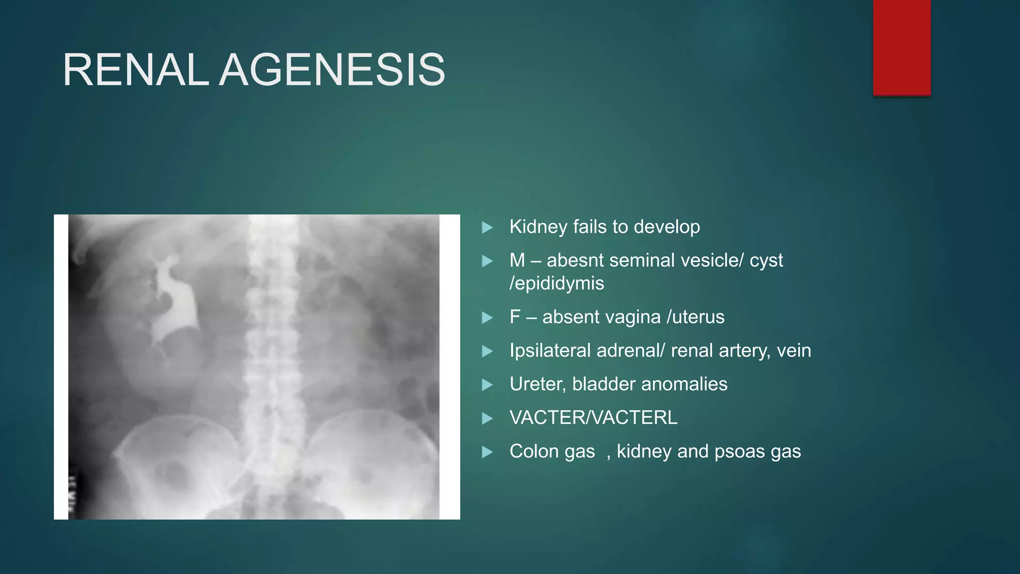
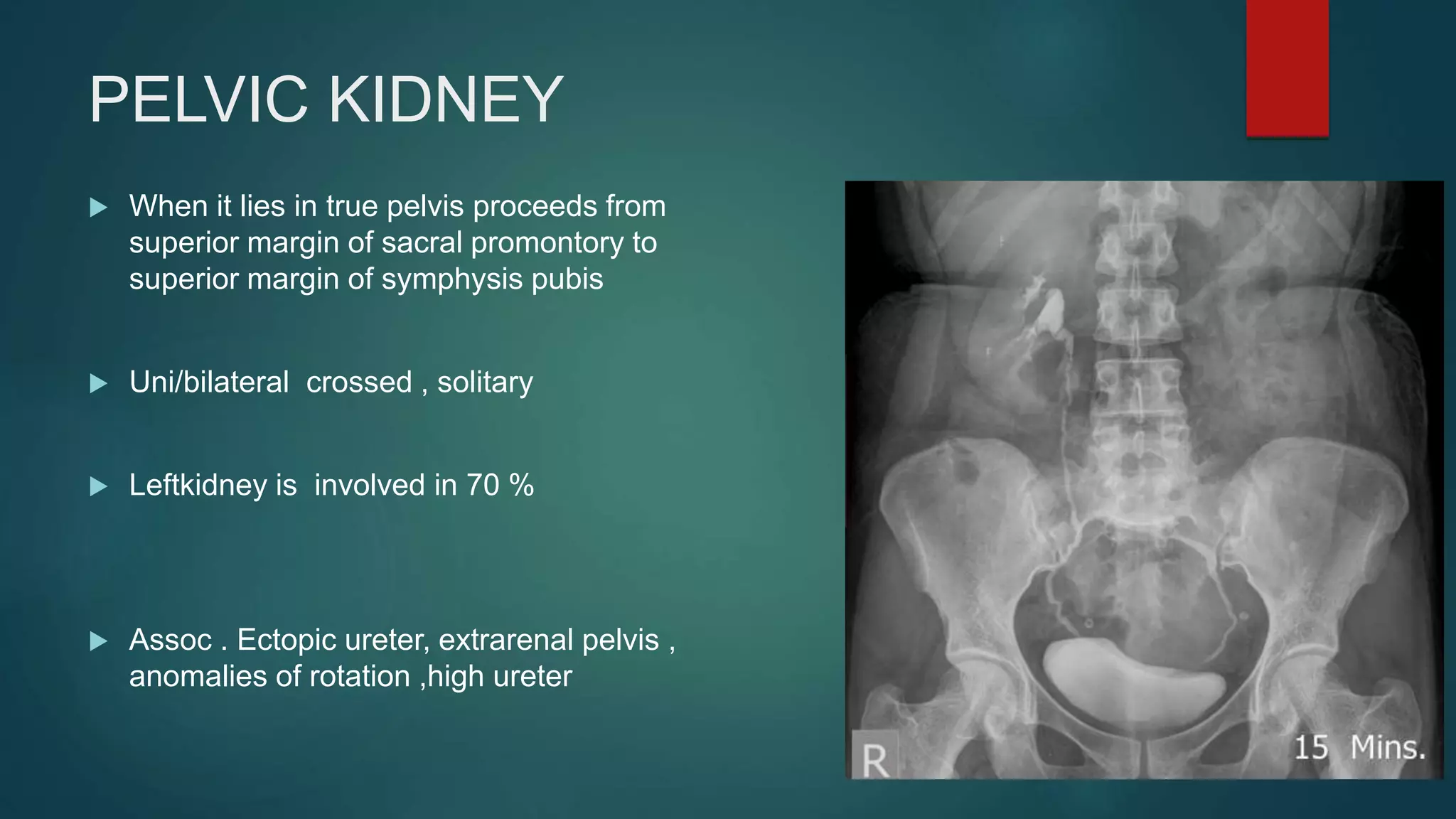
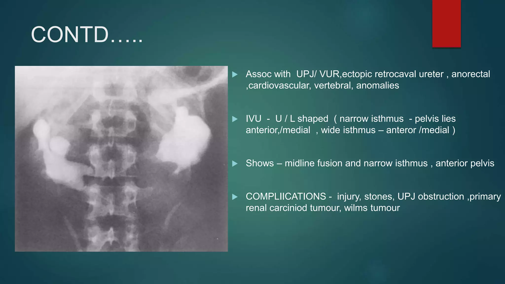
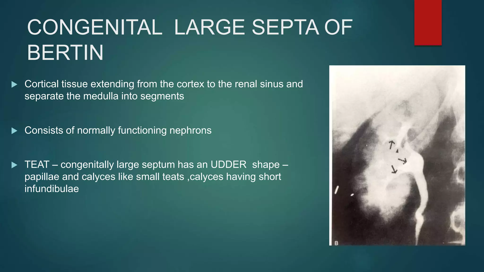
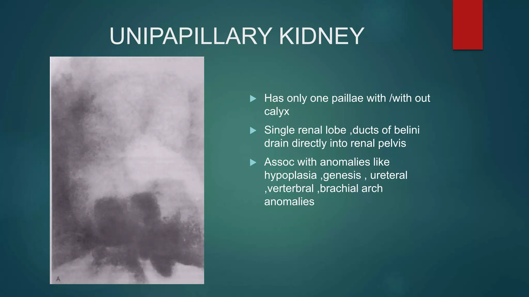
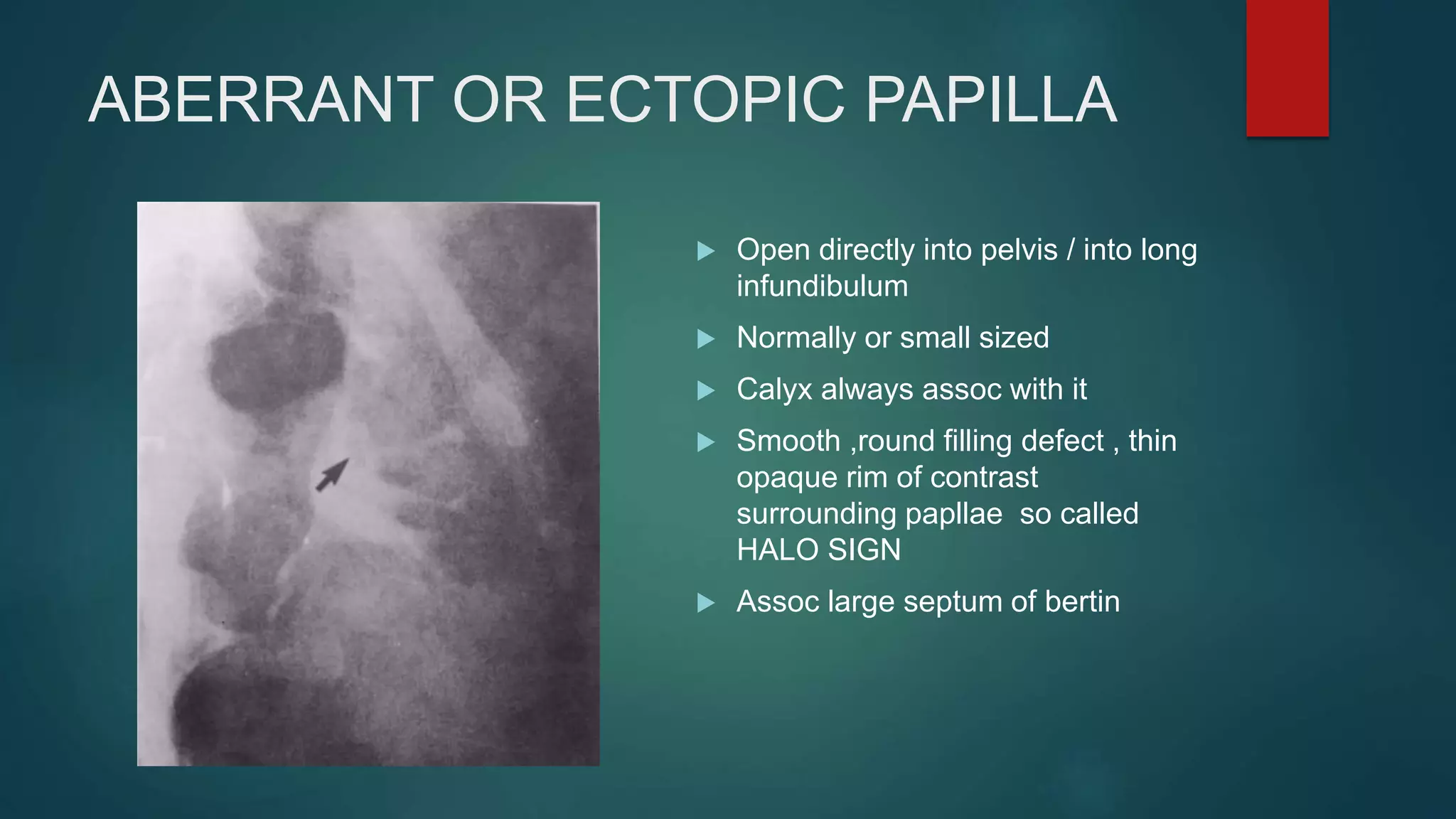
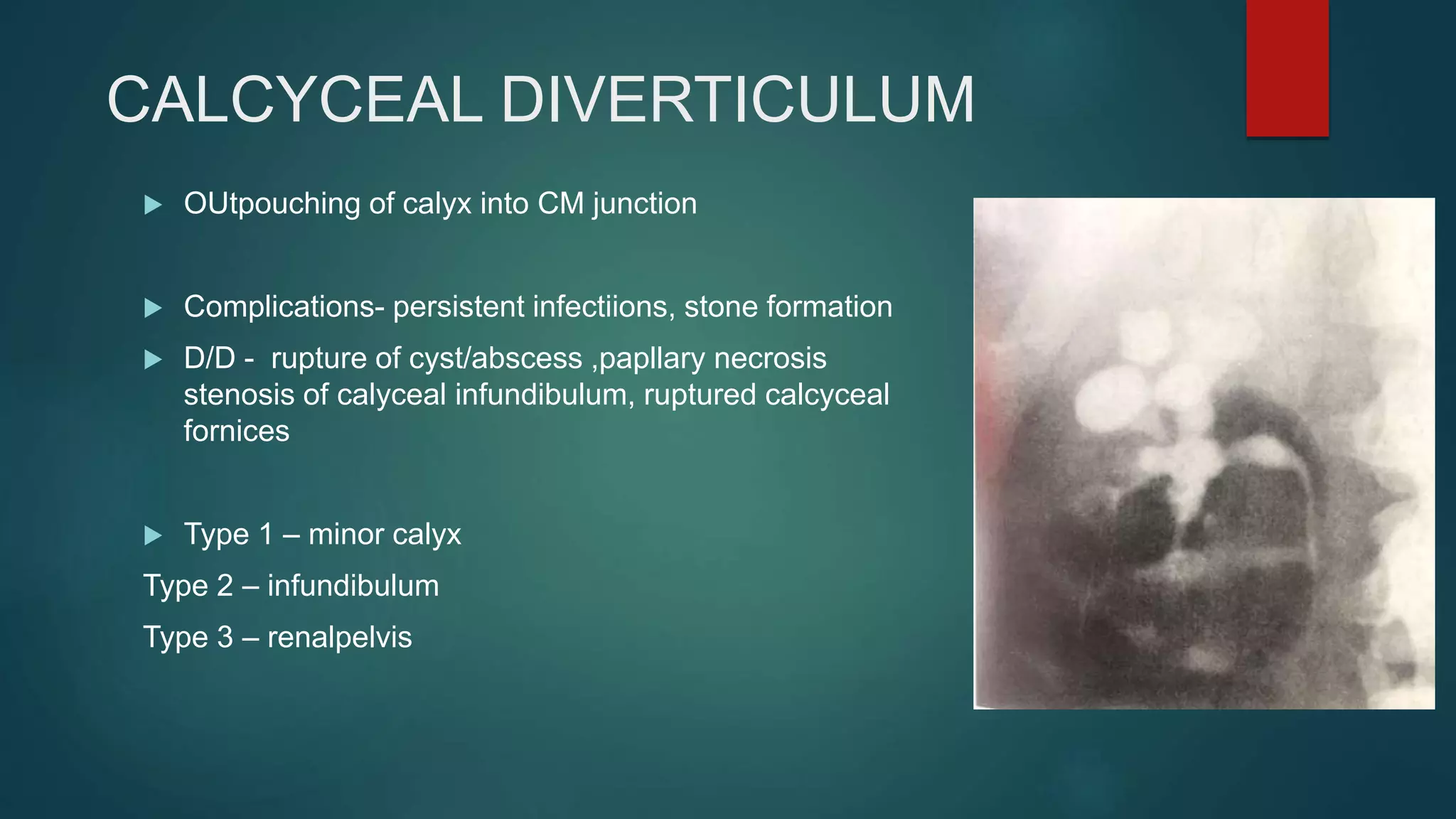
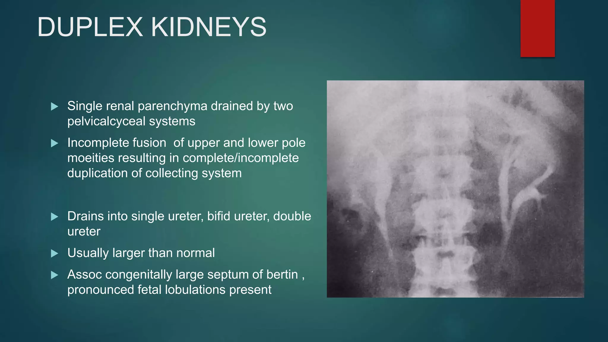
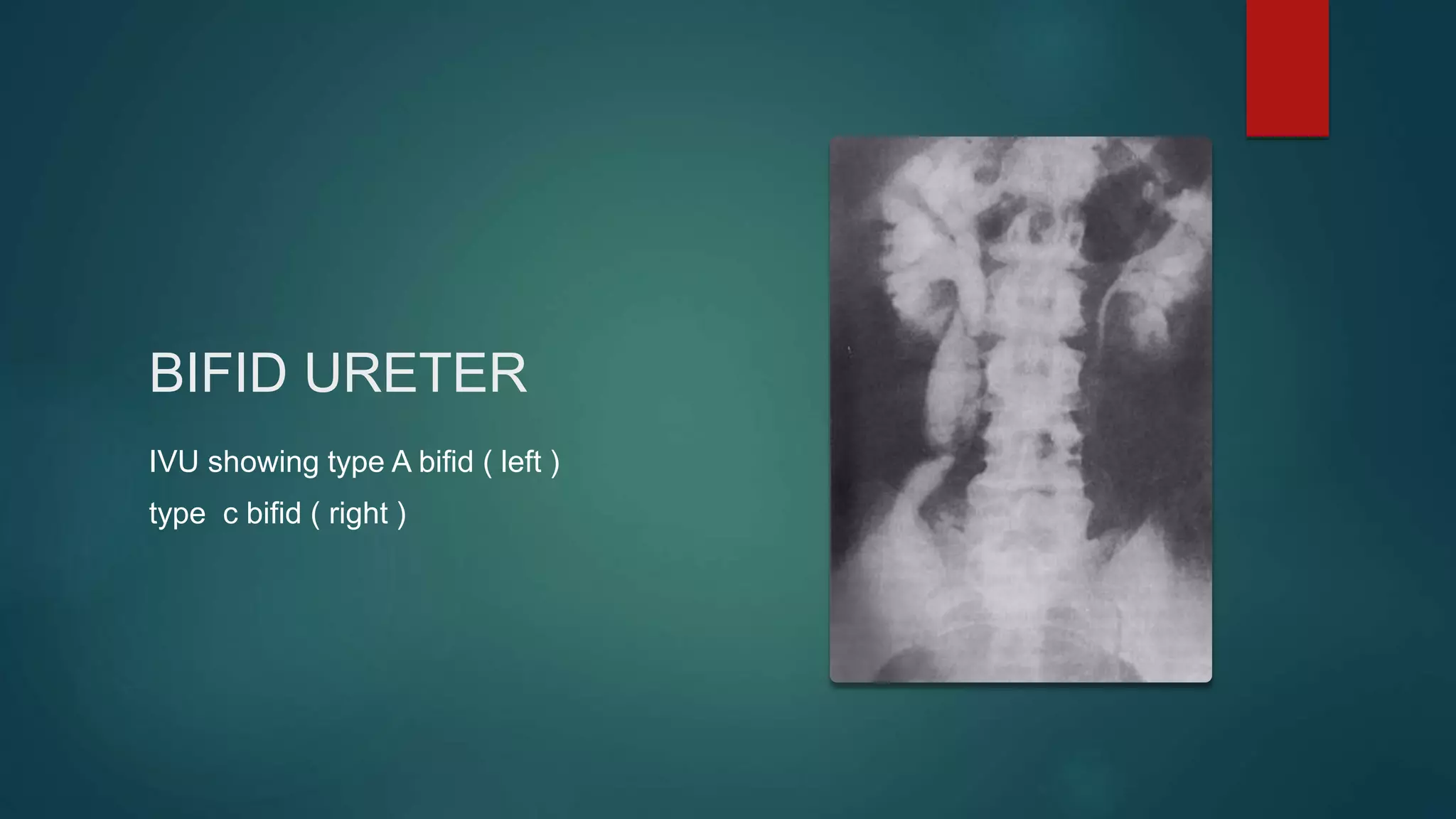
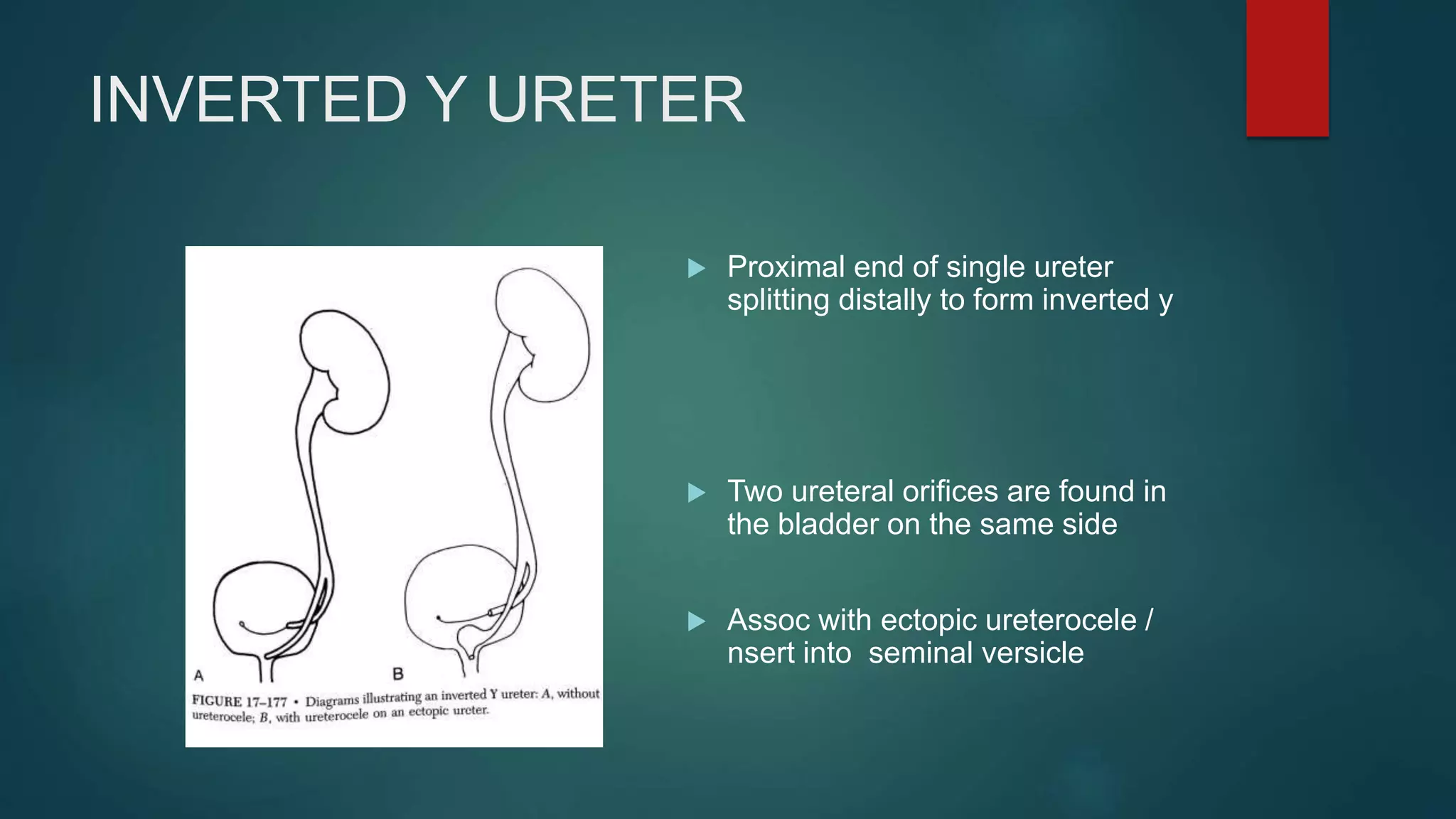
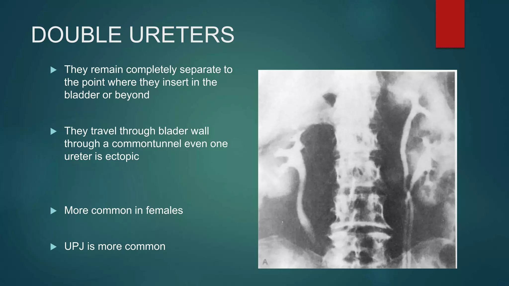
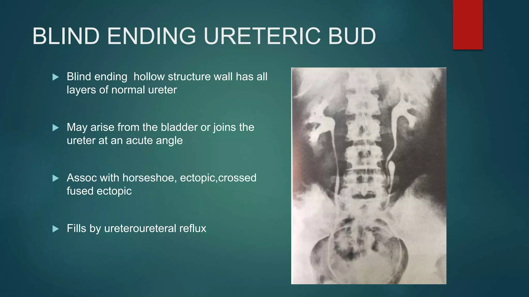
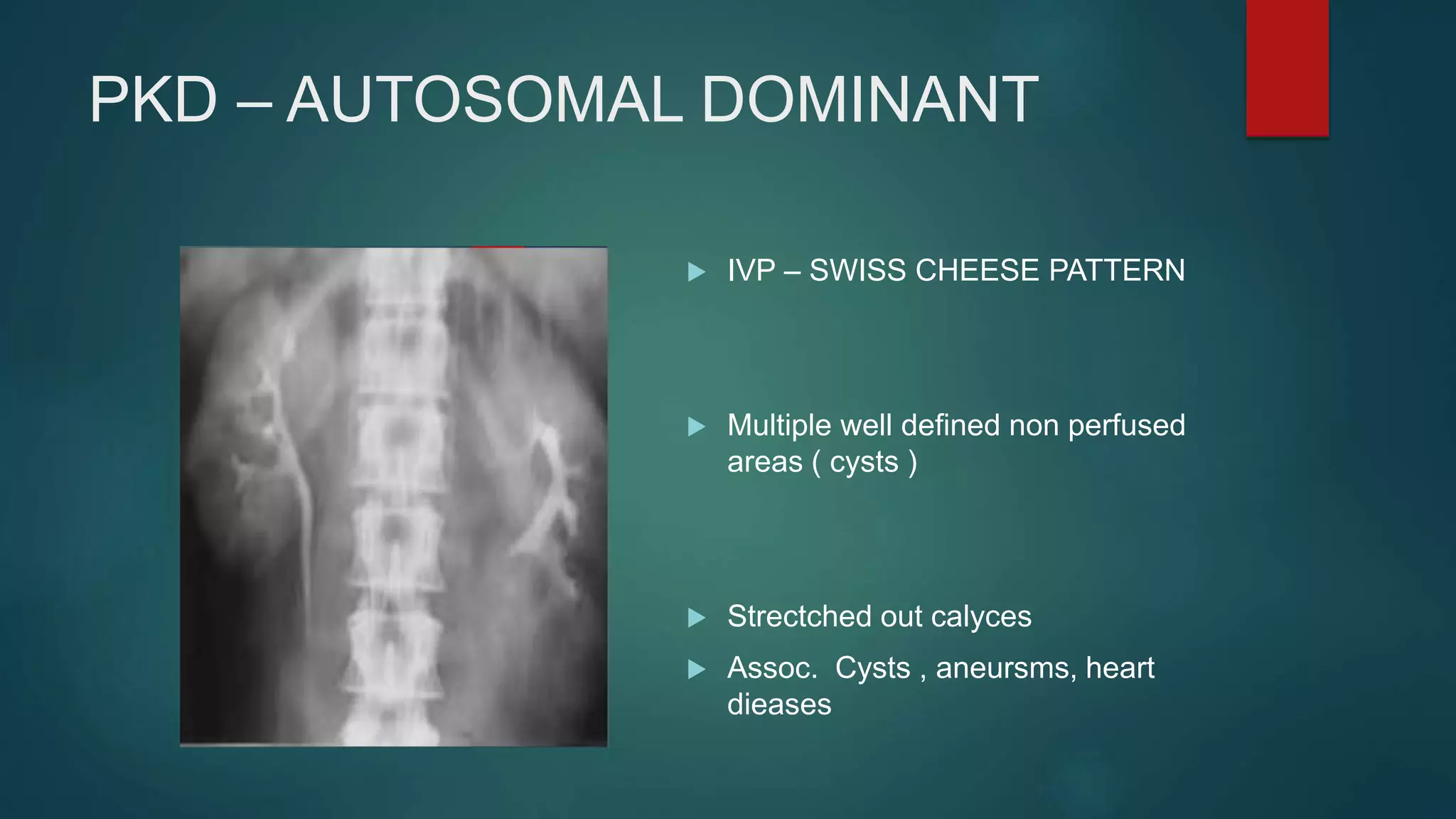
INTRAVENOUS
UROGRAM ( IVU )
DR .LEELA KRISHNA
OVERVIEW
 Congenital anomalies
 Inflammatory conditions
 Injuries
 Malignant condtions
SUPERNUMERARY KIDNEY
 Undersized/ malrotated but normal
 Total number exceeds more than 2
 Functioning ,kidney shaped
 Always smaller than normal ,
 Lies above ,below infront or behind the
usual kidney
RENAL AGENESIS
 Kidney fails to develop
 M – abesnt seminal vesicle/ cyst
/epididymis
 F – absent vagina /uterus
 Ipsilateral adrenal/ renal artery, vein
 Ureter, bladder anomalies
 VACTER/VACTERL
 Colon gas , kidney and psoas gas
RENAL HYPOPLASIA
 Congenital renal parenchymal anomaly ,in which few
nephrons are present (oligonephronia )
 Dwarf kidney (miniature version of normal kidney) with
contralateral hypertrophy
 Orthotopic, ectopic,oligomeganephronia,ask upmark
kidney
 C/S – thick walled arterioles ,clubbed
calyces,slitscars,affects single kidney ,hypertrophied
tubules ,inflammatory cells absent
 Calyces – less, glomerulus is abesnt ,
RENAL DYSPLASIA
 Congenital parenchymal malformation with abnormal nephrons and mesenchymal stroma
 1- primitive ducts , 2 – low cuboidal epithelium , 3 – cartilage ,4 – areas of loose connective tissue
 Assoc anomalies – ectopic ureter, posterior urethral valves abent abdominal muscles ,
 D/D – post obstructive atrophy, after heminephrectomy / radiation , post inflammatory changes ,high
grade renal artery stenosis, sterile VUR
ANOMALIES IN POSITION
NON ROTATION REVERSE ROTATION
RENAL ECTOPIA
 Kidney congenitally in abnormal location
 Length of ureter
 Ipsilateral - abdominal ,iliac,pelvic
 Crossed renal ectopia
 Blood supply – ectopic
 Assoc. cranial, caudal ,pelvic ectopia
 Complications – injury , obstruct labour,uvj/UPJ
obstruction,decreased functin ( 50 )
CRANIAL ECTOPIA -
( INTRATHORACIC KIDNEY )
 Partially or completely herniated through
foramen of bochdalek .
 It is not actually intrathoraic but lies below a
localized eventration of membranous part of
diaphragm
 Mistaken for mediastinal masses ,pericardial
cysts ,neuroenteric cysts, pericardial cysts
PELVIC KIDNEY
 When it lies in true pelvis proceeds from
superior margin of sacral promontory to
superior margin of symphysis pubis
 Uni/bilateral crossed , solitary
 Leftkidney is involved in 70 %
 Assoc . Ectopic ureter, extrarenal pelvis ,
anomalies of rotation ,high ureter
PELVIC KIDNEY
CROSSED ECTOPIC KIDNEY
 Kidney is located in opposite of the body
opposite the orifice of its attendant ureter
 Four varities – a) with fusion
b) With out fusion
c) Soilitary
d) Bilateral
CROSSED ECTOPIA WITH OUT
FUSION
HORSESHOE KIDNEY
 Parenchyma of each kidney lies on opposite of the kidney
and two kidneys are connected by ishmus usually at lower
poles , isthmus – normal tissue
 Ectopic.lies lower than normal kidney
 Branches from aorta,external/internal iliac,IMA
 Fuse in midline/laterallycrosses midline drain parenchyma on
opposite side
 IVU – wide istmus , right renal pelvis is lateral,left pelvis is
anterior,ureter crosses istmus ,laterally nd medially 9
FLOWER VASE APPEARANCE )
CONTD…..
 Assoc with UPJ/ VUR,ectopic retrocaval ureter , anorectal
,cardiovascular, vertebral, anomalies
 IVU - U / L shaped ( narrow isthmus - pelvis lies
anterior,/medial , wide isthmus – anteror /medial )
 Shows – midline fusion and narrow isthmus , anterior pelvis
 COMPLIICATIONS - injury, stones, UPJ obstruction ,primary
renal carciniod tumour, wilms tumour
DISC( CAKE ,LUMP KIDNEY )
 Rare variation of midline fusion when the kidney is a single disc
 Lies lower than horse shoe kidney often in pelvis ,drained by single ureter
 Seen as single confluent mass or renal tissue with renal collecting systems
lying side by side / one collecting system anterior to the other
CROSSED FUSED RENAL ECTOPIA
 Superior - kidney crosses midline ,lies superior ,pelvis rotated
anteriorly
 Sigmoid / s- crossed lies inferiorly and pelvis laterally , resident
kidney medially
 Lump -fused , irregular - pelvis anteriorly
 Unilateral L - crossed lies inferiorly ,transerversly
 Unilateral disc - fused along medial concave border , pelvis lies
anteromedially and anterolaterally
 Inferior – lies inferior , upper pole of crossed fused with lower pole
of other kidney , pelvis lies anteriorly
CONGENITAL LARGE SEPTA OF
BERTIN
 Cortical tissue extending from the cortex to the renal sinus and
separate the medulla into segments
 Consists of normally functioning nephrons
 TEAT – congenitally large septum has an UDDER shape –
papillae and calyces like small teats ,calyces having short
infundibulae
LARGE COLUMNS OF BERTIN
 Aberrant papillae - draining into
either middle of long infundibulum
/renal pelvis
 Factory siren sign – 2 or more calyces
curve around congenitally large
septum of bertin and drains into T
shaped infundibulum
UNIPAPILLARY KIDNEY
 Has only one paillae with /with out
calyx
 Single renal lobe ,ducts of belini
drain directly into renal pelvis
 Assoc with anomalies like
hypoplasia ,genesis , ureteral
,verterbral ,brachial arch
anomalies
MICROCALYX
 Presents as atiny projection of contrast material
representing an infundibulum and may be
mistaken for disease.
 Diverticula, dilated, collecting ductsand cavities
do not have tiny paillae at their termination
ABERRANT OR ECTOPIC PAPILLA
 Open directly into pelvis / into long
infundibulum
 Normally or small sized
 Calyx always assoc with it
 Smooth ,round filling defect , thin
opaque rim of contrast
surrounding papllae so called
HALO SIGN
 Assoc large septum of bertin
CALCYCEAL DIVERTICULUM
 OUtpouching of calyx into CM junction
 Complications- persistent infectiions, stone formation
 D/D - rupture of cyst/abscess ,papllary necrosis
stenosis of calyceal infundibulum, ruptured calcyceal
fornices
 Type 1 – minor calyx
Type 2 – infundibulum
Type 3 – renalpelvis
ANOMALIES IN RENAL PELVIS AND
URETER
 Duplex kidney
 Duplex ( duplicated )system
 Bifid system
 Double ureters
 Upper pole ureter
 Ectopic ureter
 Intravesical ureterocele
 Ectopic ureterocele
 Single renal parenchyma drained by two
pelvicalcyceal systems
 Incomplete fusion of upper and lower pole
moeities resulting in complete/incomplete
duplication of collecting system
 Drains into single ureter, bifid ureter, double
ureter
 Usually larger than normal
 Assoc congenitally large septum of bertin ,
pronounced fetal lobulations present
DUPLEX KIDNEYS
CONTD….
 UPJ obstruction is more common
 More in lower pole renal pelvis
 Complete ureteral duplication – hydronephrois /dysplasia ( upper pole) ,
chronic pyelonephritis ( lower pole )
 Longer than opposite normal > 3 cm,parenchymal thickness upto 1 cm
NUBBIN SIGN
 Urographic appearance of a tiny non-
functioning or poorly functioning
lower pole of a duplex kidney
 Resembles a non duplicated kidney
with the mass in lower pole
 D/D – parenchymal atrophy
secondary to vuj reflux, infection,
dysplasia secondary to aberrant
ureteral bud insertion during
development
`DROOPING LILY SIGN
 Sign of duplicated renal collecting
system
 Refers to inferolateral
displacement of opacified lower
pole moeity due to an obstructed
upper pole moiety
BIFID URETER
 Bifid ureters drain duplex kidney join to form a single ureter.
 May join extravesical(MC) or intravesical
 Y /V junctions
 YO – YO phenomenon
 Assoc with vuR, ureterocele ,stenosis,atresia
 UPJ obstruction
BIFID URETER
IVU showing type A bifid ( left )
type c bifid ( right )
INVERTED Y URETER
 Proximal end of single ureter
splitting distally to form inverted y
 Two ureteral orifices are found in
the bladder on the same side
 Assoc with ectopic ureterocele /
nsert into seminal versicle
DOUBLE URETERS
 They remain completely separate to
the point where they insert in the
bladder or beyond
 They travel through blader wall
through a commontunnel even one
ureter is ectopic
 More common in females
 UPJ is more common
URETERAL TRIPLICATION
 Type 1 - lowerpole – lateral angle of
trigone,upper pole –medial ,caudal
,ectopic – lowerpole
 Type 2 - bifid ureter , single separate
ureter
 Type 3 –trifid ureter in single orifice
BLIND ENDING URETERIC BUD
 Blind ending hollow structure wall has all
layers of normal ureter
 May arise from the bladder or joins the
ureter at an acute angle
 Assoc with horseshoe, ectopic,crossed
fused ectopic
 Fills by ureteroureteral reflux
URETERAL DIVERTICULA
 TRUE - saccular,oval ,round contains
all layers of ureteral wall
,communicates through stroma
 D/D – bladder diverticulum/ large
ureterocele /marked hydroureter
 FALSE – small / do not penentrate full
thickness , hyperplastic epithelium
 Upper,middle 1/3 rd , premalignant
ECTOPIC PATHWAY
 Upper pole ureter - opens anywhere (below
and medial to lower pole ureter) ------
WEIGERT R MEYER RULE(85%)
 Shape of shepherd cook ,circles the
superior and medial aspect ,lateral angle of
trigone –urethra
 Females – it opens post wall of urethra
/urethrovaginal septum
 Males – upper third of prostatic urethra
 More the extreme location – dysplastiic
kidney
 UTI ,pelonephritis,recurrent epidymoorchitis
ECTOPIC URETERAL ORIFICES
 Ectopic - proximal lip of bladder neck or beyond
 Obeys weigert –R meyer rule
 Normal – lateral to trigone,medial and inferior ,further
 Cranial – lateral to outerangle of trigone, outside blader
in diverticulum
 Caudal - male ( upper third of prosticurethra /vas
 Females ( posterior part of urethra/urethrovaginal
septim /wall of vagina,hymen )
URETEROCELE
 Balloning of distal end of ureter
 Assoc. single/duplex kidney
 Abnormality of mucosa
 Ectopic – defect of muscle coat of ureter and bladder wall itself (poor
detrusor backing )
INTRAVESICAL URETEROCELE
 Orifice of ureter and ureterocele –
with in bladder
 Simple ureterocele
 Opens in normal zone , - but stenotic /
non obstructed
 Adults,acquired - fibrotic narrowing
/inflammation/trauma
 Stones, persistent infection
 Superior, inferior surfaceupper ,lower
pole ureter
 IVU – COBRA HEAD SIGN /SPRING
ONION
EXTRAVESICAL URETEROCELE
 Uretrocele lies in the submucosa of bladder and some extends into
bladder neck
 Complications – obstruction, reflux, stone ,upperpole dyspalsia
 mass in upper pole of kidney, radiolucent filling defect in bladder,
POLYCYSTIC KIDNEY DISEASE
 Contrast in preserved tubules next to unopacified
tubules
 Autosomal reccesive
 Renal prarenchyma replaced by numerous tiny
cysts
 Perinatal, neonatal, infantile, juvenile
 Ologohydraminos,potters syndrome
PKD – AUTOSOMAL DOMINANT
 IVP – SWISS CHEESE PATTERN
 Multiple well defined non perfused
areas ( cysts )
 Strectched out calyces
 Assoc. Cysts , aneursms, heart
dieases
MEDULLARY SPONGE KIDNEY
 Paint brush like appaerence
 Produced by cystic dilatation of medullary
collecting ducts
 Weak assoc. wilms disease,
pheaochromocytoma,horshe shoe kidney
ACUTE BACTERIAL
PYELONEPHRITIS Renal enlargement
 Impaired excretion
 Nonhomogenous nephrogram
 Attenuation of collecting system
 Mucosal edema
 Poor definition of renal margins
 Pyeloureteral dilatation
CHRONIC PYELONEPHRITIS
 Asymmetry and Irregular renal outline
with upper pole parenchymal atrophy
 Loss of renal cortical thickness over
blunted and dilated calcyces
RENAL PAPILLARY NECROSIS
 Enlarged kidney with central paiilary necrosis with
marginal extrasavation – LOBSTER CLAW appearance
 Sequestered , sloughed papillae causing filling defect –
RING SIGN
 Multile calcyces affected – RIM like CALCIFICATION
 Tissue necrosis - blunted/ clubbed calcyces
GUTB – IVP :
Parenchymal scars & Irregularity of the papillary tips - “moth-eaten”
calices
Small cavities in the papillae communicate with the
collecting system
fibrotic reaction develops, stenosis and strictures of the caliceal
infundibula - Infundibular strictures
can lead to localized caliectasis or phantom calyx.
Scarring of renal pelvis (Kerr kink)
Earliest abnormality –
- Indistinct feathery outline
Irregularity of surface of
one or more papillae or
calyces with normal
renal size and contour.
Fuzzy & irregular calices
due to papillary necrosis.
Normal calices
MOTH EATEN APPEARANCE
CONTD…
INFUNDIBULAR STENOSIS
CAUSING PHANTOM CALYX
Phantom calix
Infundibular stenosis
PHANTOM CALYX
Decreased nephrographic opacity and nonfilling of the collecting system
elements at the lower pole of left kidney – phantom calyces
RGP
GOLF ON A TEE
Collecting system shows
contrast material
in a large papillary cavity, the
“golf ball” (∗).
Blunted calyx, the “tee,”
is adjacent (arrow).
 Cortical scarring with
dilatation & distortion of
adjoining calyces coupled
with strictures of the
pelvicaliceal system.
 Cause luminal narrowing
either directly or by causing
kinking of the renal pelvis at
the UPJ.
KERR KINK
HIKED UP PELVIS
Cephalic retraction of the inferior medial
margin of the renal pelvis at the
ureteropelvic junction (UPJ)
 Autonephrectomy.
 Diffuse, uniform, extensive
parenchymal
calcifications forming a cast
of the kidney with
autonephrectomy.
 End stage of GuTB.
PUTTY KIDNEY
If the ulcer or stricture extends to the renal pelvis or the pelvic
ureteral junction, urine outflow obstruction may
occur.
IVUmay show delayed function, clubbed calyces, or
absence of function.
Some show Hydronephrosis - irregular margins and filling
defects owing to caseous debris.
If tuberculous infection extends directly to the rest of the kidney,
the entire kidney becomes a bag of caseous
necrotic pus.
The kidney enlarges initially but subsequently may return to normal or
become atrophic.
infection may extend into peri- / pararenal space + psoas
dilatation resulting from
atony and prolonged
bacilluria
PIPE
STEM
irregular segments of
ureter due to mucosal
ulcerations
necrosis of ureteral
musculature is
accompanied by fibrosis -
stricture formation- 50%.
severe thickening of the
wall produces a rigid
shortened ureter with
narrow lumen
cork-
SCREW
TERMINAL
SEGMENT
 Rigid ureter: irregular and lacks
normal peristaltic movement,
fibrotic strictures noted.
 distortion, amputation and
irregularity of the upper pole
calices.
PIPE STEM URETER
 Fusion of multiple strictures
may create a long, irregular
narrowing. Several
nonconfluent strictures can
produce a “beaded” or
“corkscrew” ureter
BEADED / CORK SCREW
 Ulcerations causing mucosal
irregularity of ureter.
SAW TOOTH
Urinary lithiasis
 Filling defect in case of radioleucent
partial stag horn calculus
RENAL INJURIES
 Absence of unilateral excretion suggests
major vascular injury ( renal artery
avulsion )
 Disruption of pelvicalyceal system may
be seen as extravasation of opacified
urine
 Large retroperitoneal ,perinephric
hematomas – soft tissue sweling with
loss of psoas shawdow
Ureteric injury
 Excretory intravenous urography if CT is
not available: demonstrates contrast
leakage and spillage outside the course of
the urinary system.
RENAL CELL CARCINOMA
 Calcification - curvilinear ,punctate/flocculent (
central )
 Focal bulge in the renal contour such as
 Tilting of renal axis ( exophytic medial mass)
 Lower pole mass (horizantal plane)
 Displacement of adjacent calcyces ,renal pelvis
,ureter
 Absence of contrast excretion - renal vein
occlusion
Seen as filling defects in the pelvis
Surface – smooth ,stipped,serrated,frondlike
Calyceal obliteration ,fiilling defects with
distended cakyces
STIPPLESIGN –trapping of contrast material
with in interstices of tumour will give a stippled
effect ( H/S/O – tcc )
TCC PELVIS
URETER
 Tumor seen as ovoid filling defect arising from one of the walls
 GOBLET /CHAMPAGNE GLASS SIGN – dilatation below ureter
 Retrograde cathetrer may coil in infratumoral space (BERGMANN sign )
Ureteral tumours
 Due to the small caliber of the ureter, tumours are more likelyto
obstruct the kidney at small tumour size.
 Obstruction may lead to hyrdonephrosis with or without hydroureter and
may also result in a non-functioning kidney or delayed nephrogram.
Thank you

INTRAVENOUS UROGRAM ( IVU )

  • 1.
    INTRAVENOUS UROGRAM ( IVU) DR .LEELA KRISHNA
  • 2.
    OVERVIEW  Congenital anomalies Inflammatory conditions  Injuries  Malignant condtions
  • 3.
    SUPERNUMERARY KIDNEY  Undersized/malrotated but normal  Total number exceeds more than 2  Functioning ,kidney shaped  Always smaller than normal ,  Lies above ,below infront or behind the usual kidney
  • 4.
    RENAL AGENESIS  Kidneyfails to develop  M – abesnt seminal vesicle/ cyst /epididymis  F – absent vagina /uterus  Ipsilateral adrenal/ renal artery, vein  Ureter, bladder anomalies  VACTER/VACTERL  Colon gas , kidney and psoas gas
  • 5.
    RENAL HYPOPLASIA  Congenitalrenal parenchymal anomaly ,in which few nephrons are present (oligonephronia )  Dwarf kidney (miniature version of normal kidney) with contralateral hypertrophy  Orthotopic, ectopic,oligomeganephronia,ask upmark kidney  C/S – thick walled arterioles ,clubbed calyces,slitscars,affects single kidney ,hypertrophied tubules ,inflammatory cells absent  Calyces – less, glomerulus is abesnt ,
  • 6.
    RENAL DYSPLASIA  Congenitalparenchymal malformation with abnormal nephrons and mesenchymal stroma  1- primitive ducts , 2 – low cuboidal epithelium , 3 – cartilage ,4 – areas of loose connective tissue  Assoc anomalies – ectopic ureter, posterior urethral valves abent abdominal muscles ,  D/D – post obstructive atrophy, after heminephrectomy / radiation , post inflammatory changes ,high grade renal artery stenosis, sterile VUR
  • 7.
  • 8.
    ANOMALIES IN POSITION NONROTATION REVERSE ROTATION
  • 9.
    RENAL ECTOPIA  Kidneycongenitally in abnormal location  Length of ureter  Ipsilateral - abdominal ,iliac,pelvic  Crossed renal ectopia  Blood supply – ectopic  Assoc. cranial, caudal ,pelvic ectopia  Complications – injury , obstruct labour,uvj/UPJ obstruction,decreased functin ( 50 )
  • 10.
    CRANIAL ECTOPIA - (INTRATHORACIC KIDNEY )  Partially or completely herniated through foramen of bochdalek .  It is not actually intrathoraic but lies below a localized eventration of membranous part of diaphragm  Mistaken for mediastinal masses ,pericardial cysts ,neuroenteric cysts, pericardial cysts
  • 11.
    PELVIC KIDNEY  Whenit lies in true pelvis proceeds from superior margin of sacral promontory to superior margin of symphysis pubis  Uni/bilateral crossed , solitary  Leftkidney is involved in 70 %  Assoc . Ectopic ureter, extrarenal pelvis , anomalies of rotation ,high ureter
  • 12.
  • 13.
    CROSSED ECTOPIC KIDNEY Kidney is located in opposite of the body opposite the orifice of its attendant ureter  Four varities – a) with fusion b) With out fusion c) Soilitary d) Bilateral
  • 14.
  • 15.
    HORSESHOE KIDNEY  Parenchymaof each kidney lies on opposite of the kidney and two kidneys are connected by ishmus usually at lower poles , isthmus – normal tissue  Ectopic.lies lower than normal kidney  Branches from aorta,external/internal iliac,IMA  Fuse in midline/laterallycrosses midline drain parenchyma on opposite side  IVU – wide istmus , right renal pelvis is lateral,left pelvis is anterior,ureter crosses istmus ,laterally nd medially 9 FLOWER VASE APPEARANCE )
  • 16.
    CONTD…..  Assoc withUPJ/ VUR,ectopic retrocaval ureter , anorectal ,cardiovascular, vertebral, anomalies  IVU - U / L shaped ( narrow isthmus - pelvis lies anterior,/medial , wide isthmus – anteror /medial )  Shows – midline fusion and narrow isthmus , anterior pelvis  COMPLIICATIONS - injury, stones, UPJ obstruction ,primary renal carciniod tumour, wilms tumour
  • 17.
    DISC( CAKE ,LUMPKIDNEY )  Rare variation of midline fusion when the kidney is a single disc  Lies lower than horse shoe kidney often in pelvis ,drained by single ureter  Seen as single confluent mass or renal tissue with renal collecting systems lying side by side / one collecting system anterior to the other
  • 18.
    CROSSED FUSED RENALECTOPIA  Superior - kidney crosses midline ,lies superior ,pelvis rotated anteriorly  Sigmoid / s- crossed lies inferiorly and pelvis laterally , resident kidney medially  Lump -fused , irregular - pelvis anteriorly  Unilateral L - crossed lies inferiorly ,transerversly  Unilateral disc - fused along medial concave border , pelvis lies anteromedially and anterolaterally  Inferior – lies inferior , upper pole of crossed fused with lower pole of other kidney , pelvis lies anteriorly
  • 19.
    CONGENITAL LARGE SEPTAOF BERTIN  Cortical tissue extending from the cortex to the renal sinus and separate the medulla into segments  Consists of normally functioning nephrons  TEAT – congenitally large septum has an UDDER shape – papillae and calyces like small teats ,calyces having short infundibulae
  • 20.
    LARGE COLUMNS OFBERTIN  Aberrant papillae - draining into either middle of long infundibulum /renal pelvis  Factory siren sign – 2 or more calyces curve around congenitally large septum of bertin and drains into T shaped infundibulum
  • 21.
    UNIPAPILLARY KIDNEY  Hasonly one paillae with /with out calyx  Single renal lobe ,ducts of belini drain directly into renal pelvis  Assoc with anomalies like hypoplasia ,genesis , ureteral ,verterbral ,brachial arch anomalies
  • 22.
    MICROCALYX  Presents asatiny projection of contrast material representing an infundibulum and may be mistaken for disease.  Diverticula, dilated, collecting ductsand cavities do not have tiny paillae at their termination
  • 23.
    ABERRANT OR ECTOPICPAPILLA  Open directly into pelvis / into long infundibulum  Normally or small sized  Calyx always assoc with it  Smooth ,round filling defect , thin opaque rim of contrast surrounding papllae so called HALO SIGN  Assoc large septum of bertin
  • 24.
    CALCYCEAL DIVERTICULUM  OUtpouchingof calyx into CM junction  Complications- persistent infectiions, stone formation  D/D - rupture of cyst/abscess ,papllary necrosis stenosis of calyceal infundibulum, ruptured calcyceal fornices  Type 1 – minor calyx Type 2 – infundibulum Type 3 – renalpelvis
  • 25.
    ANOMALIES IN RENALPELVIS AND URETER  Duplex kidney  Duplex ( duplicated )system  Bifid system  Double ureters  Upper pole ureter  Ectopic ureter  Intravesical ureterocele  Ectopic ureterocele
  • 26.
     Single renalparenchyma drained by two pelvicalcyceal systems  Incomplete fusion of upper and lower pole moeities resulting in complete/incomplete duplication of collecting system  Drains into single ureter, bifid ureter, double ureter  Usually larger than normal  Assoc congenitally large septum of bertin , pronounced fetal lobulations present DUPLEX KIDNEYS
  • 27.
    CONTD….  UPJ obstructionis more common  More in lower pole renal pelvis  Complete ureteral duplication – hydronephrois /dysplasia ( upper pole) , chronic pyelonephritis ( lower pole )  Longer than opposite normal > 3 cm,parenchymal thickness upto 1 cm
  • 28.
    NUBBIN SIGN  Urographicappearance of a tiny non- functioning or poorly functioning lower pole of a duplex kidney  Resembles a non duplicated kidney with the mass in lower pole  D/D – parenchymal atrophy secondary to vuj reflux, infection, dysplasia secondary to aberrant ureteral bud insertion during development
  • 29.
    `DROOPING LILY SIGN Sign of duplicated renal collecting system  Refers to inferolateral displacement of opacified lower pole moeity due to an obstructed upper pole moiety
  • 30.
    BIFID URETER  Bifidureters drain duplex kidney join to form a single ureter.  May join extravesical(MC) or intravesical  Y /V junctions  YO – YO phenomenon  Assoc with vuR, ureterocele ,stenosis,atresia  UPJ obstruction
  • 31.
    BIFID URETER IVU showingtype A bifid ( left ) type c bifid ( right )
  • 32.
    INVERTED Y URETER Proximal end of single ureter splitting distally to form inverted y  Two ureteral orifices are found in the bladder on the same side  Assoc with ectopic ureterocele / nsert into seminal versicle
  • 33.
    DOUBLE URETERS  Theyremain completely separate to the point where they insert in the bladder or beyond  They travel through blader wall through a commontunnel even one ureter is ectopic  More common in females  UPJ is more common
  • 34.
    URETERAL TRIPLICATION  Type1 - lowerpole – lateral angle of trigone,upper pole –medial ,caudal ,ectopic – lowerpole  Type 2 - bifid ureter , single separate ureter  Type 3 –trifid ureter in single orifice
  • 35.
    BLIND ENDING URETERICBUD  Blind ending hollow structure wall has all layers of normal ureter  May arise from the bladder or joins the ureter at an acute angle  Assoc with horseshoe, ectopic,crossed fused ectopic  Fills by ureteroureteral reflux
  • 36.
    URETERAL DIVERTICULA  TRUE- saccular,oval ,round contains all layers of ureteral wall ,communicates through stroma  D/D – bladder diverticulum/ large ureterocele /marked hydroureter  FALSE – small / do not penentrate full thickness , hyperplastic epithelium  Upper,middle 1/3 rd , premalignant
  • 37.
    ECTOPIC PATHWAY  Upperpole ureter - opens anywhere (below and medial to lower pole ureter) ------ WEIGERT R MEYER RULE(85%)  Shape of shepherd cook ,circles the superior and medial aspect ,lateral angle of trigone –urethra  Females – it opens post wall of urethra /urethrovaginal septum  Males – upper third of prostatic urethra  More the extreme location – dysplastiic kidney  UTI ,pelonephritis,recurrent epidymoorchitis
  • 38.
    ECTOPIC URETERAL ORIFICES Ectopic - proximal lip of bladder neck or beyond  Obeys weigert –R meyer rule  Normal – lateral to trigone,medial and inferior ,further  Cranial – lateral to outerangle of trigone, outside blader in diverticulum  Caudal - male ( upper third of prosticurethra /vas  Females ( posterior part of urethra/urethrovaginal septim /wall of vagina,hymen )
  • 39.
    URETEROCELE  Balloning ofdistal end of ureter  Assoc. single/duplex kidney  Abnormality of mucosa  Ectopic – defect of muscle coat of ureter and bladder wall itself (poor detrusor backing )
  • 40.
    INTRAVESICAL URETEROCELE  Orificeof ureter and ureterocele – with in bladder  Simple ureterocele  Opens in normal zone , - but stenotic / non obstructed  Adults,acquired - fibrotic narrowing /inflammation/trauma  Stones, persistent infection  Superior, inferior surfaceupper ,lower pole ureter  IVU – COBRA HEAD SIGN /SPRING ONION
  • 41.
    EXTRAVESICAL URETEROCELE  Uretrocelelies in the submucosa of bladder and some extends into bladder neck  Complications – obstruction, reflux, stone ,upperpole dyspalsia  mass in upper pole of kidney, radiolucent filling defect in bladder,
  • 42.
    POLYCYSTIC KIDNEY DISEASE Contrast in preserved tubules next to unopacified tubules  Autosomal reccesive  Renal prarenchyma replaced by numerous tiny cysts  Perinatal, neonatal, infantile, juvenile  Ologohydraminos,potters syndrome
  • 43.
    PKD – AUTOSOMALDOMINANT  IVP – SWISS CHEESE PATTERN  Multiple well defined non perfused areas ( cysts )  Strectched out calyces  Assoc. Cysts , aneursms, heart dieases
  • 44.
    MEDULLARY SPONGE KIDNEY Paint brush like appaerence  Produced by cystic dilatation of medullary collecting ducts  Weak assoc. wilms disease, pheaochromocytoma,horshe shoe kidney
  • 45.
    ACUTE BACTERIAL PYELONEPHRITIS Renalenlargement  Impaired excretion  Nonhomogenous nephrogram  Attenuation of collecting system  Mucosal edema  Poor definition of renal margins  Pyeloureteral dilatation
  • 46.
    CHRONIC PYELONEPHRITIS  Asymmetryand Irregular renal outline with upper pole parenchymal atrophy  Loss of renal cortical thickness over blunted and dilated calcyces
  • 47.
    RENAL PAPILLARY NECROSIS Enlarged kidney with central paiilary necrosis with marginal extrasavation – LOBSTER CLAW appearance  Sequestered , sloughed papillae causing filling defect – RING SIGN  Multile calcyces affected – RIM like CALCIFICATION  Tissue necrosis - blunted/ clubbed calcyces
  • 48.
    GUTB – IVP: Parenchymal scars & Irregularity of the papillary tips - “moth-eaten” calices Small cavities in the papillae communicate with the collecting system fibrotic reaction develops, stenosis and strictures of the caliceal infundibula - Infundibular strictures can lead to localized caliectasis or phantom calyx. Scarring of renal pelvis (Kerr kink)
  • 49.
    Earliest abnormality – -Indistinct feathery outline Irregularity of surface of one or more papillae or calyces with normal renal size and contour. Fuzzy & irregular calices due to papillary necrosis. Normal calices MOTH EATEN APPEARANCE
  • 50.
  • 51.
    INFUNDIBULAR STENOSIS CAUSING PHANTOMCALYX Phantom calix Infundibular stenosis
  • 52.
    PHANTOM CALYX Decreased nephrographicopacity and nonfilling of the collecting system elements at the lower pole of left kidney – phantom calyces RGP
  • 53.
    GOLF ON ATEE Collecting system shows contrast material in a large papillary cavity, the “golf ball” (∗). Blunted calyx, the “tee,” is adjacent (arrow).
  • 54.
     Cortical scarringwith dilatation & distortion of adjoining calyces coupled with strictures of the pelvicaliceal system.  Cause luminal narrowing either directly or by causing kinking of the renal pelvis at the UPJ. KERR KINK
  • 55.
    HIKED UP PELVIS Cephalicretraction of the inferior medial margin of the renal pelvis at the ureteropelvic junction (UPJ)
  • 56.
     Autonephrectomy.  Diffuse,uniform, extensive parenchymal calcifications forming a cast of the kidney with autonephrectomy.  End stage of GuTB. PUTTY KIDNEY
  • 57.
    If the ulceror stricture extends to the renal pelvis or the pelvic ureteral junction, urine outflow obstruction may occur. IVUmay show delayed function, clubbed calyces, or absence of function. Some show Hydronephrosis - irregular margins and filling defects owing to caseous debris. If tuberculous infection extends directly to the rest of the kidney, the entire kidney becomes a bag of caseous necrotic pus. The kidney enlarges initially but subsequently may return to normal or become atrophic. infection may extend into peri- / pararenal space + psoas
  • 58.
    dilatation resulting from atonyand prolonged bacilluria PIPE STEM irregular segments of ureter due to mucosal ulcerations necrosis of ureteral musculature is accompanied by fibrosis - stricture formation- 50%. severe thickening of the wall produces a rigid shortened ureter with narrow lumen cork- SCREW TERMINAL SEGMENT
  • 59.
     Rigid ureter:irregular and lacks normal peristaltic movement, fibrotic strictures noted.  distortion, amputation and irregularity of the upper pole calices. PIPE STEM URETER
  • 60.
     Fusion ofmultiple strictures may create a long, irregular narrowing. Several nonconfluent strictures can produce a “beaded” or “corkscrew” ureter BEADED / CORK SCREW
  • 61.
     Ulcerations causingmucosal irregularity of ureter. SAW TOOTH
  • 62.
    Urinary lithiasis  Fillingdefect in case of radioleucent partial stag horn calculus
  • 63.
    RENAL INJURIES  Absenceof unilateral excretion suggests major vascular injury ( renal artery avulsion )  Disruption of pelvicalyceal system may be seen as extravasation of opacified urine  Large retroperitoneal ,perinephric hematomas – soft tissue sweling with loss of psoas shawdow
  • 64.
    Ureteric injury  Excretoryintravenous urography if CT is not available: demonstrates contrast leakage and spillage outside the course of the urinary system.
  • 65.
    RENAL CELL CARCINOMA Calcification - curvilinear ,punctate/flocculent ( central )  Focal bulge in the renal contour such as  Tilting of renal axis ( exophytic medial mass)  Lower pole mass (horizantal plane)  Displacement of adjacent calcyces ,renal pelvis ,ureter  Absence of contrast excretion - renal vein occlusion
  • 66.
    Seen as fillingdefects in the pelvis Surface – smooth ,stipped,serrated,frondlike Calyceal obliteration ,fiilling defects with distended cakyces STIPPLESIGN –trapping of contrast material with in interstices of tumour will give a stippled effect ( H/S/O – tcc ) TCC PELVIS
  • 67.
    URETER  Tumor seenas ovoid filling defect arising from one of the walls  GOBLET /CHAMPAGNE GLASS SIGN – dilatation below ureter  Retrograde cathetrer may coil in infratumoral space (BERGMANN sign )
  • 68.
    Ureteral tumours  Dueto the small caliber of the ureter, tumours are more likelyto obstruct the kidney at small tumour size.  Obstruction may lead to hyrdonephrosis with or without hydroureter and may also result in a non-functioning kidney or delayed nephrogram.
  • 69.