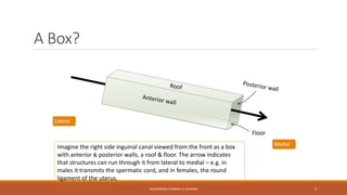
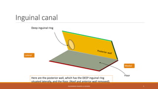
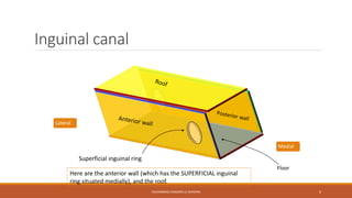
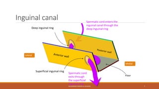
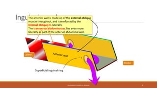
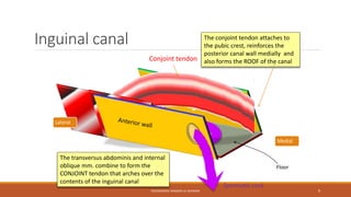
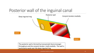
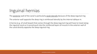
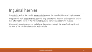
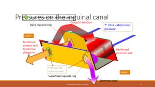
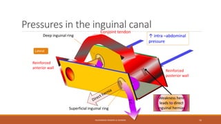
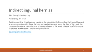
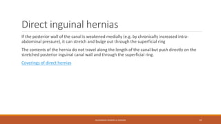
MUHAMMAD RAMZAN UL REHMAN 1
Inguinal Cannal 
MUHAMMAD RAMZAN UL REHMAN 
MUHAMMAD RAMZAN UL REHMAN 2
Inguinal canals – why have them? 
Allow contents of the scrotum to communicate with intra-abdominal contents 
Prevent mobile intra-abdominal contents (e.g. intestine) from entering the scrotum and possibly 
becoming damaged, while at the same time permitting blood vessels, nerves, lymphatics, vas 
deferens etc. to supply the scrotal contents 
MUHAMMAD RAMZAN UL REHMAN 3
A Box? 
Floor 
Imagine the right side inguinal canal viewed from the front as a box 
with anterior & posterior walls, a roof & floor. The arrow indicates 
that structures can run through it from lateral to medial – e.g. in 
males it transmits the spermatic cord, and in females, the round 
ligament of the uterus. 
Medial 
MUHAMMAD RAMZAN UL REHMAN 4 
Lateral
Inguinal canal 
Medial 
Floor 
Here are the posterior wall, which has the DEEP inguinal ring 
situated laterally, and the floor. (Roof and anterior wall removed). 
MUHAMMAD RAMZAN UL REHMAN 5 
Deep inguinal ring 
Lateral
Inguinal canal 
Medial 
Floor 
Here are the anterior wall (which has the SUPERFICIAL inguinal 
ring situated medially), and the roof. 
MUHAMMAD RAMZAN UL REHMAN 6 
Superficial inguinal ring 
Lateral
Inguinal canal 
Medial 
Floor 
Spermatic cord enters the 
inguinal canal through the 
Deep inguinal ring deep inguinal ring 
Spermatic cord 
exits through 
the superficial 
inguinal ring 
MUHAMMAD RAMZAN UL REHMAN 7 
Superficial inguinal ring 
Lateral
Inguinal canal 
Medial 
The anterior wall is made up of the external oblique 
muscle throughout, and is reinforced by the 
internal oblique m. laterally. 
The transversus abdominus m. lies even more 
laterally as part of the anterior abdominal wall. 
MUHAMMAD RAMZAN UL REHMAN 8 
Superficial inguinal ring 
Lateral
Inguinal canal 
Medial 
Floor 
Spermatic cord 
MUHAMMAD RAMZAN UL REHMAN 9 
Lateral 
The transversus abdominis and internal 
oblique mm. combine to form the 
CONJOINT tendon that arches over the 
contents of the inguinal canal 
The conjoint tendon attaches to 
the pubic crest, reinforces the 
posterior canal wall medially and 
Conjoint tendon also forms the ROOF of the canal
Posterior wall of the inguinal canal 
Conjoint tendon medially 
Posterior wall 
MUHAMMAD RAMZAN UL REHMAN 10 
Deep inguinal ring 
Medial 
Lateral 
The posterior wall is formed by transversalis fascia (orange) 
throughout and the conjoint tendon (red) medially. The wall is 
particularly weak over the deep inguinal ring
Floor of the inguinal canal 
Medial 
Floor 
The floor is formed by an incurving of the inguinal ligament, which 
is part of the external oblique muscle, forming a gutter. (Medially 
it forms the lacunar ligament which is not illustrated). 
MUHAMMAD RAMZAN UL REHMAN 11 
Lateral
Roof and anterior wall of the inguinal 
canal 
Medial 
The anterior wall of the canal is formed by external oblique muscle 
(orange) throughout and by internal oblique muscles (red/black/white) 
laterally. This wall is weak medially because of the “hole” in the external 
oblique muscle (= superficial inguinal ring). 
MUHAMMAD RAMZAN UL REHMAN 12 
Lateral 
Superficial inguinal ring
Inguinal hernias 
The posterior wall of the canal is particularly weak laterally because of the deep inguinal ring 
The anterior wall opposite the deep ring is reinforced laterally by the internal oblique m. 
A hernia (e.g. of small bowel) that comes through the deep inguinal ring will have to travel along 
the inguinal canal as it cannot push into the reinforced layers of muscle in the anterior wall of 
the canal directly opposite the deep inguinal ring 
MUHAMMAD RAMZAN UL REHMAN 13
Inguinal hernias 
The anterior wall of the canal is weak medially where the superficial inguinal ring is situated 
The posterior wall, opposite the superficial ring, is reinforced medially by the conjoint tendon 
that is formed by fibres of the internal oblique and transversus abdominis muscles 
Abdominal contents cannot normally force themselves through the superficial ring directly 
because of the reinforced posterior wall medially 
MUHAMMAD RAMZAN UL REHMAN 14
Pressures = areas where reinforcement on the is present 
inguinal canal 
Conjoint tendon 
Deep inguinal ring ↑ intra –abdominal 
MUHAMMAD RAMZAN UL REHMAN 15 
Lateral 
Medial 
pressure 
Spermatic cord 
Superficial inguinal ring 
Reinforced 
anterior wall 
by internal 
oblique m. 
Reinforced 
posterior wall 
Pressure on 
anterior wall
Pressures in the inguinal canal 
Conjoint tendon 
Deep inguinal ring ↑ intra –abdominal 
MUHAMMAD RAMZAN UL REHMAN 16 
Lateral 
pressure 
Superficial inguinal ring 
Reinforced 
anterior wall 
Reinforced 
posterior wall 
Weakness here 
leads to direct 
inguinal hernias 
S.C.
Indirect inguinal hernias 
Pass through the deep ring 
Travel along the canal 
Exit the superficial ring above and medial to the pubic tubercle (remember the inguinal ligament 
attaches to the tubercle). Since the incurved inguinal ligament forms the floor of the canal, the 
contents of the canal could not emerge below or lateral to the public tubercle (useful in surgical 
diagnosis). An example is congenital inguinal hernia. 
Coverings of indirect hernias 
MUHAMMAD RAMZAN UL REHMAN 17
Coverings of indirect hernias 
Peritoneum 
Internal spermatic fascia 
(from transversalis fascia) 
Cremaster muscle & fascia 
(from transversus abdominis and 
internal oblique mm.) 
External spermatic fascia 
(from external oblique m.) 
Superficial fascia 
Skin 
This is a list that you 
can reason out 
yourself. Work out 
the covering layers 
based on the 
abdominal wall 
layers. 
MUHAMMAD RAMZAN UL REHMAN 18
Direct inguinal hernias 
If the posterior wall of the canal is weakened medially (e.g. by chronically increased intra-abdominal 
pressure), it can stretch and bulge out through the superficial ring 
The contents of the hernia do not travel along the length of the canal but push directly on the 
stretched posterior inguinal canal wall and through the superficial ring. 
Coverings of direct hernias 
MUHAMMAD RAMZAN UL REHMAN 19
Coverings of direct hernias 
Peritoneum 
Transversalis fascia 
Conjoint tendon 
External oblique aponeurosis 
Superficial fascia 
Skin 
This is a list that you 
can reason out 
yourself. Work out 
the layers based on 
the anatomy. This 
will facilitate your 
understanding. 
MUHAMMAD RAMZAN UL REHMAN 20

Inguinal cannal

  • 1.
  • 2.
    Inguinal Cannal MUHAMMADRAMZAN UL REHMAN MUHAMMAD RAMZAN UL REHMAN 2
  • 3.
    Inguinal canals –why have them? Allow contents of the scrotum to communicate with intra-abdominal contents Prevent mobile intra-abdominal contents (e.g. intestine) from entering the scrotum and possibly becoming damaged, while at the same time permitting blood vessels, nerves, lymphatics, vas deferens etc. to supply the scrotal contents MUHAMMAD RAMZAN UL REHMAN 3
  • 4.
    A Box? Floor Imagine the right side inguinal canal viewed from the front as a box with anterior & posterior walls, a roof & floor. The arrow indicates that structures can run through it from lateral to medial – e.g. in males it transmits the spermatic cord, and in females, the round ligament of the uterus. Medial MUHAMMAD RAMZAN UL REHMAN 4 Lateral
  • 5.
    Inguinal canal Medial Floor Here are the posterior wall, which has the DEEP inguinal ring situated laterally, and the floor. (Roof and anterior wall removed). MUHAMMAD RAMZAN UL REHMAN 5 Deep inguinal ring Lateral
  • 6.
    Inguinal canal Medial Floor Here are the anterior wall (which has the SUPERFICIAL inguinal ring situated medially), and the roof. MUHAMMAD RAMZAN UL REHMAN 6 Superficial inguinal ring Lateral
  • 7.
    Inguinal canal Medial Floor Spermatic cord enters the inguinal canal through the Deep inguinal ring deep inguinal ring Spermatic cord exits through the superficial inguinal ring MUHAMMAD RAMZAN UL REHMAN 7 Superficial inguinal ring Lateral
  • 8.
    Inguinal canal Medial The anterior wall is made up of the external oblique muscle throughout, and is reinforced by the internal oblique m. laterally. The transversus abdominus m. lies even more laterally as part of the anterior abdominal wall. MUHAMMAD RAMZAN UL REHMAN 8 Superficial inguinal ring Lateral
  • 9.
    Inguinal canal Medial Floor Spermatic cord MUHAMMAD RAMZAN UL REHMAN 9 Lateral The transversus abdominis and internal oblique mm. combine to form the CONJOINT tendon that arches over the contents of the inguinal canal The conjoint tendon attaches to the pubic crest, reinforces the posterior canal wall medially and Conjoint tendon also forms the ROOF of the canal
  • 10.
    Posterior wall ofthe inguinal canal Conjoint tendon medially Posterior wall MUHAMMAD RAMZAN UL REHMAN 10 Deep inguinal ring Medial Lateral The posterior wall is formed by transversalis fascia (orange) throughout and the conjoint tendon (red) medially. The wall is particularly weak over the deep inguinal ring
  • 11.
    Floor of theinguinal canal Medial Floor The floor is formed by an incurving of the inguinal ligament, which is part of the external oblique muscle, forming a gutter. (Medially it forms the lacunar ligament which is not illustrated). MUHAMMAD RAMZAN UL REHMAN 11 Lateral
  • 12.
    Roof and anteriorwall of the inguinal canal Medial The anterior wall of the canal is formed by external oblique muscle (orange) throughout and by internal oblique muscles (red/black/white) laterally. This wall is weak medially because of the “hole” in the external oblique muscle (= superficial inguinal ring). MUHAMMAD RAMZAN UL REHMAN 12 Lateral Superficial inguinal ring
  • 13.
    Inguinal hernias Theposterior wall of the canal is particularly weak laterally because of the deep inguinal ring The anterior wall opposite the deep ring is reinforced laterally by the internal oblique m. A hernia (e.g. of small bowel) that comes through the deep inguinal ring will have to travel along the inguinal canal as it cannot push into the reinforced layers of muscle in the anterior wall of the canal directly opposite the deep inguinal ring MUHAMMAD RAMZAN UL REHMAN 13
  • 14.
    Inguinal hernias Theanterior wall of the canal is weak medially where the superficial inguinal ring is situated The posterior wall, opposite the superficial ring, is reinforced medially by the conjoint tendon that is formed by fibres of the internal oblique and transversus abdominis muscles Abdominal contents cannot normally force themselves through the superficial ring directly because of the reinforced posterior wall medially MUHAMMAD RAMZAN UL REHMAN 14
  • 15.
    Pressures = areaswhere reinforcement on the is present inguinal canal Conjoint tendon Deep inguinal ring ↑ intra –abdominal MUHAMMAD RAMZAN UL REHMAN 15 Lateral Medial pressure Spermatic cord Superficial inguinal ring Reinforced anterior wall by internal oblique m. Reinforced posterior wall Pressure on anterior wall
  • 16.
    Pressures in theinguinal canal Conjoint tendon Deep inguinal ring ↑ intra –abdominal MUHAMMAD RAMZAN UL REHMAN 16 Lateral pressure Superficial inguinal ring Reinforced anterior wall Reinforced posterior wall Weakness here leads to direct inguinal hernias S.C.
  • 17.
    Indirect inguinal hernias Pass through the deep ring Travel along the canal Exit the superficial ring above and medial to the pubic tubercle (remember the inguinal ligament attaches to the tubercle). Since the incurved inguinal ligament forms the floor of the canal, the contents of the canal could not emerge below or lateral to the public tubercle (useful in surgical diagnosis). An example is congenital inguinal hernia. Coverings of indirect hernias MUHAMMAD RAMZAN UL REHMAN 17
  • 18.
    Coverings of indirecthernias Peritoneum Internal spermatic fascia (from transversalis fascia) Cremaster muscle & fascia (from transversus abdominis and internal oblique mm.) External spermatic fascia (from external oblique m.) Superficial fascia Skin This is a list that you can reason out yourself. Work out the covering layers based on the abdominal wall layers. MUHAMMAD RAMZAN UL REHMAN 18
  • 19.
    Direct inguinal hernias If the posterior wall of the canal is weakened medially (e.g. by chronically increased intra-abdominal pressure), it can stretch and bulge out through the superficial ring The contents of the hernia do not travel along the length of the canal but push directly on the stretched posterior inguinal canal wall and through the superficial ring. Coverings of direct hernias MUHAMMAD RAMZAN UL REHMAN 19
  • 20.
    Coverings of directhernias Peritoneum Transversalis fascia Conjoint tendon External oblique aponeurosis Superficial fascia Skin This is a list that you can reason out yourself. Work out the layers based on the anatomy. This will facilitate your understanding. MUHAMMAD RAMZAN UL REHMAN 20