• The activityof proteins, including enzymes,
often must be regulated so that they function
at the proper time and place.
• The biological activity of proteins is regulated
in four principal ways
3.
1. Allosteric control-
•Allosteric proteins contain distinct regulatory sites and
multiple functional sites.
• Regulation by small signal molecules is a significant
means of controlling the activity of many proteins.
• The binding of these regulatory molecules at sites
distinct from the active site triggers conformational
changes that are transmitted to the active site.
• Moreover, allosteric proteins show the property of
cooperativity: activity at one functional site affects the
activity at others.
• As a consequence, a slight change in substrate
concentration can produce substantial changes in
activity.
4.
• Proteins displayingallosteric control are thus information
transducers: their activity can be modified in response to
signal molecules or to information shared among active sites.
• This chapter examines two of the best-understood allosteric
proteins: the enzyme aspartate transcarbamoylase (ATCase)
and the oxygen-carrying protein hemoglobin.
1. Catalysis by aspartate transcarbamoylase (ATCase) of the
first step in pyrimidine biosynthesis is inhibited by cytidine
triphosphate, the final product of that biosynthesis, in an
example of feedback inhibition.
2. The binding of O2 by hemoglobin is cooperative and is
regulated by H+, CO2 and 2,3-bisphosphoglycerate (2,3-
BPG).
5.
2. Multiple formsof enzymes.
• Isozymes, or isoenzymes, provide an avenue for
varying regulation of the same reaction at
distinct locations or times.
• Isozymes are homologous enzymes within a
single organism that catalyze the same reaction
but differ slightly in structure and more obviously
in Km and Vmax values, as well as regulatory
properties.
• Often, isozymes are expressed in a distinct tissue
or organelle or at a distinct stage of development.
6.
3. Reversible covalentmodification.
The catalytic properties of many enzymes are
markedly altered by the covalent attachment of
a modifying group, most commonly a phosphoryl
group.
• ATP serves as the phosphoryl donor in these
reactions, which are catalyzed by protein kinases.
• The removal of phosphoryl groups by hydrolysis is
catalyzed by protein phosphatases.
• example control of protein kinase A (PKA), a
ubiquitous eukaryotic enzyme that regulates
diverse target proteins.
7.
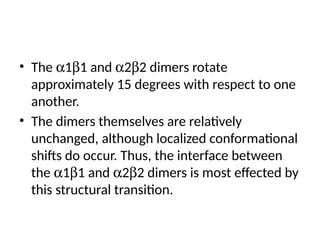
4. Proteolytic activation-
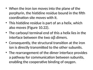
Theenzymes controlled proteolytic activation cycle between active
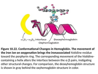
and inactive states.
• A different regulatory motif is used to irreversibly convert an inactive
enzyme into an active one.
• Many enzymes are activated by the hydrolysis of a few or even one
peptide bond in inactive precursors called zymogens or proenzymes.
• This regulatory mechanism generates digestive enzymes such as
chymotrypsin, trypsin, and pepsin.
• Caspases, which are proteolytic enzymes that are the executioners in
programmed cell death, or apoptosis are proteolytically activated
from the procaspase form.
• Blood clotting is due to a remarkable cascade of zymogen activations.
• Active digestive and clotting enzymes are switched off by the
irreversible binding of specific inhibitory proteins that are irresistible
lures to their molecular prey.
8.
• The principlesof allostery is discussed by
examining two proteins:
• the enzyme aspartate transcarbamoylase and
• the oxygen-transporting protein hemoglobin.
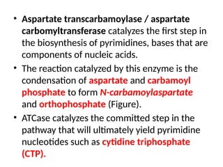
• Aspartate transcarbamoylase/ aspartate
carbomyltransferase catalyzes the first step in
the biosynthesis of pyrimidines, bases that are
components of nucleic acids.
• The reaction catalyzed by this enzyme is the
condensation of aspartate and carbamoyl
phosphate to form N-carbamoylaspartate
and orthophosphate (Figure).
• ATCase catalyzes the committed step in the
pathway that will ultimately yield pyrimidine
nucleotides such as cytidine triphosphate
(CTP).
12.
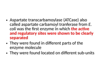
• Aspartate transcarbamoylase(ATCase) also
called aspartate carbamoyl tranferase from E.
coli was the first enzyme in which the active
and regulatory sites were shown to be clearly
separated
• They were found in different parts of the
enzyme molecule
• They were found located on different sub-units
13.
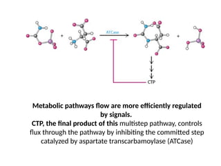
Metabolic pathways floware more efficiently regulated
by signals.
CTP, the final product of this multistep pathway, controls
flux through the pathway by inhibiting the committed step
catalyzed by aspartate transcarbamoylase (ATCase)
14.
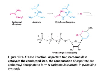
Figure 10.1. ATCaseReaction. Aspartate transcarbamoylase
catalyzes the committed step, the condensation of aspartate and
carbamoyl phosphate to form N-carbamoylaspartate, in pyrimidine
synthesis
15.
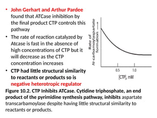
• John Gerhartand Arthur Pardee
found that ATCase inhibition by
the final product CTP controls this
pathway
• The rate of reaction catalyzed by
Atcase is fast in the absence of
high concentrations of CTP but it
will decrease as the CTP
concentration increases
• CTP had little structural similarity
to reactants or products so is
negative heterotropic regulator
Figure 10.2. CTP Inhibits ATCase. Cytidine triphosphate, an end
product of the pyrimidine synthesis pathway, inhibits aspartate
transcarbamoylase despite having little structural similarity to
reactants or products.
16.
• The increasedCTP molecules happends until sufficient
quantities of CTP are accumulated
• This effect of CTP on the enzyme is an example of feed
back or end-product inhibition
• CTP is structurally different from the substrates of the
reaction.
• Due to this it is emphasized that CTP must bind to a site
distinct from the active site where substrate will bind.
Such site are the allosteric sites or regulatory sites.
• So CTP is an example of alloteric inhibitor
• So in ATCase it was noted by several experiments that
catalytic sites and the regulatory sites are on separate
polypeptide chains
17.
ATCase Consists ofSeparable Catalytic and
Regulatory Subunits
• Evidence for distinct regulatory and catalytic
sites
• ATCase was literally separated into regulatory
and catalytic subunits by treatment with a
mercurial compound such as p-hydroxy
meruribenzoate.
• It reacts with sulfhydral groups (Figure)
18.
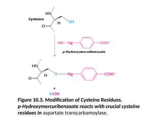
Figure 10.3. Modificationof Cysteine Residues.
p-Hydroxymercuribenzoate reacts with crucial cysteine
residues in aspartate transcarbamoylase.
19.
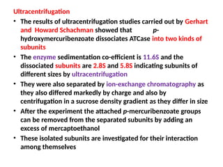
Ultracentrifugation
• The resultsof ultracentrifugation studies carried out by Gerhart
and Howard Schachman showed that p-
hydroxymercuribenzoate dissociates ATCase into two kinds of
subunits
• The enzyme sedimentation co-efficient is 11.6S and the
dissociated subunits are 2.8S and 5.8S indicating subunits of
different sizes by ultracentrifugation
• They were also separated by ion-exchange chromatography as
they also differed markedly by charge and also by
centrifugation in a sucrose density gradient as they differ in size
• After the experiment the attached p-mercuribenzoate groups
can be removed from the separated subunits by adding an
excess of mercaptoethanol
• These isolated subunits are investigated for their interaction
among themselves
20.
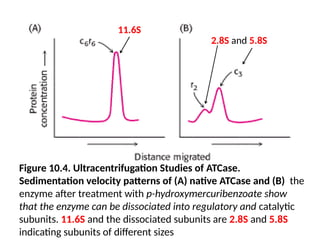
Figure 10.4. UltracentrifugationStudies of ATCase.
Sedimentation velocity patterns of (A) native ATCase and (B) the
enzyme after treatment with p-hydroxymercuribenzoate show
that the enzyme can be dissociated into regulatory and catalytic
subunits. 11.6S and the dissociated subunits are 2.8S and 5.8S
indicating subunits of different sizes
11.6S
2.8S and 5.8S
21.
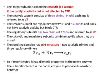
• The largersubunit is called the catalytic (c ) subunit
• It has catalytic activity but is not affected by CTP
• The catalytic subunit consists of three chains (34kda) each and is
referred to as c3
• The smaller subunit are regulatory activity (r) and r subunits and does
not have catalytic activity but binds CTP.
• The regulatory subunits has two chains of 17Kda and referred to as r2
• The catalytic and regulatory subunits combine rapidly when they are
mixed
• The resulting complex has c6r6 structure – two catalytic trimers and
three regulatory dimers
• So if reconstituted it has allosteric properties as the native enzyme
• The subunits interact in the native enzyme to produce its allosteric
behavior
22.
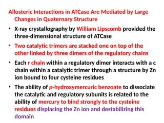
Allosteric Interactions inATCase Are Mediated by Large
Changes in Quaternary Structure
• X-ray crystallography by William Lipscomb provided the
three-dimensional structure of ATCase
• Two catalytic trimers are stacked one on top of the
other linked by three dimers of the regulatory chains
• Each r chain within a regulatory dimer interacts with a c
chain within a catalytic trimer through a structure by Zn
ion bound to four cysteine residues
• The ability of p-hydroxymercuric benzoate to dissociate
the catalytic and regulatory subunits is related to the
ability of mercury to bind strongly to the cysteine
residues displacing the Zn ion and destabilizing this
domain
23.
Figure 10.5. Structureof ATCase. (A) The quaternary structure of
aspartate transcarbamoylase as viewed from the top. The schematic
drawing at the right is a simplified representation of the relationships
between subunits. A single trimer [catalytic (c) chains, shown in orange
and yellow] is visible; in this view, the second trimer is hidden
behind the one visible. (B) A side view of the complex.
24.
Figure 10.5. Structureof ATCase. (B) ) the side view of the
complex of the quaternary structure of aspartate
transcarbamoylase as viewed from the top. The schematic
drawing at the right is a simplified representation of the
relationships between subunits. A single trimer [catalytic (c)
chains, shown in orange and yellow] is visible; in this view, the
second trimer is hidden behind the one visible.
25.
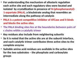
• For understandingthe mechanism of allosteric regulation
each active site and each regulatory sites were located and
isolated by crystallization in presence of N-(phosphonacetyl)-
1-aspartate (PALA), a bisubstrate analog that resembles an
intermediate along the pathway of catalysis
• PALA is a potent competitive inhibitor of ATCase and it binds
and blocks the active sites
• The PALA binding sites lies at the boundaries between pairs of
c chains within a catalytic trimer
• Key residues also include from neighboring subunits
• So it is noted that the active site are at the subunit interface,
and each catalytic trimer contributes three active sites to the
complete enzyme
• Suitable amino acid residues are available in the active sites
for the two substrates – the phosphate and carboxylate
groups
26.
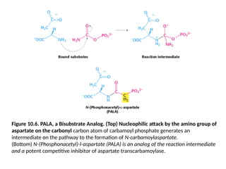
Figure 10.6. PALA,a Bisubstrate Analog. (Top) Nucleophilic attack by the amino group of
aspartate on the carbonyl carbon atom of carbamoyl phosphate generates an
intermediate on the pathway to the formation of N-carbamoylaspartate.
(Bottom) N-(Phosphonacetyl)-l-aspartate (PALA) is an analog of the reaction intermediate
and a potent competitive inhibitor of aspartate transcarbamoylase.
27.
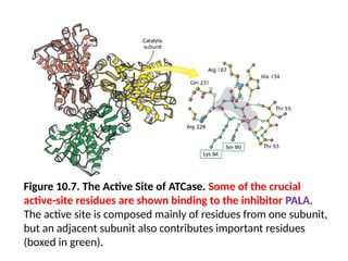
Figure 10.7. TheActive Site of ATCase. Some of the crucial
active-site residues are shown binding to the inhibitor PALA.
The active site is composed mainly of residues from one subunit,
but an adjacent subunit also contributes important residues
(boxed in green).
28.
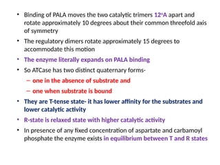
• Binding ofPALA moves the two catalytic trimers 12o
A apart and
rotate approximately 10 degrees about their common threefold axis
of symmetry
• The regulatory dimers rotate approximately 15 degrees to
accommodate this motion
• The enzyme literally expands on PALA binding
• So ATCase has two distinct quaternary forms-
– one in the absence of substrate and
– one when substrate is bound
• They are T-tense state- it has lower affinity for the substrates and
lower catalytic activity
• R-state is relaxed state with higher catalytic activity
• In presence of any fixed concentration of aspartate and carbamoyl
phosphate the enzyme exists in equilibrium between T and R states
29.
Figure 10.8. TheT-to-R State Transition in ATCase. Aspartate
transcarbamoylase exists in two conformations: a compact,
relatively inactive form called the tense (T) state and an expanded
form called the relaxed (R) state. PALA binding stabilizes the R state.
30.
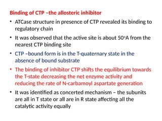
Binding of CTP–the allosteric inhibitor
• ATCase structure in presence of CTP revealed its binding to
regulatory chain
• It was observed that the active site is about 50o
A from the
nearest CTP binding site
• CTP –bound form is in the T-quaternary state in the
absence of bound substrate
• The binding of inhibitor CTP shifts the equilibrium towards
the T-state decreasing the net enzyme activity and
reducing the rate of N-carbamoyl aspartate generation
• It was identified as concerted mechanism – the subunits
are all in T state or all are in R state affecting all the
catalytic activity equally
31.
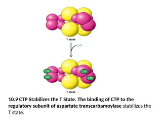
10.9 CTP Stabilizesthe T State. The binding of CTP to the
regulatory subunit of aspartate transcarbamoylase stabilizes the
T state.
32.
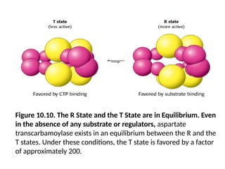
Figure 10.10. TheR State and the T State are in Equilibrium. Even
in the absence of any substrate or regulators, aspartate
transcarbamoylase exists in an equilibrium between the R and the
T states. Under these conditions, the T state is favored by a factor
of approximately 200.
33.
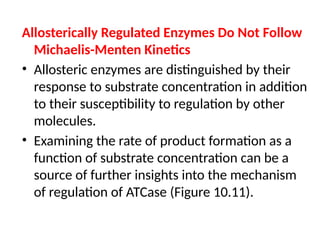
Allosterically Regulated EnzymesDo Not Follow
Michaelis-Menten Kinetics
• Allosteric enzymes are distinguished by their
response to substrate concentration in addition
to their susceptibility to regulation by other
molecules.
• Examining the rate of product formation as a
function of substrate concentration can be a
source of further insights into the mechanism
of regulation of ATCase (Figure 10.11).
34.
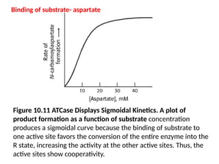
Figure 10.11 ATCaseDisplays Sigmoidal Kinetics. A plot of
product formation as a function of substrate concentration
produces a sigmoidal curve because the binding of substrate to
one active site favors the conversion of the entire enzyme into the
R state, increasing the activity at the other active sites. Thus, the
active sites show cooperativity.
Binding of substrate- aspartate
35.
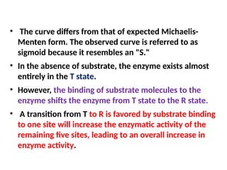
• The curvediffers from that of expected Michaelis-
Menten form. The observed curve is referred to as
sigmoid because it resembles an "S."
• In the absence of substrate, the enzyme exists almost
entirely in the T state.
• However, the binding of substrate molecules to the
enzyme shifts the enzyme from T state to the R state.
• A transition from T to R is favored by substrate binding
to one site will increase the enzymatic activity of the
remaining five sites, leading to an overall increase in
enzyme activity.
36.
• This importantproperty is called cooperativity
because the subunits cooperate with one another.
• If one subunit switches conformation, they all do.
• The sigmoid curve can be pictured as a composite
of two Michaelis-Menten curves, one
corresponding to the T state and the other to the
R state.
• An increase in substrate concentration favors a
transition from the T-state curve to the R state
curve (Figure 10.12).
37.
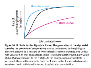
Figure 10.12. Basisfor the Sigmoidal Curve. The generation of the sigmoidal
curve by the property of cooperativity can be understood by imagining an
allosteric enzyme as a mixture of two Michaelis-Menten enzymes, one with a
high value of K m that corresponds to the T state and another with a low value
of K m that corresponds to the R state. As the concentration of substrate is
increased, the equilibrium shifts from the T state to the R state, which results
in a steep rise in activity with respect to substrate concentration.
38.
• The importanceof the changes in quaternary structure in
determining the sigmoidal curve is illustrated nicely by
studies of the isolated catalytic trimer, freed by p-hydroxy
mercuribenzoate treatment.
• The catalytic subunit shows Michaelis-Menten kinetics
with kinetic parameters that are indistinguishable from
those deduced for the R state.
• Thus, the term tense is apt: in the T state, the
regulatory dimers hold the two catalytic trimers
sufficiently close to one another that key loops on their
surfaces collide and interfere with conformational
adjustments necessary for high-affinity substrate
binding and catalysis.
39.
Allosteric Regulators Modulatethe T-to-R Equilibrium
• CTP increases the initial phase of the sigmoidal curve
(Figure 10.13).
• CTP inhibits the activity of ATCase.
• In the presence of CTP, the enzyme becomes less
responsive to the cooperative effects facilitated by
substrate binding; more substrate is required to attain a
given reaction rate.
• Interestingly, ATP, too, is an allosteric effector of ATCase.
• However, the effect of ATP is to increase the reaction
rate at a given aspartate concentration (Figure 10.14).
• At high concentrations of ATP, the kinetic profile shows a
less pronounced sigmoidal behavior.
40.
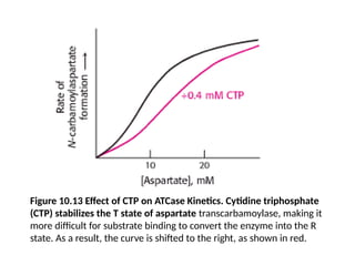
Figure 10.13 Effectof CTP on ATCase Kinetics. Cytidine triphosphate
(CTP) stabilizes the T state of aspartate transcarbamoylase, making it
more difficult for substrate binding to convert the enzyme into the R
state. As a result, the curve is shifted to the right, as shown in red.
41.
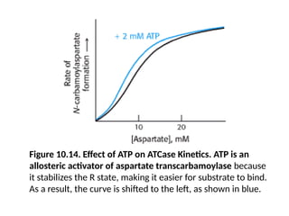
Figure 10.14. Effectof ATP on ATCase Kinetics. ATP is an
allosteric activator of aspartate transcarbamoylase because
it stabilizes the R state, making it easier for substrate to bind.
As a result, the curve is shifted to the left, as shown in blue.
42.
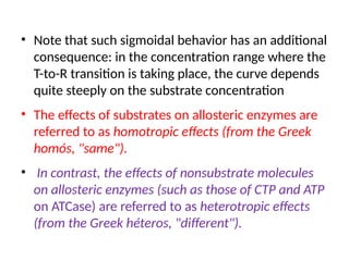
• Note thatsuch sigmoidal behavior has an additional
consequence: in the concentration range where the
T-to-R transition is taking place, the curve depends
quite steeply on the substrate concentration
• The effects of substrates on allosteric enzymes are
referred to as homotropic effects (from the Greek
homós, "same").
• In contrast, the effects of nonsubstrate molecules
on allosteric enzymes (such as those of CTP and ATP
on ATCase) are referred to as heterotropic effects
(from the Greek héteros, "different").
43.
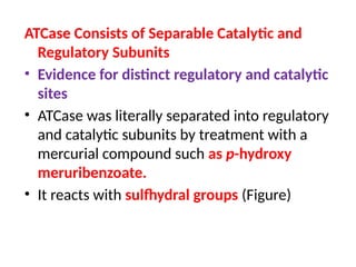
Figure 10.15. QuantitativeDescription of the MWC Model.
Fractional activity, Y, is the fraction of active sites bound to substrate and is
directly proportional to reaction velocity; a is the ratio of [S] to the
dissociation constant of S with the enzyme in the R state; L is the ratio of the
concentration of enzyme in the T state to that in the R state. The binding of
the regulators ATP and CTP to ATCase changes the value of L and thus the
response to substrate concentration.
44.
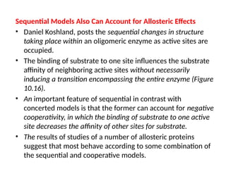
Sequential Models AlsoCan Account for Allosteric Effects
• Daniel Koshland, posts the sequential changes in structure
taking place within an oligomeric enzyme as active sites are
occupied.
• The binding of substrate to one site influences the substrate
affinity of neighboring active sites without necessarily
inducing a transition encompassing the entire enzyme (Figure
10.16).
• An important feature of sequential in contrast with
concerted models is that the former can account for negative
cooperativity, in which the binding of substrate to one active
site decreases the affinity of other sites for substrate.
• The results of studies of a number of allosteric proteins
suggest that most behave according to some combination of
the sequential and cooperative models.
45.
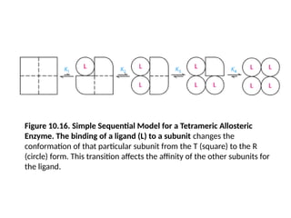
Figure 10.16. SimpleSequential Model for a Tetrameric Allosteric
Enzyme. The binding of a ligand (L) to a subunit changes the
conformation of that particular subunit from the T (square) to the R
(circle) form. This transition affects the affinity of the other subunits for
the ligand.
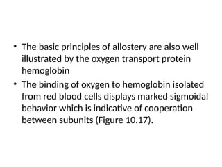
• The basicprinciples of allostery are also well
illustrated by the oxygen transport protein
hemoglobin
• The binding of oxygen to hemoglobin isolated
from red blood cells displays marked sigmoidal
behavior which is indicative of cooperation
between subunits (Figure 10.17).
48.
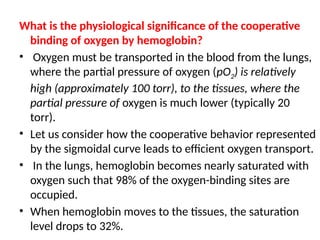
What is thephysiological significance of the cooperative
binding of oxygen by hemoglobin?
• Oxygen must be transported in the blood from the lungs,
where the partial pressure of oxygen (pO2) is relatively
high (approximately 100 torr), to the tissues, where the
partial pressure of oxygen is much lower (typically 20
torr).
• Let us consider how the cooperative behavior represented
by the sigmoidal curve leads to efficient oxygen transport.
• In the lungs, hemoglobin becomes nearly saturated with
oxygen such that 98% of the oxygen-binding sites are
occupied.
• When hemoglobin moves to the tissues, the saturation
level drops to 32%.
49.
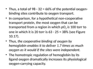
• Thus, atotal of 98 - 32 = 66% of the potential oxygen-
binding sites contribute to oxygen transport.
• In comparison, for a hypothetical non-cooperative
transport protein, the most oxygen that can be
transported from a region in which pO2 is 100 torr to
one in which it is 20 torr is 63 - 25 = 38% (see Figure
10.17).
• Thus, the cooperative binding of oxygen by
hemoglobin enables it to deliver 1.7 times as much
oxygen as it would if the sites were independent.
• The homotropic regulation of hemoglobin by its
ligand oxygen dramatically increases its physiological
oxygen-carrying capacity.
50.
Figure 10.17. CooperativityEnhances Oxygen Delivery by Hemoglobin.
Because of cooperativity between O2- binding sites, hemoglobin delivers more
O2 to tissues than would a noncooperative protein (pO2, partial pressure of
oxygen.)
51.
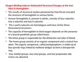
Oxygen Binding InducesSubstantial Structural Changes at the Iron
Sites in Hemoglobin
• The results of structural studies pioneered by Max Perutz revealed
the structure of hemoglobin in various forms.
• Human hemoglobin A, present in adults, consists of four subunits:
two a subunits and two b subunits.
• The a and b subunits are homologous and have similar three-
dimensional structures .
• The capacity of hemoglobin to bind oxygen depends on the presence
of a bound prosthetic group called heme.
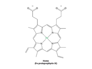
• The heme group is responsible for the distinctive red color of blood.
• The heme group consists of an organic component and a central iron
atom. The organic component, called protoporphyrin, is made up of
four pyrrole rings linked by methene bridges to form a tetrapyrrole
ring.
• Four methyl groups, two vinyl groups, and two propionate side
chains are attached.
53.
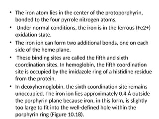
• The ironatom lies in the center of the protoporphyrin,
bonded to the four pyrrole nitrogen atoms.
• Under normal conditions, the iron is in the ferrous (Fe2+)
oxidation state.
• The iron ion can form two additional bonds, one on each
side of the heme plane.
• These binding sites are called the fifth and sixth
coordination sites. In hemoglobin, the fifth coordination
site is occupied by the imidazole ring of a histidine residue
from the protein.
• In deoxyhemoglobin, the sixth coordination site remains
unoccupied. The iron ion lies approximately 0.4 Å outside
the porphyrin plane because iron, in this form, is slightly
too large to fit into the well-defined hole within the
porphyrin ring (Figure 10.18).
54.
Figure 10.18. Positionof Iron in Deoxyhemoglobin. The iron ion
lies slightly outside the plane of the porphyrin in heme.
55.
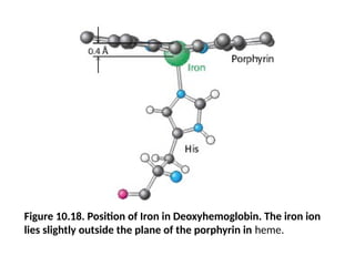
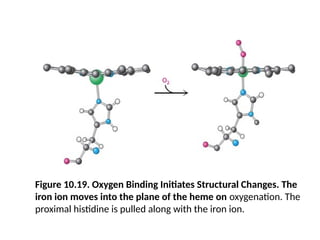
• The bindingof the oxygen molecule at the sixth
coordination site of the iron ion substantially
rearranges the electrons within the iron so that the
ion becomes effectively smaller, allowing it to move
into the plane of the porphyrin (Figure 10.19).
• This change in electronic structure is paralleled by
changes in the magnetic properties of hemoglobin,
which are the basis for functional magnetic
resonance imaging (fMRI).
• Indeed, the structural changes that take place on
oxygen binding were anticipated by Linus Pauling,
based on magnetic measurements in 1936.
56.
Figure 10.19. OxygenBinding Initiates Structural Changes. The
iron ion moves into the plane of the heme on oxygenation. The
proximal histidine is pulled along with the iron ion.
57.
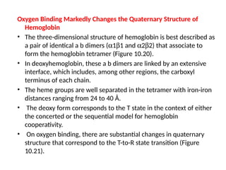
Oxygen Binding MarkedlyChanges the Quaternary Structure of
Hemoglobin
• The three-dimensional structure of hemoglobin is best described as
a pair of identical a b dimers (a1b1 and a2b2) that associate to
form the hemoglobin tetramer (Figure 10.20).
• In deoxyhemoglobin, these a b dimers are linked by an extensive
interface, which includes, among other regions, the carboxyl
terminus of each chain.
• The heme groups are well separated in the tetramer with iron-iron
distances ranging from 24 to 40 Å.
• The deoxy form corresponds to the T state in the context of either
the concerted or the sequential model for hemoglobin
cooperativity.
• On oxygen binding, there are substantial changes in quaternary
structure that correspond to the T-to-R state transition (Figure
10.21).
58.
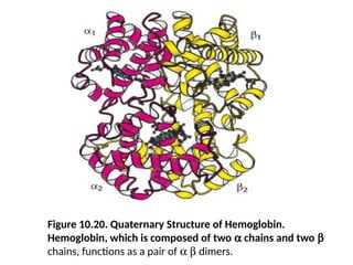
Figure 10.20. QuaternaryStructure of Hemoglobin.
Hemoglobin, which is composed of two a chains and two b
chains, functions as a pair of a b dimers.
59.
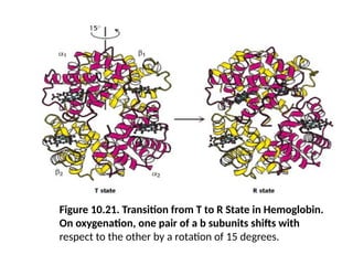
Figure 10.21. Transitionfrom T to R State in Hemoglobin.
On oxygenation, one pair of a b subunits shifts with
respect to the other by a rotation of 15 degrees.
60.
• The a1b1and a2b2 dimers rotate
approximately 15 degrees with respect to one
another.
• The dimers themselves are relatively
unchanged, although localized conformational
shifts do occur. Thus, the interface between
the a1b1 and a2b2 dimers is most effected by
this structural transition.
61.
• When theiron ion moves into the plane of the
porphyrin, the histidine residue bound in the fifth
coordination site moves with it.
• This histidine residue is part of an a helix, which
also moves (Figure 10.22).
• The carboxyl terminal end of this a helix lies in the
interface between the two ab dimers.
• Consequently, the structural transition at the iron
ion is directly transmitted to the other subunits.
• The rearrangement of the dimer interface provides
a pathway for communication between subunits,
enabling the cooperative binding of oxygen.
62.
Figure 10.22. ConformationalChanges in Hemoglobin. The movement of
the iron ion on oxygenation brings the ironassociated histidine residue
toward the porphyrin ring. The corresponding movement of the histidine-
containing a helix alters the interface between the a b pairs, instigating
other structural changes. For comparison, the deoxyhemoglobin structure
is shown in gray behind the oxyhemoglobin structure in color.
63.
• A combinedmodel of concerted and sequential is required to describe
binding of oxygen to haemoglobin.
• Hemoglobin behavior is concerted in that hemoglobin with three sites
occupied by oxygen is in the quaternary structure associated with the
R state.
• The remaining open binding site has an affinity for oxygen more than
20-fold as great as that of fully deoxygenated hemoglobin binding its
first oxygen.
• However, the behavior is not fully concerted, because hemoglobin
with oxygen bound to only one of four sites remains in the T-state
quaternary structure.
• Yet, this molecule binds oxygen 3 times as strongly as does fully
deoxygenated hemoglobin, an observation consistent only with a
sequential model.
• These results highlight the fact that the concerted and sequential
models represent idealized limiting cases, which real systems may
approach but rarely attain.
64.
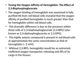
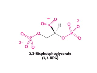
• Tuning theOxygen Affinity of Hemoglobin: The Effect of
2,3-Bisphosphoglycerate
• The oxygen binding of hemoglobin was examined in fully
purified Hb from red blood cells revealed that the oxygen
affinity of purified hemoglobin is much greater than that
for hemoglobin within red blood cells.
• This dramatic difference is due to the presence within
these cells of 2,3-bisphosphoglycerate (2,3-BPG) (also
known as 2,3-diphosphoglycerate or 2,3-DPG).
• This highly anionic compound is present in red blood cells
at approximately the same concentration as that of
hemoglobin (~ 2 mM).
• Without 2,3-BPG, hemoglobin would be an extremely
inefficient oxygen transporter, releasing only 8% of its
cargo in the tissues.
66.
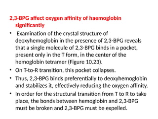
2,3-BPG affect oxygenaffinity of haemoglobin
significantly
• Examination of the crystal structure of
deoxyhemoglobin in the presence of 2,3-BPG reveals
that a single molecule of 2,3-BPG binds in a pocket,
present only in the T form, in the center of the
hemoglobin tetramer (Figure 10.23).
• On T-to-R transition, this pocket collapses.
• Thus, 2,3-BPG binds preferentially to deoxyhemoglobin
and stabilizes it, effectively reducing the oxygen affinity.
• In order for the structural transition from T to R to take
place, the bonds between hemoglobin and 2,3-BPG
must be broken and 2,3-BPG must be expelled.
67.
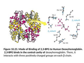
Figure 10.23. Modeof Binding of 2,3-BPG to Human Deoxyhemoglobin.
2,3-BPG binds in the central cavity of deoxyhemoglobin. There, it
interacts with three positively charged groups on each b chain.
68.
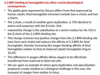
2,3-BPG binding tohemoglobin has other crucial physiological
consequences.
• The globin gene expressed by fetuses differs from that expressed by
human adults; fetal hemoglobin tetramers include two a chains and two
g chains.
• The g chain, a result of another gene duplication, is 72% identical in
amino acid sequence with the b chain. One
• noteworthy change is the substitution of a serine residue for His 143 in
the b chain of the 2,3-BPG-binding site.
• This change removes two positive charges from the 2,3-BPG-binding site
(one from each chain) and reduces the affinity of 2,3-BPG for fetal
hemoglobin, thereby increasing the oxygen-binding affinity of fetal
hemoglobin relative to that of maternal (adult) hemoglobin (Figure
10.24).
• This difference in oxygen affinity allows oxygen to be effectively
transferred from maternal to fetal red cells.
• We see again an example of where gene duplication and specialization
produced a ready solution to a biological challenge in this case, the
transport of oxygen from mother to fetus.
69.
Figure 10.24. OxygenAffinity of Fetal Red Blood Cells. Fetal red
blood cells have a higher oxygen affinity than that of maternal red
blood cells because fetal hemoglobin does not bind 2,3-BPG as well as
maternal hemoglobin does.
70.
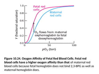
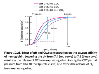
The Bohr Effect:Hydrogen Ions and Carbon Dioxide Promote
the Release of Oxygen
• Rapidly metabolizing tissues, such as contracting muscle, have
a high need for oxygen and generate large amounts of
hydrogen ions and carbon dioxide as well
• Both of these species are heterotropic effectors of hemoglobin
that enhance oxygen release.
• The oxygen affinity of hemoglobin decreases as pH decreases
from the value of 7.4 found in the lungs (Figure 10.25).
• Thus, as hemoglobin moves into a region of low pH, its
tendency to release oxygen increases.
• For example, transport from the lungs, with pH 7.4 and an
oxygen partial pressure of 100 torr, to active muscle, with a pH
of 7.2 and an oxygen partial pressure of 20 torr, results in a
release of oxygen amounting to 77% of total carrying capacity.
71.
Figure 10.25. Effectof pH and CO2 concentration on the oxygen affinity
of hemoglobin. Lowering the pH from 7.4 (red curve) to 7.2 (blue curve)
results in the release of O2 from oxyhemoglobin. Raising the CO2 partial
pressure from 0 to 40 torr (purple curve) also favors the release of O2
from oxyhemoglobin.
72.
• Recall thatonly 66% of the oxygen would be released
in the absence of any change in pH.
• In addition, hemoglobin responds to carbon dioxide
with a decrease in oxygen affinity, thus facilitating the
release of oxygen in tissues with a high carbon dioxide
concentration. In the presence of carbon dioxide at a
partial pressure of 40 torr, the amount of oxygen
released approaches 90% of the maximum carrying
capacity.
• Thus, the heterotropic regulation of hemoglobin by
hydrogen ions and carbon dioxide further increases
the oxygen-transporting efficiency of this
magnificent allosteric protein.
73.
• The regulationof oxygen binding by hydrogen ions and
carbon dioxide is called the Bohr effect after Christian
Bohr, who described this phenomenon in 1904.
• The results of structural and chemical studies have
revealed much about the chemical basis of the Bohr
effect.
• At least two sets of chemical groups are responsible for
the effect of protons: the amino termini and the side
chains of histidines b146 and a122, which have pKa
values near pH 7.
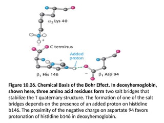
• Consider histidine b146. In deoxyhemoglobin, the
terminal carboxylate group of b146 forms a salt bridge
with a lysine residue in the a subunit of the other ab
dimer.
74.
• This interactionlocks the side chain of histidine b146 in a
position where it can participate in a salt bridge with
negatively charged aspartate 94 in the same chain, provided
that the imidazole group of the histidine residue is
protonated (Figure 10.26).
• At high pH, the side chain of histidine b146 is not
protonated and the salt bridge does not form.
• As the pH drops, however, the side chain of histidine b146
becomes protonated, the salt bridge with aspartate b94
forms, and the quaternary structure characteristic of
deoxyhemoglobin is stabilized, leading to a greater tendency
for oxygen to be released at actively metabolizing tissues.
• No significant change takes place in oxyhemoglobin over the
same pH range.
75.
Figure 10.26. ChemicalBasis of the Bohr Effect. In deoxyhemoglobin,
shown here, three amino acid residues form two salt bridges that
stabilize the T quaternary structure. The formation of one of the salt
bridges depends on the presence of an added proton on histidine
b146. The proximity of the negative charge on aspartate 94 favors
protonation of histidine b146 in deoxyhemoglobin.
76.
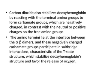
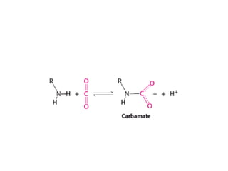
• Carbon dioxidealso stabilizes deoxyhemoglobin
by reacting with the terminal amino groups to
form carbamate groups, which are negatively
charged, in contrast with the neutral or positive
charges on the free amino groups.
• The amino termini lie at the interface between
the a b dimers, and these negatively charged
carbamate groups participate in saltbridge
interactions, characteristic of the T-state
structure, which stabilize deoxyhemoglobin's
structure and favor the release of oxygen.
77.
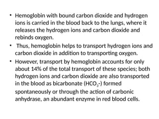
• Hemoglobin withbound carbon dioxide and hydrogen
ions is carried in the blood back to the lungs, where it
releases the hydrogen ions and carbon dioxide and
rebinds oxygen.
• Thus, hemoglobin helps to transport hydrogen ions and
carbon dioxide in addition to transporting oxygen.
• However, transport by hemoglobin accounts for only
about 14% of the total transport of these species; both
hydrogen ions and carbon dioxide are also transported
in the blood as bicarbonate (HCO3-) formed
spontaneously or through the action of carbonic
anhydrase, an abundant enzyme in red blood cells.
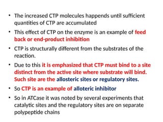
![Figure 10.5. Structure of ATCase. (A) The quaternary structure of
aspartate transcarbamoylase as viewed from the top. The schematic
drawing at the right is a simplified representation of the relationships
between subunits. A single trimer [catalytic (c) chains, shown in orange
and yellow] is visible; in this view, the second trimer is hidden
behind the one visible. (B) A side view of the complex.](https://image.slidesharecdn.com/unit4class2sigmoidalkineticsatcase-250510051701-670837ed/85/unit-4-class-2-Sigmoidal-kinetics-ATCase-pptx-23-320.jpg)
![Figure 10.5. Structure of ATCase. (B) ) the side view of the
complex of the quaternary structure of aspartate
transcarbamoylase as viewed from the top. The schematic
drawing at the right is a simplified representation of the
relationships between subunits. A single trimer [catalytic (c)
chains, shown in orange and yellow] is visible; in this view, the
second trimer is hidden behind the one visible.](https://image.slidesharecdn.com/unit4class2sigmoidalkineticsatcase-250510051701-670837ed/85/unit-4-class-2-Sigmoidal-kinetics-ATCase-pptx-24-320.jpg)
![Figure 10.15. Quantitative Description of the MWC Model.
Fractional activity, Y, is the fraction of active sites bound to substrate and is
directly proportional to reaction velocity; a is the ratio of [S] to the
dissociation constant of S with the enzyme in the R state; L is the ratio of the
concentration of enzyme in the T state to that in the R state. The binding of
the regulators ATP and CTP to ATCase changes the value of L and thus the
response to substrate concentration.](https://image.slidesharecdn.com/unit4class2sigmoidalkineticsatcase-250510051701-670837ed/85/unit-4-class-2-Sigmoidal-kinetics-ATCase-pptx-43-320.jpg)