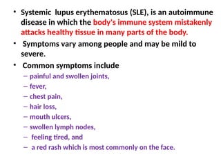
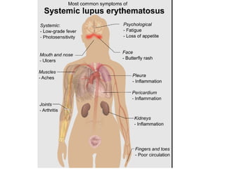
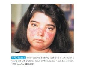
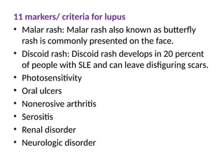
Systemic Lupus Erythomatosus
It is one of the best examples of autoimmune diseases
• Systemic lupus erythematosus (SLE), is an autoimmune
disease in which the body's immune system mistakenly
attacks healthy tissue in many parts of the body.
• Symptoms vary among people and may be mild to
severe.
• Common symptoms include
– painful and swollen joints,
– fever,
– chest pain,
– hair loss,
– mouth ulcers,
– swollen lymph nodes,
– feeling tired, and
– a red rash which is most commonly on the face.
• It typically appears in women between 20 and 40 years of
age;
• The ratio of female to male patients is 10:1.
• Lupus is more frequent in African-American and Hispanic
women than in Caucasians, although it is not known why
this is so.
• Affected individuals may produce autoantibodies to a vast
array of tissue antigens, such as DNA, histones, RBCs,
platelets, leukocytes, and clotting factors; interaction of
these auto-antibodies with their specific antigens produces
various symptoms.
• Auto-antibody specific for RBCs and platelets, for example,
can lead to complement-mediated lysis, resulting in
hemolytic anemia and thrombocytopenia, respectively.
• When immune complexes of auto-antibodies
with various nuclear antigens are deposited
along the walls of small blood vessels, a type
III hypersensitive reaction develops.
• The complexes activate the complement
system and generate membrane-attack
complexes and complement split products
that damage the wall of the blood vessel,
resulting in vasculitis and glomerulonephritis.
• Excessive complement activation in patients with
severe SLE produces elevated serum levels of the
complement split products C3a and C5a, which may be
three to four times higher than normal.
• C5a induces increased expression of the type 3
complement receptor (CR3) on neutrophils, facilitating
neutrophil aggregation and attachment to the vascular
endothelium.
• As neutrophils attach to small blood vessels, the
number of circulating neutrophils declines
(neutropenia) and various occlusions of the small blood
vessels develop (vasculitis).
• These occlusions can lead to widespread tissue
damage.
11 markers/ criteria for lupus
• Malar rash: Malar rash also known as butterfly
rash is commonly presented on the face.
• Discoid rash: Discoid rash develops in 20 percent
of people with SLE and can leave disfiguring scars.
• Photosensitivity
• Oral ulcers
• Nonerosive arthritis
• Serositis
• Renal disorder
• Neurologic disorder
• Laboratory diagnosis of SLE focuses on the
characteristic antinuclear antibodies, which
are directed against double stranded or
single-stranded DNA, nucleoprotein,
histones, and nucleolar RNA.
• Indirect immunofluorescent staining with
serum from SLE patients produces various
characteristic nucleus-staining patterns.
• Diagnosis
• Complete blood count. This test measures the
number of red blood cells, white blood cells and
platelets as well as the amount of hemoglobin, a
protein in red blood cells. ...
• Erythrocyte sedimentation rate. ...
• Kidney and liver assessment. ...
• Urinalysis. ...
• Antinuclear antibody (ANA) test.
• Tests performed
• The doctor may refer the patient to a
dermatologist for a skin biopsy, the analysis of
a small sample of skin.
• He or she may perform a “lupus band test,”
which checks for lupus-related antibodies.
Treatments for more severe SLE may include:
• High-dose corticosteroids.
• Immunosuppressive medicines - the medicines
suppress the immune system
• Medicines most commonly used include
mycophenolate,
• azathioprine,
• cyclophosphamide, and
• valcosporin

class 20 unit 4Systemic Lupus Erythomatosus.pptx

  • 1.
    Systemic Lupus Erythomatosus Itis one of the best examples of autoimmune diseases
  • 2.
    • Systemic lupuserythematosus (SLE), is an autoimmune disease in which the body's immune system mistakenly attacks healthy tissue in many parts of the body. • Symptoms vary among people and may be mild to severe. • Common symptoms include – painful and swollen joints, – fever, – chest pain, – hair loss, – mouth ulcers, – swollen lymph nodes, – feeling tired, and – a red rash which is most commonly on the face.
  • 5.
    • It typicallyappears in women between 20 and 40 years of age; • The ratio of female to male patients is 10:1. • Lupus is more frequent in African-American and Hispanic women than in Caucasians, although it is not known why this is so. • Affected individuals may produce autoantibodies to a vast array of tissue antigens, such as DNA, histones, RBCs, platelets, leukocytes, and clotting factors; interaction of these auto-antibodies with their specific antigens produces various symptoms. • Auto-antibody specific for RBCs and platelets, for example, can lead to complement-mediated lysis, resulting in hemolytic anemia and thrombocytopenia, respectively.
  • 6.
    • When immunecomplexes of auto-antibodies with various nuclear antigens are deposited along the walls of small blood vessels, a type III hypersensitive reaction develops. • The complexes activate the complement system and generate membrane-attack complexes and complement split products that damage the wall of the blood vessel, resulting in vasculitis and glomerulonephritis.
  • 7.
    • Excessive complementactivation in patients with severe SLE produces elevated serum levels of the complement split products C3a and C5a, which may be three to four times higher than normal. • C5a induces increased expression of the type 3 complement receptor (CR3) on neutrophils, facilitating neutrophil aggregation and attachment to the vascular endothelium. • As neutrophils attach to small blood vessels, the number of circulating neutrophils declines (neutropenia) and various occlusions of the small blood vessels develop (vasculitis). • These occlusions can lead to widespread tissue damage.
  • 8.
    11 markers/ criteriafor lupus • Malar rash: Malar rash also known as butterfly rash is commonly presented on the face. • Discoid rash: Discoid rash develops in 20 percent of people with SLE and can leave disfiguring scars. • Photosensitivity • Oral ulcers • Nonerosive arthritis • Serositis • Renal disorder • Neurologic disorder
  • 9.
    • Laboratory diagnosisof SLE focuses on the characteristic antinuclear antibodies, which are directed against double stranded or single-stranded DNA, nucleoprotein, histones, and nucleolar RNA. • Indirect immunofluorescent staining with serum from SLE patients produces various characteristic nucleus-staining patterns.
  • 10.
    • Diagnosis • Completeblood count. This test measures the number of red blood cells, white blood cells and platelets as well as the amount of hemoglobin, a protein in red blood cells. ... • Erythrocyte sedimentation rate. ... • Kidney and liver assessment. ... • Urinalysis. ... • Antinuclear antibody (ANA) test.
  • 11.
    • Tests performed •The doctor may refer the patient to a dermatologist for a skin biopsy, the analysis of a small sample of skin. • He or she may perform a “lupus band test,” which checks for lupus-related antibodies.
  • 12.
    Treatments for moresevere SLE may include: • High-dose corticosteroids. • Immunosuppressive medicines - the medicines suppress the immune system • Medicines most commonly used include mycophenolate, • azathioprine, • cyclophosphamide, and • valcosporin