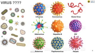
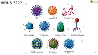
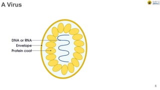
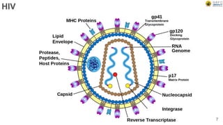
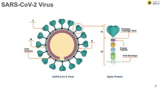
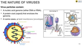
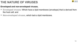
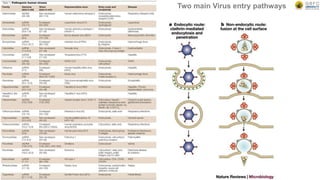
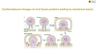
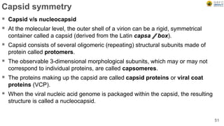
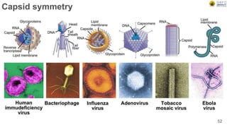
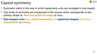
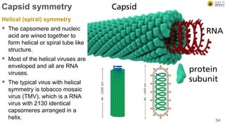
SlideShare a Scribd company logo
By – Dr. Mafatlal M. Kher
Unit 1: Nature and Properties of Viruses
Date & Time : Monday, 27 September 2021
Semester : V
Program : B.Sc. Biotechnology
School : School of Science
Subject code : BSBO502 (Unit I)
1
VIRUS ????
2
It’s just a piece of bad news wrapped up in protein.
— Nobel laureate Peter Medawar (1915–1987) describing a virus
VIRUS ????
4
VIRUS ????
5
A Virus
6
HIV
7
SARS-CoV-2 Virus
8
Koch’s postulates
 The agent must be present in every case of the disease.
 The agent must be isolated from the host and grown in vitro.
 The disease must be reproduced when a pure culture of the agent is inoculated
into a healthy susceptible host.
 The same agent must be recovered once again from the experimentally infected
host.
9
Virus
 Martinus Beijerinck is often called the
Father of Virology.
10
Martinus W. Beijerinck
Discovery of the first virus?
 In 1892, Dmitri Ivanovsky to show that sap from a
diseased tobacco plant remained infectious to
healthy tobacco plants despite having been
filtered.
 Martinus Beijerinck called the filtered, infectious
substance a "virus" and this discovery is
considered to be the beginning of virology.
11
Dmitri Ivanovsky
DEFINITION OF VIRUSES
 Viruses are submicroscopic, obligate intracellular parasites. Most are too small to
be seen by optical microscopes, and they have no choice but to replicate inside
host cells.
12
DEFINITION OF VIRUSES
How viruses are UNIQUE?
 Virus particles are produced from the assembly of preformed components, while
other biological agents grow from an increase in the integrated sum of their
components and reproduce by division.
 Virus particles (virions) do not grow or undergo division.
 Viruses lack the genetic information that encodes the tools necessary for the
generation of metabolic energy or for protein synthesis (ribosomes).
13
THE NATURE OF VIRUSES
14
Virus particles contain:
 A nucleic acid genome (either DNA or RNA).
 A protein coat (capsid) that encloses the
genome.
 In some cases, a lipid membrane (envelope)
THE NATURE OF VIRUSES
Enveloped and non-enveloped viruses.
 Enveloped viruses: Which have a lipid membrane (envelope) that is derived from
the host cell; and
 Non-enveloped viruses, which lack a lipid membrane.
15
Two main Virus entry pathways
Conformational changes of viral fusion proteins leading to membrane fusion.
THE NATURE OF VIRUSES
 The infectious virus particle is called a virion.
 Virus particles are very small: between 20 and 500 nanometers (nm) in
diameter.
 Viruses are obligatory intracellular parasites. (Only demonstrate life inside the
host cell).
 Viruses multiply inside cells by expressing and replicating their genomes.
18
THE NATURE OF VIRUSES
Viruses need the following machinery provided by cells:
 Enzyme systems that synthesize amino acids, nucleotides, carbohydrates, and
lipids.
 Enzyme systems that generate useable chemical energy in the form of ATP.
 Ribosomes, tRNAs, and enzymes used in protein synthesis.
 Membranes that concentrate cellular macromolecules, small molecules, and ions.
19
THE NATURE OF VIRUSES
Viruses consist of a nucleic acid
genome packaged in a protein coat.
 Viruses are the smallest and simplest
forms of life on Earth.
 They consist of a set of nucleic acid
genes enclosed in a protein coat,
called a capsid, which in some cases
is surrounded by or encloses a lipid
membrane, called an envelope.
 The viral genome encodes proteins
that enable it to replicate and to be
transmitted from one cell to another,
and from one organism to another.
 The complete, infectious virus particle
is called a virion.
20
Figure: Schematic diagram of virus particles. Illustrated
are the two most common capsid morphologies: a roughly
spherical shell (left) and a tubular rod (right). Some virus
particles have an envelope (left) and some do not (right).
Nucleic acid genomes are shown as black curved lines,
capsid proteins as green spheres, and envelope proteins
as orange knobbed spikes. Source: Acheson (2011)
THE NATURE OF VIRUSES
Viruses are dependent on living cells for their replication
 Viruses can replicate only within living cells because they are obligatory
intracellular parasites.
21
THE NATURE OF VIRUSES
Viruses are dependent on living cells for their REPLICATION: Viruses depend on
cells for their replication because they lack the following basic elements required for
growth and replication, which are present in all living cells:
 Enzyme systems that produce the basic chemical building blocks of life:
nucleotides, amino acids, carbohydrates, and lipids.
 Enzyme systems that generate useable chemical energy, usually in the form of
adenosine triphosphate (ATP), by photosynthesis or by metabolism of sugars and
other small molecules.
 Ribosomes, transfer RNAs, and the associated enzymatic machinery that directs
protein synthesis.
 Membranes that localize and concentrate in a defined space these cellular
macromolecules, the small organic molecules involved in growth and metabolism,
and specific inorganic ions.
22
VIRUS REPLICATION
 The virion binds to cell surface receptors.
 The virion or viral genome enters the cell;
the viral genome is uncoated.
 Early viral genes are expressed.
 Early viral proteins direct replication of the
viral genome.
 Late viral genes are expressed from newly
replicated viral genomes.
 Late viral proteins package genomes and
assemble progeny virus particles.
 Virions are released from the host cell
23
THE NATURE OF VIRUSES
Virus particles breakdown and release their genomes inside the cell
 Viruses are not the only obligatory intracellular parasites known.
 A number of small unicellular organisms including chlamydiae and rickettsiae,
certain other bacterial species, and some protozoa can multiply only inside other
host cells.
24
THE NATURE OF VIRUSES
Virus particles break down and release their genomes inside the cell.
Viruses replicate by a pathway that is very different from the mode of
replication of other intracellular parasites:
 Virus replication begins with at least partial disintegration of the virus particle, and
release (uncoating) of the viral genome within the cell.
 Once uncoated, the viral genome can be used as a template for synthesis of
messenger RNAs, which in turn synthesize viral proteins using the enzyme
systems, energy, ribosomes, and molecular building blocks that are present in the
cell.
 These viral proteins then direct replication of the viral genome.
 Viral structural proteins encapsidate the newly replicated genomes to form
progeny virus particles.
25
THE NATURE OF VIRUSES
Virus particles break down and release their genomes inside the cell.
Multiplication of other intracellular parasites in host cell:
 Unicellular organisms that replicate inside other cells invariably remain intact and
retain their genomes within their own cellular membranes.
 They replicate not by disintegration and reassembly, but by growth and division
into daughter cells.
 Such cellular parasites always contain their own ribosomes and protein synthetic
machinery, and their genes code for enzymes that direct many of the basic
metabolic pathways.
26
THE NATURE OF VIRUSES
Virus genomes are either RNA or DNA, but not both
 All viruses contain genomes made of one and only one type of nucleic acid.
 The smallest known viruses are 20 nanometers1 (nm) in diameter; their genomes
contain fewer than 2000 nucleotides, and they code for as few as 2 proteins. The
largest known viruses are some 500 nm in diameter; their genomes are as large
as 1.2 million nucleotides, and they code for over 1200 proteins.
 Depending on the virus, the genome can be either RNA or DNA, and it can be
either single-stranded or double-stranded.
 Some viral genomes are circular and others are linear
27
THE NATURE OF VIRUSES
Virus genomes are either RNA or DNA, but not both
 Viruses are the only known forms of life that can have genomes made of RNA.
 All cellular organisms store the information required to sustain life, to grow, and to
reproduce exclusively in DNA molecules, and all RNA molecules in these
organisms are transcribed from DNA sequences.
 RNA-containing viruses are therefore unique, and they face two related
problems as a result of their RNA genomes:
They must synthesize messenger RNAs from an RNA template, and
They must replicate their genome RNA.
 Most RNA viruses encode their own RNA-dependent RNA polymerases to
completed both these functions.
28
WHY STUDY VIRUSES?
 Viruses are important disease-causing agents.
 Probably all different forms of life can be infected by viruses.
 Viruses can transfer genes between organisms.
 Viruses are important players in the regulation of the Earth’s ecology.
 Viruses can be engineered to prevent and cure disease.
 Study of viruses reveals basic mechanisms of gene expression, cell physiology,
and intracellular signaling pathways.
29
WHY STUDY VIRUSES?
 Smallpox, influenza, yellow fever,
COVID-19 and AIDS (acquired
immunodeficiency syndrome).
 Viruses are responsible for many
cases of human encephalitis,
meningitis, pneumonia, hepatitis,
and cervical cancer, as well as warts
and the common cold.
 Viruses causing respiratory
infections, gastroenteritis, and
diarrhea in young children lead to
millions of deaths each year in less-
developed countries
30
Viruses are important disease-causing agents
WHY STUDY VIRUSES?
Viruses can infect all forms of life
 Viruses also infect animals, plants, and insects.
 Outbreaks of virus diseases in domesticated animals can lead to destruction of
thousands or millions of animals to avoid even more widespread epidemics.
 These diseases include avian influenza; foot-and-mouth disease of cattle;
infectious gastroenteritis and bronchitis in pigs, cattle, and chickens; sheep lung
tumors caused by a retrovirus; canine distemper; and feline immunodeficiency
disease.
 Virus diseases affecting domesticated plants such as potatoes, tomatoes, tobacco,
coconut trees, and citrus trees are common and widespread.
 Insect viruses that kill silkworms, used for centuries in Asia and Europe to produce
silk, have plagued that industry over the ages.
 Viruses can also infect and kill bacteria, archaea, algae, fungi, and protozoa. 31
WHY STUDY VIRUSES?
Viruses are the most abundant form of life on Earth
 Recent studies of soil and seawater have revealed
that bacterial viruses, also called bacteriophages, are
much more numerous than previously imagined.
 There are 10–50 million bacteriophages on average
per mL of seawater, and even more in many soils.
 Given the enormous volume of the oceans, scientists
have calculated that there may be as many as 1031
bacteriophages in the world.
 This is about 10-fold greater than the estimated
number of bacteria. In terms of mass, this many
phages would weigh about 100 million tons, or the
equivalent of 1 million blue whales (the largest animal
on Earth).
 More astonishing, these 1031 phages, if lined up
head-to-tail, would stretch some 200 million light
years into space—that is, far into the universe
beyond many of our known neighboring galaxies. 32
Scientists estimate that there are approximately 1031
tailed bacteriophages on Earth. Each phage
measures approximately 200 nm (0.2 µm) in length
from top of head to base of tail. Aligned head to tail,
these phages would therefore cover the following
distance:
1031 × 0.2 µm = 0.2 × 1025 meters = 2 × 1024 meters
= 2 × 10²¹ kilometers.
Because 1 light year (the distance travelled by light
in one year) =1013 kilometers,
2 × 10²¹ kilometers = 2 × 10²¹ /1013 light years
= 2 × 10⁸ light years (200 million light years).
Note that our Milky Way galaxy measures
approximately 100,000 light years edge to edge, and
the furthest visible galaxies in the universe are
approximately 10 billion (10 × 109) light years distant.
Phages lined up through the universe
WHY STUDY VIRUSES?
Role of virus in Ecology
 More important is the ecological role played by bacteriophages and viruses that
infect unicellular eukaryotic organisms such as algae and cyanobacteria.
 From 95 to 98% of the biomass in the oceans is microbial (the remaining 2–5%
being made up of all other forms of life, including fish, marine invertebrates,
marine mammals, birds, and plants), and roughly half of the oxygen in the Earth’s
atmosphere is generated by photosynthetic activity of marine microbes.
 It has been estimated that 20% of the microbes in the Earth’s oceans are
destroyed each day by virus infections.
 Therefore, these viruses play a major role in the carbon and oxygen cycles that
regulate our atmosphere and help feed the world’s population.
33
WHY STUDY VIRUSES?
Role of virus in Ecology
 More important is the ecological role played by bacteriophages and viruses that
infect unicellular eukaryotic organisms such as algae and cyanobacteria.
 From 95 to 98% of the biomass in the oceans is microbial (the remaining 2–5%
being made up of all other forms of life, including fish, marine invertebrates,
marine mammals, birds, and plants), and roughly half of the oxygen in the Earth’s
atmosphere is generated by photosynthetic activity of marine microbes.
 It has been estimated that 20% of the microbes in the Earth’s oceans are
destroyed each day by virus infections.
 Therefore, these viruses play a major role in the carbon and oxygen cycles that
regulate our atmosphere and help feed the world’s population.
34
WHY STUDY VIRUSES?
Some viruses are useful
 Sources of enzymes. A number of enzymes used in molecular biology are virus enzymes.
Examples include reverse transcriptases from retroviruses and RNA polymerases from
phages.
 Pesticides. Some insect pests are control led with baculoviruses, and myxoma virus has
been used to control rabbits.
 Anti-bacterial agents. In the mid-twentieth century phages were used to treat some
bacterial infections in humans. Interest waned with the discovery of antibiotics, but has
been renewed with the emergence of anti biotic-resistant strains of bacteria.
 Anti-cancer agents. Genetically modified strains of viruses, such as herpes simplex virus
and vaccinia virus, are being investigated for treatment of cancers. These strains have
been modified so that they are able to infect and destroy specific tumor cells, but are
unable to infect normal cells.
 Gene vectors for protein production. Viruses, such as certain baculoviruses and
adenoviruses, are used as vectors to take genes into an i mal cells growing in culture.
This technology is used to make cells produce useful proteins, such as vaccine
components. Some genetically modified cells are used for mass production of proteins
35
WHY STUDY VIRUSES?
The study of viruses has led to numerous discoveries in molecular and cell
biology
 Because viruses replicate within cells but express a limited number of viral genes,
they are ideal tools for understanding the biology of cellular processes.
 Research on animal, insect, and plant viruses has shed light on the functioning of
these organisms, their diseases, and molecular mechanisms of replication, cell
division, and signaling pathways.
 The intensive study of bacteriophages led to discovery of some of the
fundamental principles of molecular biology and genetics.
36
WHY STUDY VIRUSES?
The study of viruses has led to numerous discoveries in molecular and cell
biology: Common examples
 Study of gene expression in small DNA viruses led to the identification of
promoters for eukaryotic RNA polymerases.
 Research on the replication of bacteriophage and animal virus DNAs laid the
foundations for understanding the enzymes involved in cellular DNA replication.
 RNA splicing in eukaryotic cells was first discovered by studying messenger RNAs
of DNA viruses.
 Study of cancer-producing viruses led to the isolation of numerous cellular
oncogenes and the understanding that cancer is caused by their mutation or
unregulated expression.
37
WHY STUDY VIRUSES?
The study of viruses has led to numerous discoveries in molecular and cell
biology: Common examples
 A famous experiment carried by Alfred Hershey and Martha Chase, and published
in 1952, used phage T2 and E. coli to provi1de strong1 evidence that genetic
material is composed of DNA.
 The first enhancers to be characterized were in genes of simian virus 40 (SV40).
 Introns were discovered during studies of adenovirus transcription.
 The first internal ribosome entry site to be discovered was found in the RNA of
poliovirus
38
WHY STUDY VIRUSES?: Case studies
Evidence that DNA is the genetic material of bacteriophage T2. The Hershey-
Chase experiment
 Bacteriophage T2 was grown in E. coli in the presence of 35S (as sulphate) to label
the protein moiety, or 32P (as phosphate) to mainly label the nucleic acid. Purified,
labelled phages were allowed to attach to sensitive host cells and then given time
for the infection to commence.
 The phages, still on the outside of the cell, were then subjected to the shearing
forces of a Waring blender.Such treatment removes any phage components
attached to the outside of the cell but does not affect cell viability. Moreover, the
cells are still able to produce infectious progeny virus.
 When the cells were separated from the medium, it was observed that 75% of the
35S (i.e. phage protein) had been removed from the cells by blending but only 15%
of the 32P (i.e. phage nucleic acid) had been removed. Thus, after infection, the
bulk of the phage protein appeared to have no further function and this suggested
(but does not prove – that had to await more rigorous experiments with purified
nucleic acid genomes) that the nucleic acid is the carrier of viral heredity.
 The transfer of the phage nucleic acid from its protein coat to the bacterial cell upon
infection also accounts for the existence of the eclipse period during the early
stages of intracellular virus development, since the nucleic acid on its own cannot
normally infect a cell.
39
Source: Dimmock et al. (2017)
WHY STUDY VIRUSES?: Case studies
The experiment of Fraenkel-Conrat and Singer which proved that RNA is the
genetic material of tobacco mosaic virus
 Conrat and Singer (1957) were able to confirm by a different means the hereditary
role of viral RNA.
 Their experiment was based on the earlier discovery that particles of tobacco
mosaic virus can be dissociated into their protein and RNA components, and then
reassembled to give particles which are morphologically mature and fully infectious.
 When particles of two different strains (differing in the symptoms produced in the
host plant) were each disassociated and the RNA of one reassociated with the
protein of the other, and vice versa, the properties of the virus which was
propagated when the resulting ‘hybrid’ particles were used to infect host plants were
always those of the parent virus from which the RNA was derived.
 The ultimate proof that viral nucleic acid is the genetic material came from
numerous observations that, under special circumstances, purified viral nucleic acid
is capable of initiating infection, albeit with a reduced efficiency. For example, in
1956 Gierer and Schramm, and Fraenkel-Conrat independently showed that the
purified RNA of tobacco mosaic virus can be infectious, provided precautions are
taken to protect it from inactivation by ribonuclease. An extreme example is the
causative agent of potato spindle tuber disease which lacks any protein component
and consists solely of RNA. Because such agents have no protein coat, they cannot
be called viruses and are referred to as viroids
40
Source: Dimmock et al. (2017)
THEORIES OF VIRAL ORIGIN
The regressive, or reduction or Degenerate cells hypothesis
 Just as fleas are descended from flies by loss of wings, viruses may be derived
from pro- or eukaryotic cells that have dispensed with many of their cellular
functions (degeneracy).
 Viruses began as small cells that, much like bacteria such as Chlamydia, infect
larger cells. These pre-virus cells then lost their metabolic and most of their
reproductive abilities, and became inert outside of a cellular environment, and
reliant on cellular pathways for reproduction.
41
THEORIES OF VIRAL ORIGIN
The escape, or cellular, hypothesis
 Mobile elements such as retrotransposons obtained genes encoding capsid
proteins and enzymes, and, much like plasmids we know today, were able to
escape from their original cellular environment and move to, and replicate in, other
cells.
 Alternatively, some nucleic acid might have been transferred accidentally into a
cell of a different species (e.g. through a wound or by sexual contact) and, instead
of being degraded as would normally be the case, might have survived and
replicated (escape).
42
THEORIES OF VIRAL ORIGIN
Co-evolution, or virus-first, hypothesis
 Both cells and viruses evolved alongside each other.
 The co-evolution hypothesis predicts that viruses for the three domains of life –
Archaea, Bacteria, and Eucaryota – would have some genetic similarity to their
hosts, and differ from viruses infecting other.
43
THEORIES OF VIRAL ORIGIN
 Despite decades of discussion and argument there are no firm indications if either,
or of these theories are correct.
 Rapid sequencing of viral and cellular genomes is now providing data for computer
analysis that are giving an ever-better understanding of the relatedness of different
viruses.
 However, while such analyses may identify, or more commonly infer, the
progenitors of a virus, they cannot decide between degeneracy, escape or co-
evolution.
 It is unlikely that all currently-known viruses have evolved from a single progenitor.
Rather, viruses have probably arisen numerous times in the past by any of the
mechanisms discussed so far.
44
Concept of viroids, virusoids,
satellite viruses and Prions
45
Viroids
 Viroids are infectious agents that consist only of RNA.
 They cause over 20 different plant diseases, including potato spindle-tuber
disease, exocortis disease of citrus trees, and chrysanthemum stunt disease.
 Viroids are covalently closed, circular ssRNAs, about 250 to 370 nucleotides long.
 The RNA of viroids does not encode any gene products, so they cannot replicate
themselves.
 Viroid is replicated by a host cell enzyme called a DNA-dependent RNA
polymerase.
 This enzyme normally functions in the host to synthesize RNA using DNA as the
template during transcription.
 However, when infected by a viroid, the host polymerase evidently uses the viroid
RNA as a template for RNA synthesis, rather than its own DNA.
 The host polymerase synthesizes a complementary RNA molecule, which then
serves as the template for synthesis of new viroid RNAs.
46
Satellite viruses/virusoids
 Virusoids possess linear or circular RNA
as genetic material.
 Virusoids can not replicate
autonomously they require the cells
infected with a virus that function as a
helper for replication.
47
Satellite viruses/virusoids
 Satellites are similar to viroids in that they also consist only of a nucleic acid (either
DNA or RNA).
 They differ from viroids in that they may encode one or more gene products and
need a helper virus to replicate and infect host cells.
 There is no homology between the genome of the satellite and its helper virus.
 Satellites are further divided into three types:
Satellite viruses,
Satellite RNAs, and
Satellite DNAs.
 Satellite viruses encode their own capsid proteins, whereas satellite RNAs and
DNAs do not.
 Most satellites use plant viruses as their helper viruses.
48
Prions
 Prions (proteinaceous infectious particle:
misfolded protein which is infectious in
nature) cause a variety of neuro
degenerative diseases in humans and other
animals.
 Prions are encoded by host chromosomes.
 Prion protein triggers normal protein to fold
abnormally which causes disease.
 Scrapie in sheep, Bovine spongiform
encephalopathy (BSE or “mad cow disease”):
Cow and Humans. Human diseases kuru, fatal
familial insomnia, Creutzfeldt-Jakob disease
(CJD), and Gerstmann-Strassler-Scheinker
syndrome (GSS). All result in progressive
degeneration of the brain and eventual death.
At present, no effective treatment exists.
 The best-studied prion is the scrapie prion.
Researchers have shown that scrapie is
caused by an abnormal form of a cellular
protein.The abnormal form is called PrPsc (for
scrapie-associated prion protein), and the
normal cellular form is called PrPc. 49
Reference: Wiley et al 2020 chapter 6
Viriods, Satellites and Prions
50
Viriod like sat RNA Linear sat RNA Viriods
Host Plants Plants Plants
Helper virus SNMV CMV None
Genome ssRNA ssRNA ssRNA
Replication HV Replicase HV Replicase Cellular polymerase
Site of replication Cytoplasm Nucleus/cytoplasm Nucleus
Coding capacity None None None
# Encapsidation HV CP (Helper virus coat
protein)
HV CP None
# Encapsidation: The enclosure of viral nucleic acid within a capsid.
Capsid symmetry
 Capsid v/s nucleocapsid
 At the molecular level, the outer shell of a virion can be a rigid, symmetrical
container called a capsid (derived from the Latin capsa ⫽ box).
 Capsid consists of several oligomeric (repeating) structural subunits made of
protein called protomers.
 The observable 3-dimensional morphological subunits, which may or may not
correspond to individual proteins, are called capsomeres.
 The proteins making up the capsid are called capsid proteins or viral coat
proteins (VCP).
 When the viral nucleic acid genome is packaged within the capsid, the resulting
structure is called a nucleocapsid.
51
Capsid symmetry
52
Capsid symmetry
 Symmetry refers to the way in which capsomere units are arranged in viral capsid.
 Two kinds of symmetry are recognized in the viruses which corresponds to two
primary shape ie. Rod and spherical shape of virus.
 Rod shaped virus have helical symmetry and spherical shaped virus have
icosahedral symmetry.
53
Capsid symmetry
Helical (spiral) symmetry
 The capsomere and nucleic
acid are wined together to
form helical or spiral tube like
structure.
 Most of the helical viruses are
enveloped and all are RNA
viruses.
 The typical virus with helical
symmetry is tobacco mosaic
virus (TMV), which is a RNA
virus with 2130 identical
capsomeres arranged in a
helix. 54
Capsid symmetry
Icosahedral (cubical) symmetry
 An icosahedral is a polygon with 12 vertices (corner), 20 facet (sides) and 30
edges.
 Each facet is an equilateral triange.
 Icosahedral capsid is the most stable and found in human pathogenic virus eg.
Adenovirus, Picornavirus, Papovavirus, herpes virus etc.
 Icosahedral capsid are of two types;
 Pentagon; Pentagonal capsomere at the vertices
 Hexagon; Hexagonal capsomere at the vertices
55
Capsid symmetry
Complex symmetry
 Some virus are more complex, being composed of several separate capsomere
with separate shape and symmetry.
 They do not have either icosahedral or helical symmetry due to complexity of their
capsid structure. Eg. Pox virus, Bacteriophage.
Binal symmetry: it is a type of complex symmetry
 Some viruses such as T-phage (T2,T4 etc) have compex symmetry including head
and tail
 The most complicated virus in terms of structure are some bacteriophage which
possess icosahedral head and helical tail. Such structure is called binal symmetry.
56
Cultivation and purification of viruses
 Viruses are obligate intracellular parasites so they depend on host for their
survival.
 They cannot be grown in non-living culture media or on agar plates alone, they
must require living cells to support their replication.
57
Cultivation and purification of viruses
 The diagnosis of viral diseases like the flu and a cold are usually straightforward
and do not require further laboratory confirmation.
 In the classical sense, Koch’s postulates cannot be applied to a viral disease
because, unlike bacterial cells, viruses cannot be cultivated in pure culture.
58
Cultivation and purification of viruses
Rivers’ postulates
 Thomas M. Rivers in 1937 expanded Koch’s postulates to include viruses.
 He proposed filtrates of the infectious material isolated from the diseased host
shown not to contain bacterial or other cultivatable organisms must produce the
same disease as found in the original host; or, the filtrates must produce specific
antibodies in appropriate animals.
 This concept has come to be known as Rivers’ postulates
59
Cultivation and purification of viruses
Virus require living cells as their medium
 In vivo: Laboratory bred animals and embryonic bird tissues.
 In Vitro: Cell or Tissue culture Methods
60
Cultivation and purification of viruses
The Objective of Virus cultivations:-
 To isolate and identify viruses in clinical samples.
 To do research on virus structure, replications, genetics, and effects on host cell.
 To prepare viruses for vaccine production.
61
Cultivation and purification of viruses
The primary purpose of virus cultivation is:
 To isolate and identify viruses in clinical samples.
 To do research on viral structure, replication, genetics and effects on host cell.
 To prepare viruses for vaccine production.
62
Cultivation and purification of viruses
Cultivation of viruses can be
discussed under following
headings:
 Animal Inoculation
 Inoculation into embryonated
egg
 Cell Culture
63
Cultivation and purification of viruses
Animal Inoculation
 Viruses which are not cultivated in embryonated egg and tissue culture are cultivated in
laboratory animals such as mice, guinea pig, hamster, rabbits and primates are used.
 The selected animals should be healthy and free from any communicable diseases.
 Suckling mice(less than 48 hours old) are most commonly used.
 Suckling mice are susceptible to togavirus and coxsackie virues, which are inoculated by
intracerebral and intranasal route.
 Viruses can also be inoculated by intraperitoneal and subcutaneous route.
 After inoculation, virus multiply in host and develops disease. The animals are observed
for symptoms of disease and death.
 Then the virus is isolated and purified from the tissue of these animals.
 Live inoculation was first used on human volunteers for the study of yellow fever virus.
64
Cultivation and purification of viruses
Advantages of Animal Inoculation
 Diagnosis, Pathogenesis and clinical symptoms are determined.
 Production of antibodies can be identified.
 Primary isolation of certain viruses.
 Mice provide a reliable model for studying viral replication.
 Used for the study of immune responses, epidemiology and oncogenesis.
65
Cultivation and purification of viruses
Limitations of Animal Inoculation
 Expensive and difficulties in maintenance of animals.
 Difficulty in choosing of animals for particular virus
 Some human viruses cannot be grown in animals, or can be grown but do not
cause disease.
 Mice do not provide models for vaccine development.
 It will lead to generation of escape mutants
 Issues related to animal welfare systems.
66
Cultivation and purification of viruses
Inoculation into embryonated egg
 Good pasture in 1931 first used the embryonated hen’s egg for
the cultivation of virus.
 The process of cultivation of viruses in embryonated eggs
depends on the type of egg which is used.
 Viruses are inoculated into chick embryo of 7-12 days old.
 For inoculation, eggs are first prepared for cultivation, the shell
surface is first disinfected with iodine and penetrated with a
small sterile drill.
 After inoculation, the opening is sealed with gelatin or paraffin
and incubated at 36°c for 2-3 days.
 After incubation, the egg is broken and virus is isolated from
tissue of egg.
 Viral growth and multiplication in the egg embryo is indicated by
the death of the embryo, by embryo cell damage, or by the
formation of typical pocks or lesions on the egg membranes
 Viruses can be cultivated in various parts of egg like
chorioallantoic membrane, allantoic cavity, amniotic sac and
yolk sac.
67
Cultivation and purification of viruses
Inoculation into embryonated egg
Chorioallantoic Membrane (CAM):
 Inoculation is mainly for growing poxvirus.
 After incubation and incubation, visible lesions called pocks are observed, which is
grey white area in transparent CAM.
 Herpes simplex virus is also grown.
 Single virus gives single pocks
 This method is suitable for plaque studies.
68
Cultivation and purification of viruses
Inoculation into embryonated egg
Allantoic cavity:
 Inoculation is mainly done for production of vaccine of influenza virus, yellow fever,
rabies.
 Most of avian viruses can be isolated using this method.
69
Cultivation and purification of viruses
Inoculation into embryonated
egg
Amniotic sac:
 Inoculation is mainly done for
primary isolation of influenza virus
and the mumps virus.
 Growth and replication of virus in
egg embryo can be detected by
haemagglutination assay.
70
Cultivation and purification of viruses
Inoculation into embryonated egg
Yolk sac inoculation:
 It is also a simplest method for growth and multiplication of virus.
 It is inoculated for cultivation of some viruses and some bacteria (Chlamydia,
Rickettsiae)
 Immune interference mechanism can be detected in most of avian viruses.
71
Cultivation and purification of viruses
Advantages of Inoculation into embryonated egg:
 Widely used method for the isolation of virus and growth.
 Ideal substrate for the viral growth and replication.
 Isolation and cultivation of many avian and few mammalian viruses.
 Cost effective and maintenance is much easier.
 Less labor is needed.
 The embryonated eggs are readily available.
 Sterile and wide range of tissues and fluids
 They are free from contaminating bacteria and many latent viruses.
 Specific and non specific factors of defense are not involved in embryonated eggs.
 Widely used method to grow virus for some vaccine production.
72
Cultivation and purification of viruses
Limitations of Inoculation into embryonated egg:
 The site of inoculation for varies with different virus i.e. each virus have different
sites for their growth and replication.
73
Cultivation and purification of viruses
Tissue/cell Culture: There are three types of tissue culture; organ culture, explant
culture and cell culture.
 Organ cultures are mainly done for highly specialized parasites of certain organs
e.g. tracheal ring culture is done for isolation of coronavirus.
 Explant culture is rarely done.
 Cell culture is mostly used for identification and cultivation of viruses.
74
Cultivation and purification of viruses
Cell culture is mostly used for identification and cultivation of viruses.
 Cell culture is the process by which cells are grown under controlled conditions.
 Cells are grown in vitro on glass or a treated plastic surface in a suitable growth
medium.
 At first growth medium, usually balanced salt solution containing 13 amino acids,
sugar, proteins, salts, calf serum, buffer, antibiotics and phenol red are taken and
the host tissue or cell is inoculated.
 On incubation the cell divide and spread out on the glass surface to form a
confluent monolayer.
75
Cultivation and purification of viruses
Types of cell culture.
Primary cell culture:
 These are normal cells derived from animal or human cells.
 They are able to grow only for limited time and cannot be maintained in serial
culture.
 They are used for the primary isolation of viruses and production of vaccine.
 Examples: Monkey kidney cell culture, Human amnion cell culture
76
Cultivation and purification of viruses
Types of cell culture.
Diploid cell culture (Semi-continuous cell lines):
 They are diploid and contain the same number of chromosomes as the parent
cells.
 They can be sub-cultured up to 50 times by serial transfer following senescence
and the cell strain is lost.
 They are used for the isolation of some fastidious viruses and production of viral
vaccines.
 Examples: Human embryonic lung strain, Rhesus embryo cell strain
77
Cultivation and purification of viruses
Types of cell culture.
Heteroploid cultures (Continuous cell lines):
 They are derived from cancer cells.
 They can be serially cultured indefinitely so named as continuous cell lines
 They can be maintained either by serial subculture or by storing in deep freeze at -
70°c.
 Due to derivation from cancer cells they are not useful for vaccine production.
 Examples: HeLa (Human Carcinoma of cervix cell line), HEP-2 (Humman
Epithelioma of larynx cell line), Vero (Vervet monkey) kidney cell lines, BHK-21
(Baby Hamster Kidney cell line).
78
Cultivation and purification of viruses
Types of cell culture.
Susceptible Cell Lines:
 Herpes Simplex Vero Hep-2, human
diploid (HEK and HEL),human amnion
 VZV human diploid (HEL, HEK)
 CMV human diploid fibroblasts
 Adenovirus Hep2, HEK,
 Poliovirus MK, BGM, LLC-MK2,
human diploid, Vero, Hep-
2,Rhadomyosarcoma
 Coxsackie B MK, BGM, LLC-MK2,
vero, hep-2
 Echo MK, BGM, LLC-MK2, human
diploid, Rd
 Influenza A MK, LLC-MK2, MDCK
 Influenza B MK, LLC-MK2, MDCK
 Parainfluenza MK, LLC-MK2
 Mumps MK, LLC-MK2, HEK, Vero
 RSV Hep-2, Vero
 Rhinovirus human diploid (HEK, HEL)
 Measles MK, HEK
 Rubella Vero, RK13
79
Cultivation and purification of viruses
Advantages of cell culture
 Relative ease,
 Broad spectrum,
 Cheaper and
 Sensitivity
80
Cultivation and purification of viruses
Limitations of cell culture
 The process requires trained technicians with experience in working on a full time
basis.
 State health laboratories and hospital laboratories do not isolate and identify
viruses in clinical work.
 Tissue or serum for analysis is sent to central laboratories to identify virus.
81
Cultivation and purification of viruses
Cultivation of plant viruses
 There are some methods of cultivation of plant viruses such as plant tissue
cultures, cultures of separated cells, or cultures of protoplasts, etc. viruses can be
grown in whole plants.
 Leaves are mechanically inoculated by rubbing with a mixture of viruses and an
abrasive. When the cell wall is broken by the abrasive, the viruses directly contact
the plasma membrane and infect the exposed host cells.
 A localized necrotic lesion often develops due to the rapid death of cells in the
infected area.
 Some plant viruses can be transmitted only if a diseased part is grafted onto a
healthy plant.
82
Cultivation and purification of viruses
Cultivation of bacteriophages
 Bacteriophages are cultivated in either broth or agar cultures of young, actively
growing bacterial cells.
83
Purification of Viruses
Ultracentrifugation
 Ultracentrifugation is used to separate macromolecules into different component parts based on the size of the
particles in the mixture.
 Different kinds of particles such as viruses, bacteria, and organelles have different sizes and will be driven away
from the centrifugal axis in the equipment to different locations accordingly. This is called velocity sedimentation.
 A viscous liquid such as glycerol, sucrose, or cellufine sulfate, is used in the process to help control the rate of
migration of the different kinds of particles or else the rate of their migration away from the axis will be too fast. A
slower rate of migration helps to better purify the mixture.
 An alternative form of centrifugation called isopycnic density can also be used , which separates molecules
based on their buoyancy density in the viscous fluid. The fluid has a greater density at the bottom than at the top.
When the mixture is centrifuged, the particles, including the virus molecules, migrate to the level of the liquid which
relates to their density. A combination of both velocity sedimentation and isopycnic density centrifugation can be a
particularly useful form of purification
84
Purification of Viruses
Chromatography
 Chromatography is useful for purifying both enveloped and non-enveloped viruses. Most viruses are enveloped which
mean that they have their nucleic acid (DNA and RNA) covered in a protein cover called the capsid which further has a
membrane envelope on it.
 Examples of enveloped viruses are the chickenpox virus and the influenza virus. Non-enveloped viruses do not have
the envelope. Examples of these non-enveloped viruses are parvovirus and adeno virus. Non-enveloped viruses are not
impacted by heat, drying or acids while enveloped viruses can be affected by these.
 The level of purification varies from one virus to the next. Pore size has an impact on how much of the virus is removed
and so does the kind of resin, protein solution, and buffer.
 It is also more difficult to remove smaller viruses fully with this method.
85
Purification of Plant Viruses
 Purification refers to the separation of virus particles
from host components in a biologically active state.
 Purified virus is required for the production of
antibodies, physical, biochemical and molecular
characterization of virus isolates.
 Purification of virus involves several steps such as
propagation of the virus in the host, extraction of sap,
clarification, concentration and further purification.
Purity of purified preparation can be checked through
UV absorption spectra and its infectivity by
inoculating to a susceptible host under optimal
environmental conditions in an insect-proof
glasshouse. Purification methods vary with different
viruses, and there are no universal methods of virus
purification. Procedures that are effective for one
virus may not work with the other. Stable viruses that
reach high concentration in their propagation hosts
are easy to purify compared to viruses that are less
stable and occur in low concentration in their hosts. 86
Classification of viruses
 Classification on the basis of disease
 Classification on the basis of host organism
 Classification on the basis of virus particle morphology
 Classification on the basis of viral nucleic acids
 Classification on the basis of taxonomy
 Satellites, viroids and prions
87
References
 Acheson NH (2011) Fundamentals of molecular virology (Second edition). Wiley-
Blackwell, Oxford, UK
 Cann AJ (2016) Principles of molecular virology. Academic Press + Elsevier Inc
 Carter J, Saunders V (2013) Virology: Principles and Applications (Second
edition). Wiley-Blackwell, Oxford, UK
 Dimmock NJ, Easton AJ, Leppard KN (2017) Introduction to modern virology
(Seventh edition). Wiley-Blackwell, Oxford, UK
 Korsman SNJ, Van Zyl GU, Nutt L, Andersson MI, Preiser W (2012) Virology: An
illustrated colour text. Churchill Livingstone Elsevier, Edinburgh, UK
 Willey J, Sherwood L, Woolverton C (2020) Prescott’s Microbiology (Eleventh
edition): Chapter six, McGraw-Hill Education, New York, USA
88

More Related Content

What's hot

Viruses lecture 1
Viruses lecture 1Viruses lecture 1
Viruses lecture 1
AnzaDar3
 
Phi X 174 - 2.pptx
Phi X 174 - 2.pptxPhi X 174 - 2.pptx
Phi X 174 - 2.pptx
mohamedfaisal447872
 
33. introduction to virology
33. introduction to virology33. introduction to virology
33. introduction to virology
sulochan_lohani
 
Bacterial virus (Bacteriophage)
Bacterial virus (Bacteriophage)Bacterial virus (Bacteriophage)
Bacterial virus (Bacteriophage)
ahmedadel697
 
Cauliflower mosaic virus
Cauliflower mosaic virus Cauliflower mosaic virus
Cauliflower mosaic virus
Dr. Pawan Kumar Kanaujia
 
3. structure of viruses
3. structure  of viruses3. structure  of viruses
3. structure of viruses
Digambarrao Bindu ACS College, Bhokar
 
Virus structure: classification: replication
Virus structure: classification: replicationVirus structure: classification: replication
Virus structure: classification: replication
Marilen Parungao
 
Animal viruses
Animal virusesAnimal viruses
Animal viruses
Monika K Raut
 
Picorna viruses
Picorna viruses  Picorna viruses
Picorna viruses
Rachna Tewari
 
Nomenclature and classification of viruses
Nomenclature and classification of virusesNomenclature and classification of viruses
Nomenclature and classification of viruses
N.H. Shankar Reddy
 
Viruses ppt
Viruses pptViruses ppt
Virology
VirologyVirology
Viruses
VirusesViruses
Viruses
Rinaldo John
 
Cultivation of viruses uhf copy - copy
Cultivation of  viruses uhf   copy - copyCultivation of  viruses uhf   copy - copy
Cultivation of viruses uhf copy - copy
heena thakur
 
Introduction to viruses
Introduction to virusesIntroduction to viruses
5. introduction to viruses
5. introduction to viruses5. introduction to viruses
5. introduction to viruses
Vivi Yunisa
 
Viral structure & properaties
Viral structure & properatiesViral structure & properaties
Viral structure & properaties
Student
 
Virus and its classification
Virus and its classificationVirus and its classification
Virus and its classification
DrJayKathiriya
 
Lect 1 introduction to medical virology
Lect 1 introduction to medical virologyLect 1 introduction to medical virology
Lect 1 introduction to medical virology
Dr. Riaz Ahmad Bhutta
 
Virion
VirionVirion

What's hot (20)

Viruses lecture 1
Viruses lecture 1Viruses lecture 1
Viruses lecture 1
 
Phi X 174 - 2.pptx
Phi X 174 - 2.pptxPhi X 174 - 2.pptx
Phi X 174 - 2.pptx
 
33. introduction to virology
33. introduction to virology33. introduction to virology
33. introduction to virology
 
Bacterial virus (Bacteriophage)
Bacterial virus (Bacteriophage)Bacterial virus (Bacteriophage)
Bacterial virus (Bacteriophage)
 
Cauliflower mosaic virus
Cauliflower mosaic virus Cauliflower mosaic virus
Cauliflower mosaic virus
 
3. structure of viruses
3. structure  of viruses3. structure  of viruses
3. structure of viruses
 
Virus structure: classification: replication
Virus structure: classification: replicationVirus structure: classification: replication
Virus structure: classification: replication
 
Animal viruses
Animal virusesAnimal viruses
Animal viruses
 
Picorna viruses
Picorna viruses  Picorna viruses
Picorna viruses
 
Nomenclature and classification of viruses
Nomenclature and classification of virusesNomenclature and classification of viruses
Nomenclature and classification of viruses
 
Viruses ppt
Viruses pptViruses ppt
Viruses ppt
 
Virology
VirologyVirology
Virology
 
Viruses
VirusesViruses
Viruses
 
Cultivation of viruses uhf copy - copy
Cultivation of  viruses uhf   copy - copyCultivation of  viruses uhf   copy - copy
Cultivation of viruses uhf copy - copy
 
Introduction to viruses
Introduction to virusesIntroduction to viruses
Introduction to viruses
 
5. introduction to viruses
5. introduction to viruses5. introduction to viruses
5. introduction to viruses
 
Viral structure & properaties
Viral structure & properatiesViral structure & properaties
Viral structure & properaties
 
Virus and its classification
Virus and its classificationVirus and its classification
Virus and its classification
 
Lect 1 introduction to medical virology
Lect 1 introduction to medical virologyLect 1 introduction to medical virology
Lect 1 introduction to medical virology
 
Virion
VirionVirion
Virion
 

Similar to Unit 1 virology

Viruses-size,shape & types
Viruses-size,shape & typesViruses-size,shape & types
Viruses-size,shape & types
Vidya Kalaivani Rajkumar
 
Study of viruses
Study of virusesStudy of viruses
Virology_Unit-I (complete Unit).pdf
Virology_Unit-I (complete Unit).pdfVirology_Unit-I (complete Unit).pdf
Virology_Unit-I (complete Unit).pdf
Sapna307933
 
Virus, Viroid, Bacteriophage by BNP.pdf
Virus, Viroid, Bacteriophage by BNP.pdfVirus, Viroid, Bacteriophage by BNP.pdf
Virus, Viroid, Bacteriophage by BNP.pdf
College of pharmaceutical sciences
 
Viruses
VirusesViruses
Viruses
KerOro SUn
 
Applied virology converted
Applied virology convertedApplied virology converted
Applied virology converted
Nistarini College, Purulia (W.B) India
 
1 lecture virology 2021
1 lecture virology 20211 lecture virology 2021
1 lecture virology 2021
ssuser958c39
 
Chapter 13 viruse
Chapter 13 viruseChapter 13 viruse
Chapter 13 viruse
BilalHoushaymi
 
Virus & virus like organisms
Virus & virus like organismsVirus & virus like organisms
Virus & virus like organisms
Khaled AlKhodari
 
Virus
VirusVirus
Presentation 9
Presentation 9 Presentation 9
01- General structure and classification of viruses1.pptx
01- General structure and classification of viruses1.pptx01- General structure and classification of viruses1.pptx
01- General structure and classification of viruses1.pptx
MaiBarakat8
 
Virsus
VirsusVirsus
The topic is presented by sakura khan
The topic is presented by sakura khanThe topic is presented by sakura khan
The topic is presented by sakura khan
meena khan
 
Viruses – Morphology & Classification.pptx
Viruses – Morphology & Classification.pptxViruses – Morphology & Classification.pptx
Viruses – Morphology & Classification.pptx
Sanket Ratnaparkhi
 
7.-Virology.pdf
7.-Virology.pdf7.-Virology.pdf
7.-Virology.pdf
metti007
 
Biology 11 Federal.pdf
Biology 11 Federal.pdfBiology 11 Federal.pdf
Biology 11 Federal.pdf
Unidad de Emprendimiento ambulante
 
viruses-190413061820.pdf
viruses-190413061820.pdfviruses-190413061820.pdf
viruses-190413061820.pdf
Benson803707
 
VIRUSES CLASSIFICATION , LIFE CYCLE OF VIRUSES. CHARACTERISTICS OF VIRUSES
VIRUSES CLASSIFICATION , LIFE CYCLE OF VIRUSES. CHARACTERISTICS OF VIRUSES VIRUSES CLASSIFICATION , LIFE CYCLE OF VIRUSES. CHARACTERISTICS OF VIRUSES
VIRUSES CLASSIFICATION , LIFE CYCLE OF VIRUSES. CHARACTERISTICS OF VIRUSES
Shylesh M
 
Presentation 0n virus (09 12-2014)
Presentation 0n virus (09 12-2014)Presentation 0n virus (09 12-2014)
Presentation 0n virus (09 12-2014)
Asraful Islam Rayhan
 

Similar to Unit 1 virology (20)

Viruses-size,shape & types
Viruses-size,shape & typesViruses-size,shape & types
Viruses-size,shape & types
 
Study of viruses
Study of virusesStudy of viruses
Study of viruses
 
Virology_Unit-I (complete Unit).pdf
Virology_Unit-I (complete Unit).pdfVirology_Unit-I (complete Unit).pdf
Virology_Unit-I (complete Unit).pdf
 
Virus, Viroid, Bacteriophage by BNP.pdf
Virus, Viroid, Bacteriophage by BNP.pdfVirus, Viroid, Bacteriophage by BNP.pdf
Virus, Viroid, Bacteriophage by BNP.pdf
 
Viruses
VirusesViruses
Viruses
 
Applied virology converted
Applied virology convertedApplied virology converted
Applied virology converted
 
1 lecture virology 2021
1 lecture virology 20211 lecture virology 2021
1 lecture virology 2021
 
Chapter 13 viruse
Chapter 13 viruseChapter 13 viruse
Chapter 13 viruse
 
Virus & virus like organisms
Virus & virus like organismsVirus & virus like organisms
Virus & virus like organisms
 
Virus
VirusVirus
Virus
 
Presentation 9
Presentation 9 Presentation 9
Presentation 9
 
01- General structure and classification of viruses1.pptx
01- General structure and classification of viruses1.pptx01- General structure and classification of viruses1.pptx
01- General structure and classification of viruses1.pptx
 
Virsus
VirsusVirsus
Virsus
 
The topic is presented by sakura khan
The topic is presented by sakura khanThe topic is presented by sakura khan
The topic is presented by sakura khan
 
Viruses – Morphology & Classification.pptx
Viruses – Morphology & Classification.pptxViruses – Morphology & Classification.pptx
Viruses – Morphology & Classification.pptx
 
7.-Virology.pdf
7.-Virology.pdf7.-Virology.pdf
7.-Virology.pdf
 
Biology 11 Federal.pdf
Biology 11 Federal.pdfBiology 11 Federal.pdf
Biology 11 Federal.pdf
 
viruses-190413061820.pdf
viruses-190413061820.pdfviruses-190413061820.pdf
viruses-190413061820.pdf
 
VIRUSES CLASSIFICATION , LIFE CYCLE OF VIRUSES. CHARACTERISTICS OF VIRUSES
VIRUSES CLASSIFICATION , LIFE CYCLE OF VIRUSES. CHARACTERISTICS OF VIRUSES VIRUSES CLASSIFICATION , LIFE CYCLE OF VIRUSES. CHARACTERISTICS OF VIRUSES
VIRUSES CLASSIFICATION , LIFE CYCLE OF VIRUSES. CHARACTERISTICS OF VIRUSES
 
Presentation 0n virus (09 12-2014)
Presentation 0n virus (09 12-2014)Presentation 0n virus (09 12-2014)
Presentation 0n virus (09 12-2014)
 

More from Dr. Mafatlal Kher

Cleavage: Definition, types, and mechanism
Cleavage: Definition, types, and mechanismCleavage: Definition, types, and mechanism
Cleavage: Definition, types, and mechanism
Dr. Mafatlal Kher
 
Micropropagation
MicropropagationMicropropagation
Micropropagation
Dr. Mafatlal Kher
 
0.1 plant tissue culture step by step guide
0.1 plant tissue culture step by step guide0.1 plant tissue culture step by step guide
0.1 plant tissue culture step by step guide
Dr. Mafatlal Kher
 
Gametogenesis, fertilization and types of eggs
Gametogenesis, fertilization and types of eggsGametogenesis, fertilization and types of eggs
Gametogenesis, fertilization and types of eggs
Dr. Mafatlal Kher
 
Translation: Protein synthesis
Translation: Protein synthesisTranslation: Protein synthesis
Translation: Protein synthesis
Dr. Mafatlal Kher
 
Unit 1 transcription
Unit 1 transcriptionUnit 1 transcription
Unit 1 transcription
Dr. Mafatlal Kher
 
Unit 2 plant tissue culture lab introduction
Unit 2 plant tissue culture lab introductionUnit 2 plant tissue culture lab introduction
Unit 2 plant tissue culture lab introduction
Dr. Mafatlal Kher
 
Unit 2 plant tissue culture applications, advantages and limitations
Unit 2 plant tissue culture applications, advantages and limitationsUnit 2 plant tissue culture applications, advantages and limitations
Unit 2 plant tissue culture applications, advantages and limitations
Dr. Mafatlal Kher
 
Unit 3.1 case study banana tissue culture
Unit 3.1 case study banana tissue cultureUnit 3.1 case study banana tissue culture
Unit 3.1 case study banana tissue culture
Dr. Mafatlal Kher
 
Unit 3.1 case study bamboo tissue culture
Unit 3.1 case study bamboo tissue cultureUnit 3.1 case study bamboo tissue culture
Unit 3.1 case study bamboo tissue culture
Dr. Mafatlal Kher
 
Unit 3.0 introduction and history of plant tissue culture
Unit 3.0 introduction and history of plant tissue cultureUnit 3.0 introduction and history of plant tissue culture
Unit 3.0 introduction and history of plant tissue culture
Dr. Mafatlal Kher
 
Unit 2 plant tissue culture lab ms media preparation
Unit 2 plant tissue culture lab ms media preparationUnit 2 plant tissue culture lab ms media preparation
Unit 2 plant tissue culture lab ms media preparation
Dr. Mafatlal Kher
 

More from Dr. Mafatlal Kher (12)

Cleavage: Definition, types, and mechanism
Cleavage: Definition, types, and mechanismCleavage: Definition, types, and mechanism
Cleavage: Definition, types, and mechanism
 
Micropropagation
MicropropagationMicropropagation
Micropropagation
 
0.1 plant tissue culture step by step guide
0.1 plant tissue culture step by step guide0.1 plant tissue culture step by step guide
0.1 plant tissue culture step by step guide
 
Gametogenesis, fertilization and types of eggs
Gametogenesis, fertilization and types of eggsGametogenesis, fertilization and types of eggs
Gametogenesis, fertilization and types of eggs
 
Translation: Protein synthesis
Translation: Protein synthesisTranslation: Protein synthesis
Translation: Protein synthesis
 
Unit 1 transcription
Unit 1 transcriptionUnit 1 transcription
Unit 1 transcription
 
Unit 2 plant tissue culture lab introduction
Unit 2 plant tissue culture lab introductionUnit 2 plant tissue culture lab introduction
Unit 2 plant tissue culture lab introduction
 
Unit 2 plant tissue culture applications, advantages and limitations
Unit 2 plant tissue culture applications, advantages and limitationsUnit 2 plant tissue culture applications, advantages and limitations
Unit 2 plant tissue culture applications, advantages and limitations
 
Unit 3.1 case study banana tissue culture
Unit 3.1 case study banana tissue cultureUnit 3.1 case study banana tissue culture
Unit 3.1 case study banana tissue culture
 
Unit 3.1 case study bamboo tissue culture
Unit 3.1 case study bamboo tissue cultureUnit 3.1 case study bamboo tissue culture
Unit 3.1 case study bamboo tissue culture
 
Unit 3.0 introduction and history of plant tissue culture
Unit 3.0 introduction and history of plant tissue cultureUnit 3.0 introduction and history of plant tissue culture
Unit 3.0 introduction and history of plant tissue culture
 
Unit 2 plant tissue culture lab ms media preparation
Unit 2 plant tissue culture lab ms media preparationUnit 2 plant tissue culture lab ms media preparation
Unit 2 plant tissue culture lab ms media preparation
 

Recently uploaded

Pollock and Snow "DEIA in the Scholarly Landscape, Session One: Setting Expec...
Pollock and Snow "DEIA in the Scholarly Landscape, Session One: Setting Expec...Pollock and Snow "DEIA in the Scholarly Landscape, Session One: Setting Expec...
Pollock and Snow "DEIA in the Scholarly Landscape, Session One: Setting Expec...
National Information Standards Organization (NISO)
 
ANATOMY AND BIOMECHANICS OF HIP JOINT.pdf
ANATOMY AND BIOMECHANICS OF HIP JOINT.pdfANATOMY AND BIOMECHANICS OF HIP JOINT.pdf
ANATOMY AND BIOMECHANICS OF HIP JOINT.pdf
Priyankaranawat4
 
The basics of sentences session 5pptx.pptx
The basics of sentences session 5pptx.pptxThe basics of sentences session 5pptx.pptx
The basics of sentences session 5pptx.pptx
heathfieldcps1
 
South African Journal of Science: Writing with integrity workshop (2024)
South African Journal of Science: Writing with integrity workshop (2024)South African Journal of Science: Writing with integrity workshop (2024)
South African Journal of Science: Writing with integrity workshop (2024)
Academy of Science of South Africa
 
Pride Month Slides 2024 David Douglas School District
Pride Month Slides 2024 David Douglas School DistrictPride Month Slides 2024 David Douglas School District
Pride Month Slides 2024 David Douglas School District
David Douglas School District
 
The simplified electron and muon model, Oscillating Spacetime: The Foundation...
The simplified electron and muon model, Oscillating Spacetime: The Foundation...The simplified electron and muon model, Oscillating Spacetime: The Foundation...
The simplified electron and muon model, Oscillating Spacetime: The Foundation...
RitikBhardwaj56
 
Film vocab for eal 3 students: Australia the movie
Film vocab for eal 3 students: Australia the movieFilm vocab for eal 3 students: Australia the movie
Film vocab for eal 3 students: Australia the movie
Nicholas Montgomery
 
CACJapan - GROUP Presentation 1- Wk 4.pdf
CACJapan - GROUP Presentation 1- Wk 4.pdfCACJapan - GROUP Presentation 1- Wk 4.pdf
CACJapan - GROUP Presentation 1- Wk 4.pdf
camakaiclarkmusic
 
The basics of sentences session 6pptx.pptx
The basics of sentences session 6pptx.pptxThe basics of sentences session 6pptx.pptx
The basics of sentences session 6pptx.pptx
heathfieldcps1
 
Smart-Money for SMC traders good time and ICT
Smart-Money for SMC traders good time and ICTSmart-Money for SMC traders good time and ICT
Smart-Money for SMC traders good time and ICT
simonomuemu
 
Chapter 4 - Islamic Financial Institutions in Malaysia.pptx
Chapter 4 - Islamic Financial Institutions in Malaysia.pptxChapter 4 - Islamic Financial Institutions in Malaysia.pptx
Chapter 4 - Islamic Financial Institutions in Malaysia.pptx
Mohd Adib Abd Muin, Senior Lecturer at Universiti Utara Malaysia
 
ISO/IEC 27001, ISO/IEC 42001, and GDPR: Best Practices for Implementation and...
ISO/IEC 27001, ISO/IEC 42001, and GDPR: Best Practices for Implementation and...ISO/IEC 27001, ISO/IEC 42001, and GDPR: Best Practices for Implementation and...
ISO/IEC 27001, ISO/IEC 42001, and GDPR: Best Practices for Implementation and...
PECB
 
World environment day ppt For 5 June 2024
World environment day ppt For 5 June 2024World environment day ppt For 5 June 2024
World environment day ppt For 5 June 2024
ak6969907
 
Liberal Approach to the Study of Indian Politics.pdf
Liberal Approach to the Study of Indian Politics.pdfLiberal Approach to the Study of Indian Politics.pdf
Liberal Approach to the Study of Indian Politics.pdf
WaniBasim
 
PCOS corelations and management through Ayurveda.
PCOS corelations and management through Ayurveda.PCOS corelations and management through Ayurveda.
PCOS corelations and management through Ayurveda.
Dr. Shivangi Singh Parihar
 
Digital Artefact 1 - Tiny Home Environmental Design
Digital Artefact 1 - Tiny Home Environmental DesignDigital Artefact 1 - Tiny Home Environmental Design
Digital Artefact 1 - Tiny Home Environmental Design
amberjdewit93
 
A Independência da América Espanhola LAPBOOK.pdf
A Independência da América Espanhola LAPBOOK.pdfA Independência da América Espanhola LAPBOOK.pdf
A Independência da América Espanhola LAPBOOK.pdf
Jean Carlos Nunes Paixão
 
Top five deadliest dog breeds in America
Top five deadliest dog breeds in AmericaTop five deadliest dog breeds in America
Top five deadliest dog breeds in America
Bisnar Chase Personal Injury Attorneys
 
Your Skill Boost Masterclass: Strategies for Effective Upskilling
Your Skill Boost Masterclass: Strategies for Effective UpskillingYour Skill Boost Masterclass: Strategies for Effective Upskilling
Your Skill Boost Masterclass: Strategies for Effective Upskilling
Excellence Foundation for South Sudan
 
Introduction to AI for Nonprofits with Tapp Network
Introduction to AI for Nonprofits with Tapp NetworkIntroduction to AI for Nonprofits with Tapp Network
Introduction to AI for Nonprofits with Tapp Network
TechSoup
 

Recently uploaded (20)

Pollock and Snow "DEIA in the Scholarly Landscape, Session One: Setting Expec...
Pollock and Snow "DEIA in the Scholarly Landscape, Session One: Setting Expec...Pollock and Snow "DEIA in the Scholarly Landscape, Session One: Setting Expec...
Pollock and Snow "DEIA in the Scholarly Landscape, Session One: Setting Expec...
 
ANATOMY AND BIOMECHANICS OF HIP JOINT.pdf
ANATOMY AND BIOMECHANICS OF HIP JOINT.pdfANATOMY AND BIOMECHANICS OF HIP JOINT.pdf
ANATOMY AND BIOMECHANICS OF HIP JOINT.pdf
 
The basics of sentences session 5pptx.pptx
The basics of sentences session 5pptx.pptxThe basics of sentences session 5pptx.pptx
The basics of sentences session 5pptx.pptx
 
South African Journal of Science: Writing with integrity workshop (2024)
South African Journal of Science: Writing with integrity workshop (2024)South African Journal of Science: Writing with integrity workshop (2024)
South African Journal of Science: Writing with integrity workshop (2024)
 
Pride Month Slides 2024 David Douglas School District
Pride Month Slides 2024 David Douglas School DistrictPride Month Slides 2024 David Douglas School District
Pride Month Slides 2024 David Douglas School District
 
The simplified electron and muon model, Oscillating Spacetime: The Foundation...
The simplified electron and muon model, Oscillating Spacetime: The Foundation...The simplified electron and muon model, Oscillating Spacetime: The Foundation...
The simplified electron and muon model, Oscillating Spacetime: The Foundation...
 
Film vocab for eal 3 students: Australia the movie
Film vocab for eal 3 students: Australia the movieFilm vocab for eal 3 students: Australia the movie
Film vocab for eal 3 students: Australia the movie
 
CACJapan - GROUP Presentation 1- Wk 4.pdf
CACJapan - GROUP Presentation 1- Wk 4.pdfCACJapan - GROUP Presentation 1- Wk 4.pdf
CACJapan - GROUP Presentation 1- Wk 4.pdf
 
The basics of sentences session 6pptx.pptx
The basics of sentences session 6pptx.pptxThe basics of sentences session 6pptx.pptx
The basics of sentences session 6pptx.pptx
 
Smart-Money for SMC traders good time and ICT
Smart-Money for SMC traders good time and ICTSmart-Money for SMC traders good time and ICT
Smart-Money for SMC traders good time and ICT
 
Chapter 4 - Islamic Financial Institutions in Malaysia.pptx
Chapter 4 - Islamic Financial Institutions in Malaysia.pptxChapter 4 - Islamic Financial Institutions in Malaysia.pptx
Chapter 4 - Islamic Financial Institutions in Malaysia.pptx
 
ISO/IEC 27001, ISO/IEC 42001, and GDPR: Best Practices for Implementation and...
ISO/IEC 27001, ISO/IEC 42001, and GDPR: Best Practices for Implementation and...ISO/IEC 27001, ISO/IEC 42001, and GDPR: Best Practices for Implementation and...
ISO/IEC 27001, ISO/IEC 42001, and GDPR: Best Practices for Implementation and...
 
World environment day ppt For 5 June 2024
World environment day ppt For 5 June 2024World environment day ppt For 5 June 2024
World environment day ppt For 5 June 2024
 
Liberal Approach to the Study of Indian Politics.pdf
Liberal Approach to the Study of Indian Politics.pdfLiberal Approach to the Study of Indian Politics.pdf
Liberal Approach to the Study of Indian Politics.pdf
 
PCOS corelations and management through Ayurveda.
PCOS corelations and management through Ayurveda.PCOS corelations and management through Ayurveda.
PCOS corelations and management through Ayurveda.
 
Digital Artefact 1 - Tiny Home Environmental Design
Digital Artefact 1 - Tiny Home Environmental DesignDigital Artefact 1 - Tiny Home Environmental Design
Digital Artefact 1 - Tiny Home Environmental Design
 
A Independência da América Espanhola LAPBOOK.pdf
A Independência da América Espanhola LAPBOOK.pdfA Independência da América Espanhola LAPBOOK.pdf
A Independência da América Espanhola LAPBOOK.pdf
 
Top five deadliest dog breeds in America
Top five deadliest dog breeds in AmericaTop five deadliest dog breeds in America
Top five deadliest dog breeds in America
 
Your Skill Boost Masterclass: Strategies for Effective Upskilling
Your Skill Boost Masterclass: Strategies for Effective UpskillingYour Skill Boost Masterclass: Strategies for Effective Upskilling
Your Skill Boost Masterclass: Strategies for Effective Upskilling
 
Introduction to AI for Nonprofits with Tapp Network
Introduction to AI for Nonprofits with Tapp NetworkIntroduction to AI for Nonprofits with Tapp Network
Introduction to AI for Nonprofits with Tapp Network
 

Unit 1 virology

  • 1. By – Dr. Mafatlal M. Kher Unit 1: Nature and Properties of Viruses Date & Time : Monday, 27 September 2021 Semester : V Program : B.Sc. Biotechnology School : School of Science Subject code : BSBO502 (Unit I) 1
  • 3. It’s just a piece of bad news wrapped up in protein. — Nobel laureate Peter Medawar (1915–1987) describing a virus
  • 9. Koch’s postulates  The agent must be present in every case of the disease.  The agent must be isolated from the host and grown in vitro.  The disease must be reproduced when a pure culture of the agent is inoculated into a healthy susceptible host.  The same agent must be recovered once again from the experimentally infected host. 9
  • 10. Virus  Martinus Beijerinck is often called the Father of Virology. 10 Martinus W. Beijerinck
  • 11. Discovery of the first virus?  In 1892, Dmitri Ivanovsky to show that sap from a diseased tobacco plant remained infectious to healthy tobacco plants despite having been filtered.  Martinus Beijerinck called the filtered, infectious substance a "virus" and this discovery is considered to be the beginning of virology. 11 Dmitri Ivanovsky
  • 12. DEFINITION OF VIRUSES  Viruses are submicroscopic, obligate intracellular parasites. Most are too small to be seen by optical microscopes, and they have no choice but to replicate inside host cells. 12
  • 13. DEFINITION OF VIRUSES How viruses are UNIQUE?  Virus particles are produced from the assembly of preformed components, while other biological agents grow from an increase in the integrated sum of their components and reproduce by division.  Virus particles (virions) do not grow or undergo division.  Viruses lack the genetic information that encodes the tools necessary for the generation of metabolic energy or for protein synthesis (ribosomes). 13
  • 14. THE NATURE OF VIRUSES 14 Virus particles contain:  A nucleic acid genome (either DNA or RNA).  A protein coat (capsid) that encloses the genome.  In some cases, a lipid membrane (envelope)
  • 15. THE NATURE OF VIRUSES Enveloped and non-enveloped viruses.  Enveloped viruses: Which have a lipid membrane (envelope) that is derived from the host cell; and  Non-enveloped viruses, which lack a lipid membrane. 15
  • 16. Two main Virus entry pathways
  • 17. Conformational changes of viral fusion proteins leading to membrane fusion.
  • 18. THE NATURE OF VIRUSES  The infectious virus particle is called a virion.  Virus particles are very small: between 20 and 500 nanometers (nm) in diameter.  Viruses are obligatory intracellular parasites. (Only demonstrate life inside the host cell).  Viruses multiply inside cells by expressing and replicating their genomes. 18
  • 19. THE NATURE OF VIRUSES Viruses need the following machinery provided by cells:  Enzyme systems that synthesize amino acids, nucleotides, carbohydrates, and lipids.  Enzyme systems that generate useable chemical energy in the form of ATP.  Ribosomes, tRNAs, and enzymes used in protein synthesis.  Membranes that concentrate cellular macromolecules, small molecules, and ions. 19
  • 20. THE NATURE OF VIRUSES Viruses consist of a nucleic acid genome packaged in a protein coat.  Viruses are the smallest and simplest forms of life on Earth.  They consist of a set of nucleic acid genes enclosed in a protein coat, called a capsid, which in some cases is surrounded by or encloses a lipid membrane, called an envelope.  The viral genome encodes proteins that enable it to replicate and to be transmitted from one cell to another, and from one organism to another.  The complete, infectious virus particle is called a virion. 20 Figure: Schematic diagram of virus particles. Illustrated are the two most common capsid morphologies: a roughly spherical shell (left) and a tubular rod (right). Some virus particles have an envelope (left) and some do not (right). Nucleic acid genomes are shown as black curved lines, capsid proteins as green spheres, and envelope proteins as orange knobbed spikes. Source: Acheson (2011)
  • 21. THE NATURE OF VIRUSES Viruses are dependent on living cells for their replication  Viruses can replicate only within living cells because they are obligatory intracellular parasites. 21
  • 22. THE NATURE OF VIRUSES Viruses are dependent on living cells for their REPLICATION: Viruses depend on cells for their replication because they lack the following basic elements required for growth and replication, which are present in all living cells:  Enzyme systems that produce the basic chemical building blocks of life: nucleotides, amino acids, carbohydrates, and lipids.  Enzyme systems that generate useable chemical energy, usually in the form of adenosine triphosphate (ATP), by photosynthesis or by metabolism of sugars and other small molecules.  Ribosomes, transfer RNAs, and the associated enzymatic machinery that directs protein synthesis.  Membranes that localize and concentrate in a defined space these cellular macromolecules, the small organic molecules involved in growth and metabolism, and specific inorganic ions. 22
  • 23. VIRUS REPLICATION  The virion binds to cell surface receptors.  The virion or viral genome enters the cell; the viral genome is uncoated.  Early viral genes are expressed.  Early viral proteins direct replication of the viral genome.  Late viral genes are expressed from newly replicated viral genomes.  Late viral proteins package genomes and assemble progeny virus particles.  Virions are released from the host cell 23
  • 24. THE NATURE OF VIRUSES Virus particles breakdown and release their genomes inside the cell  Viruses are not the only obligatory intracellular parasites known.  A number of small unicellular organisms including chlamydiae and rickettsiae, certain other bacterial species, and some protozoa can multiply only inside other host cells. 24
  • 25. THE NATURE OF VIRUSES Virus particles break down and release their genomes inside the cell. Viruses replicate by a pathway that is very different from the mode of replication of other intracellular parasites:  Virus replication begins with at least partial disintegration of the virus particle, and release (uncoating) of the viral genome within the cell.  Once uncoated, the viral genome can be used as a template for synthesis of messenger RNAs, which in turn synthesize viral proteins using the enzyme systems, energy, ribosomes, and molecular building blocks that are present in the cell.  These viral proteins then direct replication of the viral genome.  Viral structural proteins encapsidate the newly replicated genomes to form progeny virus particles. 25
  • 26. THE NATURE OF VIRUSES Virus particles break down and release their genomes inside the cell. Multiplication of other intracellular parasites in host cell:  Unicellular organisms that replicate inside other cells invariably remain intact and retain their genomes within their own cellular membranes.  They replicate not by disintegration and reassembly, but by growth and division into daughter cells.  Such cellular parasites always contain their own ribosomes and protein synthetic machinery, and their genes code for enzymes that direct many of the basic metabolic pathways. 26
  • 27. THE NATURE OF VIRUSES Virus genomes are either RNA or DNA, but not both  All viruses contain genomes made of one and only one type of nucleic acid.  The smallest known viruses are 20 nanometers1 (nm) in diameter; their genomes contain fewer than 2000 nucleotides, and they code for as few as 2 proteins. The largest known viruses are some 500 nm in diameter; their genomes are as large as 1.2 million nucleotides, and they code for over 1200 proteins.  Depending on the virus, the genome can be either RNA or DNA, and it can be either single-stranded or double-stranded.  Some viral genomes are circular and others are linear 27
  • 28. THE NATURE OF VIRUSES Virus genomes are either RNA or DNA, but not both  Viruses are the only known forms of life that can have genomes made of RNA.  All cellular organisms store the information required to sustain life, to grow, and to reproduce exclusively in DNA molecules, and all RNA molecules in these organisms are transcribed from DNA sequences.  RNA-containing viruses are therefore unique, and they face two related problems as a result of their RNA genomes: They must synthesize messenger RNAs from an RNA template, and They must replicate their genome RNA.  Most RNA viruses encode their own RNA-dependent RNA polymerases to completed both these functions. 28
  • 29. WHY STUDY VIRUSES?  Viruses are important disease-causing agents.  Probably all different forms of life can be infected by viruses.  Viruses can transfer genes between organisms.  Viruses are important players in the regulation of the Earth’s ecology.  Viruses can be engineered to prevent and cure disease.  Study of viruses reveals basic mechanisms of gene expression, cell physiology, and intracellular signaling pathways. 29
  • 30. WHY STUDY VIRUSES?  Smallpox, influenza, yellow fever, COVID-19 and AIDS (acquired immunodeficiency syndrome).  Viruses are responsible for many cases of human encephalitis, meningitis, pneumonia, hepatitis, and cervical cancer, as well as warts and the common cold.  Viruses causing respiratory infections, gastroenteritis, and diarrhea in young children lead to millions of deaths each year in less- developed countries 30 Viruses are important disease-causing agents
  • 31. WHY STUDY VIRUSES? Viruses can infect all forms of life  Viruses also infect animals, plants, and insects.  Outbreaks of virus diseases in domesticated animals can lead to destruction of thousands or millions of animals to avoid even more widespread epidemics.  These diseases include avian influenza; foot-and-mouth disease of cattle; infectious gastroenteritis and bronchitis in pigs, cattle, and chickens; sheep lung tumors caused by a retrovirus; canine distemper; and feline immunodeficiency disease.  Virus diseases affecting domesticated plants such as potatoes, tomatoes, tobacco, coconut trees, and citrus trees are common and widespread.  Insect viruses that kill silkworms, used for centuries in Asia and Europe to produce silk, have plagued that industry over the ages.  Viruses can also infect and kill bacteria, archaea, algae, fungi, and protozoa. 31
  • 32. WHY STUDY VIRUSES? Viruses are the most abundant form of life on Earth  Recent studies of soil and seawater have revealed that bacterial viruses, also called bacteriophages, are much more numerous than previously imagined.  There are 10–50 million bacteriophages on average per mL of seawater, and even more in many soils.  Given the enormous volume of the oceans, scientists have calculated that there may be as many as 1031 bacteriophages in the world.  This is about 10-fold greater than the estimated number of bacteria. In terms of mass, this many phages would weigh about 100 million tons, or the equivalent of 1 million blue whales (the largest animal on Earth).  More astonishing, these 1031 phages, if lined up head-to-tail, would stretch some 200 million light years into space—that is, far into the universe beyond many of our known neighboring galaxies. 32 Scientists estimate that there are approximately 1031 tailed bacteriophages on Earth. Each phage measures approximately 200 nm (0.2 µm) in length from top of head to base of tail. Aligned head to tail, these phages would therefore cover the following distance: 1031 × 0.2 µm = 0.2 × 1025 meters = 2 × 1024 meters = 2 × 10²¹ kilometers. Because 1 light year (the distance travelled by light in one year) =1013 kilometers, 2 × 10²¹ kilometers = 2 × 10²¹ /1013 light years = 2 × 10⁸ light years (200 million light years). Note that our Milky Way galaxy measures approximately 100,000 light years edge to edge, and the furthest visible galaxies in the universe are approximately 10 billion (10 × 109) light years distant. Phages lined up through the universe
  • 33. WHY STUDY VIRUSES? Role of virus in Ecology  More important is the ecological role played by bacteriophages and viruses that infect unicellular eukaryotic organisms such as algae and cyanobacteria.  From 95 to 98% of the biomass in the oceans is microbial (the remaining 2–5% being made up of all other forms of life, including fish, marine invertebrates, marine mammals, birds, and plants), and roughly half of the oxygen in the Earth’s atmosphere is generated by photosynthetic activity of marine microbes.  It has been estimated that 20% of the microbes in the Earth’s oceans are destroyed each day by virus infections.  Therefore, these viruses play a major role in the carbon and oxygen cycles that regulate our atmosphere and help feed the world’s population. 33
  • 34. WHY STUDY VIRUSES? Role of virus in Ecology  More important is the ecological role played by bacteriophages and viruses that infect unicellular eukaryotic organisms such as algae and cyanobacteria.  From 95 to 98% of the biomass in the oceans is microbial (the remaining 2–5% being made up of all other forms of life, including fish, marine invertebrates, marine mammals, birds, and plants), and roughly half of the oxygen in the Earth’s atmosphere is generated by photosynthetic activity of marine microbes.  It has been estimated that 20% of the microbes in the Earth’s oceans are destroyed each day by virus infections.  Therefore, these viruses play a major role in the carbon and oxygen cycles that regulate our atmosphere and help feed the world’s population. 34
  • 35. WHY STUDY VIRUSES? Some viruses are useful  Sources of enzymes. A number of enzymes used in molecular biology are virus enzymes. Examples include reverse transcriptases from retroviruses and RNA polymerases from phages.  Pesticides. Some insect pests are control led with baculoviruses, and myxoma virus has been used to control rabbits.  Anti-bacterial agents. In the mid-twentieth century phages were used to treat some bacterial infections in humans. Interest waned with the discovery of antibiotics, but has been renewed with the emergence of anti biotic-resistant strains of bacteria.  Anti-cancer agents. Genetically modified strains of viruses, such as herpes simplex virus and vaccinia virus, are being investigated for treatment of cancers. These strains have been modified so that they are able to infect and destroy specific tumor cells, but are unable to infect normal cells.  Gene vectors for protein production. Viruses, such as certain baculoviruses and adenoviruses, are used as vectors to take genes into an i mal cells growing in culture. This technology is used to make cells produce useful proteins, such as vaccine components. Some genetically modified cells are used for mass production of proteins 35
  • 36. WHY STUDY VIRUSES? The study of viruses has led to numerous discoveries in molecular and cell biology  Because viruses replicate within cells but express a limited number of viral genes, they are ideal tools for understanding the biology of cellular processes.  Research on animal, insect, and plant viruses has shed light on the functioning of these organisms, their diseases, and molecular mechanisms of replication, cell division, and signaling pathways.  The intensive study of bacteriophages led to discovery of some of the fundamental principles of molecular biology and genetics. 36
  • 37. WHY STUDY VIRUSES? The study of viruses has led to numerous discoveries in molecular and cell biology: Common examples  Study of gene expression in small DNA viruses led to the identification of promoters for eukaryotic RNA polymerases.  Research on the replication of bacteriophage and animal virus DNAs laid the foundations for understanding the enzymes involved in cellular DNA replication.  RNA splicing in eukaryotic cells was first discovered by studying messenger RNAs of DNA viruses.  Study of cancer-producing viruses led to the isolation of numerous cellular oncogenes and the understanding that cancer is caused by their mutation or unregulated expression. 37
  • 38. WHY STUDY VIRUSES? The study of viruses has led to numerous discoveries in molecular and cell biology: Common examples  A famous experiment carried by Alfred Hershey and Martha Chase, and published in 1952, used phage T2 and E. coli to provi1de strong1 evidence that genetic material is composed of DNA.  The first enhancers to be characterized were in genes of simian virus 40 (SV40).  Introns were discovered during studies of adenovirus transcription.  The first internal ribosome entry site to be discovered was found in the RNA of poliovirus 38
  • 39. WHY STUDY VIRUSES?: Case studies Evidence that DNA is the genetic material of bacteriophage T2. The Hershey- Chase experiment  Bacteriophage T2 was grown in E. coli in the presence of 35S (as sulphate) to label the protein moiety, or 32P (as phosphate) to mainly label the nucleic acid. Purified, labelled phages were allowed to attach to sensitive host cells and then given time for the infection to commence.  The phages, still on the outside of the cell, were then subjected to the shearing forces of a Waring blender.Such treatment removes any phage components attached to the outside of the cell but does not affect cell viability. Moreover, the cells are still able to produce infectious progeny virus.  When the cells were separated from the medium, it was observed that 75% of the 35S (i.e. phage protein) had been removed from the cells by blending but only 15% of the 32P (i.e. phage nucleic acid) had been removed. Thus, after infection, the bulk of the phage protein appeared to have no further function and this suggested (but does not prove – that had to await more rigorous experiments with purified nucleic acid genomes) that the nucleic acid is the carrier of viral heredity.  The transfer of the phage nucleic acid from its protein coat to the bacterial cell upon infection also accounts for the existence of the eclipse period during the early stages of intracellular virus development, since the nucleic acid on its own cannot normally infect a cell. 39 Source: Dimmock et al. (2017)
  • 40. WHY STUDY VIRUSES?: Case studies The experiment of Fraenkel-Conrat and Singer which proved that RNA is the genetic material of tobacco mosaic virus  Conrat and Singer (1957) were able to confirm by a different means the hereditary role of viral RNA.  Their experiment was based on the earlier discovery that particles of tobacco mosaic virus can be dissociated into their protein and RNA components, and then reassembled to give particles which are morphologically mature and fully infectious.  When particles of two different strains (differing in the symptoms produced in the host plant) were each disassociated and the RNA of one reassociated with the protein of the other, and vice versa, the properties of the virus which was propagated when the resulting ‘hybrid’ particles were used to infect host plants were always those of the parent virus from which the RNA was derived.  The ultimate proof that viral nucleic acid is the genetic material came from numerous observations that, under special circumstances, purified viral nucleic acid is capable of initiating infection, albeit with a reduced efficiency. For example, in 1956 Gierer and Schramm, and Fraenkel-Conrat independently showed that the purified RNA of tobacco mosaic virus can be infectious, provided precautions are taken to protect it from inactivation by ribonuclease. An extreme example is the causative agent of potato spindle tuber disease which lacks any protein component and consists solely of RNA. Because such agents have no protein coat, they cannot be called viruses and are referred to as viroids 40 Source: Dimmock et al. (2017)
  • 41. THEORIES OF VIRAL ORIGIN The regressive, or reduction or Degenerate cells hypothesis  Just as fleas are descended from flies by loss of wings, viruses may be derived from pro- or eukaryotic cells that have dispensed with many of their cellular functions (degeneracy).  Viruses began as small cells that, much like bacteria such as Chlamydia, infect larger cells. These pre-virus cells then lost their metabolic and most of their reproductive abilities, and became inert outside of a cellular environment, and reliant on cellular pathways for reproduction. 41
  • 42. THEORIES OF VIRAL ORIGIN The escape, or cellular, hypothesis  Mobile elements such as retrotransposons obtained genes encoding capsid proteins and enzymes, and, much like plasmids we know today, were able to escape from their original cellular environment and move to, and replicate in, other cells.  Alternatively, some nucleic acid might have been transferred accidentally into a cell of a different species (e.g. through a wound or by sexual contact) and, instead of being degraded as would normally be the case, might have survived and replicated (escape). 42
  • 43. THEORIES OF VIRAL ORIGIN Co-evolution, or virus-first, hypothesis  Both cells and viruses evolved alongside each other.  The co-evolution hypothesis predicts that viruses for the three domains of life – Archaea, Bacteria, and Eucaryota – would have some genetic similarity to their hosts, and differ from viruses infecting other. 43
  • 44. THEORIES OF VIRAL ORIGIN  Despite decades of discussion and argument there are no firm indications if either, or of these theories are correct.  Rapid sequencing of viral and cellular genomes is now providing data for computer analysis that are giving an ever-better understanding of the relatedness of different viruses.  However, while such analyses may identify, or more commonly infer, the progenitors of a virus, they cannot decide between degeneracy, escape or co- evolution.  It is unlikely that all currently-known viruses have evolved from a single progenitor. Rather, viruses have probably arisen numerous times in the past by any of the mechanisms discussed so far. 44
  • 45. Concept of viroids, virusoids, satellite viruses and Prions 45
  • 46. Viroids  Viroids are infectious agents that consist only of RNA.  They cause over 20 different plant diseases, including potato spindle-tuber disease, exocortis disease of citrus trees, and chrysanthemum stunt disease.  Viroids are covalently closed, circular ssRNAs, about 250 to 370 nucleotides long.  The RNA of viroids does not encode any gene products, so they cannot replicate themselves.  Viroid is replicated by a host cell enzyme called a DNA-dependent RNA polymerase.  This enzyme normally functions in the host to synthesize RNA using DNA as the template during transcription.  However, when infected by a viroid, the host polymerase evidently uses the viroid RNA as a template for RNA synthesis, rather than its own DNA.  The host polymerase synthesizes a complementary RNA molecule, which then serves as the template for synthesis of new viroid RNAs. 46
  • 47. Satellite viruses/virusoids  Virusoids possess linear or circular RNA as genetic material.  Virusoids can not replicate autonomously they require the cells infected with a virus that function as a helper for replication. 47
  • 48. Satellite viruses/virusoids  Satellites are similar to viroids in that they also consist only of a nucleic acid (either DNA or RNA).  They differ from viroids in that they may encode one or more gene products and need a helper virus to replicate and infect host cells.  There is no homology between the genome of the satellite and its helper virus.  Satellites are further divided into three types: Satellite viruses, Satellite RNAs, and Satellite DNAs.  Satellite viruses encode their own capsid proteins, whereas satellite RNAs and DNAs do not.  Most satellites use plant viruses as their helper viruses. 48
  • 49. Prions  Prions (proteinaceous infectious particle: misfolded protein which is infectious in nature) cause a variety of neuro degenerative diseases in humans and other animals.  Prions are encoded by host chromosomes.  Prion protein triggers normal protein to fold abnormally which causes disease.  Scrapie in sheep, Bovine spongiform encephalopathy (BSE or “mad cow disease”): Cow and Humans. Human diseases kuru, fatal familial insomnia, Creutzfeldt-Jakob disease (CJD), and Gerstmann-Strassler-Scheinker syndrome (GSS). All result in progressive degeneration of the brain and eventual death. At present, no effective treatment exists.  The best-studied prion is the scrapie prion. Researchers have shown that scrapie is caused by an abnormal form of a cellular protein.The abnormal form is called PrPsc (for scrapie-associated prion protein), and the normal cellular form is called PrPc. 49 Reference: Wiley et al 2020 chapter 6
  • 50. Viriods, Satellites and Prions 50 Viriod like sat RNA Linear sat RNA Viriods Host Plants Plants Plants Helper virus SNMV CMV None Genome ssRNA ssRNA ssRNA Replication HV Replicase HV Replicase Cellular polymerase Site of replication Cytoplasm Nucleus/cytoplasm Nucleus Coding capacity None None None # Encapsidation HV CP (Helper virus coat protein) HV CP None # Encapsidation: The enclosure of viral nucleic acid within a capsid.
  • 51. Capsid symmetry  Capsid v/s nucleocapsid  At the molecular level, the outer shell of a virion can be a rigid, symmetrical container called a capsid (derived from the Latin capsa ⫽ box).  Capsid consists of several oligomeric (repeating) structural subunits made of protein called protomers.  The observable 3-dimensional morphological subunits, which may or may not correspond to individual proteins, are called capsomeres.  The proteins making up the capsid are called capsid proteins or viral coat proteins (VCP).  When the viral nucleic acid genome is packaged within the capsid, the resulting structure is called a nucleocapsid. 51
  • 53. Capsid symmetry  Symmetry refers to the way in which capsomere units are arranged in viral capsid.  Two kinds of symmetry are recognized in the viruses which corresponds to two primary shape ie. Rod and spherical shape of virus.  Rod shaped virus have helical symmetry and spherical shaped virus have icosahedral symmetry. 53
  • 54. Capsid symmetry Helical (spiral) symmetry  The capsomere and nucleic acid are wined together to form helical or spiral tube like structure.  Most of the helical viruses are enveloped and all are RNA viruses.  The typical virus with helical symmetry is tobacco mosaic virus (TMV), which is a RNA virus with 2130 identical capsomeres arranged in a helix. 54
  • 55. Capsid symmetry Icosahedral (cubical) symmetry  An icosahedral is a polygon with 12 vertices (corner), 20 facet (sides) and 30 edges.  Each facet is an equilateral triange.  Icosahedral capsid is the most stable and found in human pathogenic virus eg. Adenovirus, Picornavirus, Papovavirus, herpes virus etc.  Icosahedral capsid are of two types;  Pentagon; Pentagonal capsomere at the vertices  Hexagon; Hexagonal capsomere at the vertices 55
  • 56. Capsid symmetry Complex symmetry  Some virus are more complex, being composed of several separate capsomere with separate shape and symmetry.  They do not have either icosahedral or helical symmetry due to complexity of their capsid structure. Eg. Pox virus, Bacteriophage. Binal symmetry: it is a type of complex symmetry  Some viruses such as T-phage (T2,T4 etc) have compex symmetry including head and tail  The most complicated virus in terms of structure are some bacteriophage which possess icosahedral head and helical tail. Such structure is called binal symmetry. 56
  • 57. Cultivation and purification of viruses  Viruses are obligate intracellular parasites so they depend on host for their survival.  They cannot be grown in non-living culture media or on agar plates alone, they must require living cells to support their replication. 57
  • 58. Cultivation and purification of viruses  The diagnosis of viral diseases like the flu and a cold are usually straightforward and do not require further laboratory confirmation.  In the classical sense, Koch’s postulates cannot be applied to a viral disease because, unlike bacterial cells, viruses cannot be cultivated in pure culture. 58
  • 59. Cultivation and purification of viruses Rivers’ postulates  Thomas M. Rivers in 1937 expanded Koch’s postulates to include viruses.  He proposed filtrates of the infectious material isolated from the diseased host shown not to contain bacterial or other cultivatable organisms must produce the same disease as found in the original host; or, the filtrates must produce specific antibodies in appropriate animals.  This concept has come to be known as Rivers’ postulates 59
  • 60. Cultivation and purification of viruses Virus require living cells as their medium  In vivo: Laboratory bred animals and embryonic bird tissues.  In Vitro: Cell or Tissue culture Methods 60
  • 61. Cultivation and purification of viruses The Objective of Virus cultivations:-  To isolate and identify viruses in clinical samples.  To do research on virus structure, replications, genetics, and effects on host cell.  To prepare viruses for vaccine production. 61
  • 62. Cultivation and purification of viruses The primary purpose of virus cultivation is:  To isolate and identify viruses in clinical samples.  To do research on viral structure, replication, genetics and effects on host cell.  To prepare viruses for vaccine production. 62
  • 63. Cultivation and purification of viruses Cultivation of viruses can be discussed under following headings:  Animal Inoculation  Inoculation into embryonated egg  Cell Culture 63
  • 64. Cultivation and purification of viruses Animal Inoculation  Viruses which are not cultivated in embryonated egg and tissue culture are cultivated in laboratory animals such as mice, guinea pig, hamster, rabbits and primates are used.  The selected animals should be healthy and free from any communicable diseases.  Suckling mice(less than 48 hours old) are most commonly used.  Suckling mice are susceptible to togavirus and coxsackie virues, which are inoculated by intracerebral and intranasal route.  Viruses can also be inoculated by intraperitoneal and subcutaneous route.  After inoculation, virus multiply in host and develops disease. The animals are observed for symptoms of disease and death.  Then the virus is isolated and purified from the tissue of these animals.  Live inoculation was first used on human volunteers for the study of yellow fever virus. 64
  • 65. Cultivation and purification of viruses Advantages of Animal Inoculation  Diagnosis, Pathogenesis and clinical symptoms are determined.  Production of antibodies can be identified.  Primary isolation of certain viruses.  Mice provide a reliable model for studying viral replication.  Used for the study of immune responses, epidemiology and oncogenesis. 65
  • 66. Cultivation and purification of viruses Limitations of Animal Inoculation  Expensive and difficulties in maintenance of animals.  Difficulty in choosing of animals for particular virus  Some human viruses cannot be grown in animals, or can be grown but do not cause disease.  Mice do not provide models for vaccine development.  It will lead to generation of escape mutants  Issues related to animal welfare systems. 66
  • 67. Cultivation and purification of viruses Inoculation into embryonated egg  Good pasture in 1931 first used the embryonated hen’s egg for the cultivation of virus.  The process of cultivation of viruses in embryonated eggs depends on the type of egg which is used.  Viruses are inoculated into chick embryo of 7-12 days old.  For inoculation, eggs are first prepared for cultivation, the shell surface is first disinfected with iodine and penetrated with a small sterile drill.  After inoculation, the opening is sealed with gelatin or paraffin and incubated at 36°c for 2-3 days.  After incubation, the egg is broken and virus is isolated from tissue of egg.  Viral growth and multiplication in the egg embryo is indicated by the death of the embryo, by embryo cell damage, or by the formation of typical pocks or lesions on the egg membranes  Viruses can be cultivated in various parts of egg like chorioallantoic membrane, allantoic cavity, amniotic sac and yolk sac. 67
  • 68. Cultivation and purification of viruses Inoculation into embryonated egg Chorioallantoic Membrane (CAM):  Inoculation is mainly for growing poxvirus.  After incubation and incubation, visible lesions called pocks are observed, which is grey white area in transparent CAM.  Herpes simplex virus is also grown.  Single virus gives single pocks  This method is suitable for plaque studies. 68
  • 69. Cultivation and purification of viruses Inoculation into embryonated egg Allantoic cavity:  Inoculation is mainly done for production of vaccine of influenza virus, yellow fever, rabies.  Most of avian viruses can be isolated using this method. 69
  • 70. Cultivation and purification of viruses Inoculation into embryonated egg Amniotic sac:  Inoculation is mainly done for primary isolation of influenza virus and the mumps virus.  Growth and replication of virus in egg embryo can be detected by haemagglutination assay. 70
  • 71. Cultivation and purification of viruses Inoculation into embryonated egg Yolk sac inoculation:  It is also a simplest method for growth and multiplication of virus.  It is inoculated for cultivation of some viruses and some bacteria (Chlamydia, Rickettsiae)  Immune interference mechanism can be detected in most of avian viruses. 71
  • 72. Cultivation and purification of viruses Advantages of Inoculation into embryonated egg:  Widely used method for the isolation of virus and growth.  Ideal substrate for the viral growth and replication.  Isolation and cultivation of many avian and few mammalian viruses.  Cost effective and maintenance is much easier.  Less labor is needed.  The embryonated eggs are readily available.  Sterile and wide range of tissues and fluids  They are free from contaminating bacteria and many latent viruses.  Specific and non specific factors of defense are not involved in embryonated eggs.  Widely used method to grow virus for some vaccine production. 72
  • 73. Cultivation and purification of viruses Limitations of Inoculation into embryonated egg:  The site of inoculation for varies with different virus i.e. each virus have different sites for their growth and replication. 73
  • 74. Cultivation and purification of viruses Tissue/cell Culture: There are three types of tissue culture; organ culture, explant culture and cell culture.  Organ cultures are mainly done for highly specialized parasites of certain organs e.g. tracheal ring culture is done for isolation of coronavirus.  Explant culture is rarely done.  Cell culture is mostly used for identification and cultivation of viruses. 74
  • 75. Cultivation and purification of viruses Cell culture is mostly used for identification and cultivation of viruses.  Cell culture is the process by which cells are grown under controlled conditions.  Cells are grown in vitro on glass or a treated plastic surface in a suitable growth medium.  At first growth medium, usually balanced salt solution containing 13 amino acids, sugar, proteins, salts, calf serum, buffer, antibiotics and phenol red are taken and the host tissue or cell is inoculated.  On incubation the cell divide and spread out on the glass surface to form a confluent monolayer. 75
  • 76. Cultivation and purification of viruses Types of cell culture. Primary cell culture:  These are normal cells derived from animal or human cells.  They are able to grow only for limited time and cannot be maintained in serial culture.  They are used for the primary isolation of viruses and production of vaccine.  Examples: Monkey kidney cell culture, Human amnion cell culture 76
  • 77. Cultivation and purification of viruses Types of cell culture. Diploid cell culture (Semi-continuous cell lines):  They are diploid and contain the same number of chromosomes as the parent cells.  They can be sub-cultured up to 50 times by serial transfer following senescence and the cell strain is lost.  They are used for the isolation of some fastidious viruses and production of viral vaccines.  Examples: Human embryonic lung strain, Rhesus embryo cell strain 77
  • 78. Cultivation and purification of viruses Types of cell culture. Heteroploid cultures (Continuous cell lines):  They are derived from cancer cells.  They can be serially cultured indefinitely so named as continuous cell lines  They can be maintained either by serial subculture or by storing in deep freeze at - 70°c.  Due to derivation from cancer cells they are not useful for vaccine production.  Examples: HeLa (Human Carcinoma of cervix cell line), HEP-2 (Humman Epithelioma of larynx cell line), Vero (Vervet monkey) kidney cell lines, BHK-21 (Baby Hamster Kidney cell line). 78
  • 79. Cultivation and purification of viruses Types of cell culture. Susceptible Cell Lines:  Herpes Simplex Vero Hep-2, human diploid (HEK and HEL),human amnion  VZV human diploid (HEL, HEK)  CMV human diploid fibroblasts  Adenovirus Hep2, HEK,  Poliovirus MK, BGM, LLC-MK2, human diploid, Vero, Hep- 2,Rhadomyosarcoma  Coxsackie B MK, BGM, LLC-MK2, vero, hep-2  Echo MK, BGM, LLC-MK2, human diploid, Rd  Influenza A MK, LLC-MK2, MDCK  Influenza B MK, LLC-MK2, MDCK  Parainfluenza MK, LLC-MK2  Mumps MK, LLC-MK2, HEK, Vero  RSV Hep-2, Vero  Rhinovirus human diploid (HEK, HEL)  Measles MK, HEK  Rubella Vero, RK13 79
  • 80. Cultivation and purification of viruses Advantages of cell culture  Relative ease,  Broad spectrum,  Cheaper and  Sensitivity 80
  • 81. Cultivation and purification of viruses Limitations of cell culture  The process requires trained technicians with experience in working on a full time basis.  State health laboratories and hospital laboratories do not isolate and identify viruses in clinical work.  Tissue or serum for analysis is sent to central laboratories to identify virus. 81
  • 82. Cultivation and purification of viruses Cultivation of plant viruses  There are some methods of cultivation of plant viruses such as plant tissue cultures, cultures of separated cells, or cultures of protoplasts, etc. viruses can be grown in whole plants.  Leaves are mechanically inoculated by rubbing with a mixture of viruses and an abrasive. When the cell wall is broken by the abrasive, the viruses directly contact the plasma membrane and infect the exposed host cells.  A localized necrotic lesion often develops due to the rapid death of cells in the infected area.  Some plant viruses can be transmitted only if a diseased part is grafted onto a healthy plant. 82
  • 83. Cultivation and purification of viruses Cultivation of bacteriophages  Bacteriophages are cultivated in either broth or agar cultures of young, actively growing bacterial cells. 83
  • 84. Purification of Viruses Ultracentrifugation  Ultracentrifugation is used to separate macromolecules into different component parts based on the size of the particles in the mixture.  Different kinds of particles such as viruses, bacteria, and organelles have different sizes and will be driven away from the centrifugal axis in the equipment to different locations accordingly. This is called velocity sedimentation.  A viscous liquid such as glycerol, sucrose, or cellufine sulfate, is used in the process to help control the rate of migration of the different kinds of particles or else the rate of their migration away from the axis will be too fast. A slower rate of migration helps to better purify the mixture.  An alternative form of centrifugation called isopycnic density can also be used , which separates molecules based on their buoyancy density in the viscous fluid. The fluid has a greater density at the bottom than at the top. When the mixture is centrifuged, the particles, including the virus molecules, migrate to the level of the liquid which relates to their density. A combination of both velocity sedimentation and isopycnic density centrifugation can be a particularly useful form of purification 84
  • 85. Purification of Viruses Chromatography  Chromatography is useful for purifying both enveloped and non-enveloped viruses. Most viruses are enveloped which mean that they have their nucleic acid (DNA and RNA) covered in a protein cover called the capsid which further has a membrane envelope on it.  Examples of enveloped viruses are the chickenpox virus and the influenza virus. Non-enveloped viruses do not have the envelope. Examples of these non-enveloped viruses are parvovirus and adeno virus. Non-enveloped viruses are not impacted by heat, drying or acids while enveloped viruses can be affected by these.  The level of purification varies from one virus to the next. Pore size has an impact on how much of the virus is removed and so does the kind of resin, protein solution, and buffer.  It is also more difficult to remove smaller viruses fully with this method. 85
  • 86. Purification of Plant Viruses  Purification refers to the separation of virus particles from host components in a biologically active state.  Purified virus is required for the production of antibodies, physical, biochemical and molecular characterization of virus isolates.  Purification of virus involves several steps such as propagation of the virus in the host, extraction of sap, clarification, concentration and further purification. Purity of purified preparation can be checked through UV absorption spectra and its infectivity by inoculating to a susceptible host under optimal environmental conditions in an insect-proof glasshouse. Purification methods vary with different viruses, and there are no universal methods of virus purification. Procedures that are effective for one virus may not work with the other. Stable viruses that reach high concentration in their propagation hosts are easy to purify compared to viruses that are less stable and occur in low concentration in their hosts. 86
  • 87. Classification of viruses  Classification on the basis of disease  Classification on the basis of host organism  Classification on the basis of virus particle morphology  Classification on the basis of viral nucleic acids  Classification on the basis of taxonomy  Satellites, viroids and prions 87
  • 88. References  Acheson NH (2011) Fundamentals of molecular virology (Second edition). Wiley- Blackwell, Oxford, UK  Cann AJ (2016) Principles of molecular virology. Academic Press + Elsevier Inc  Carter J, Saunders V (2013) Virology: Principles and Applications (Second edition). Wiley-Blackwell, Oxford, UK  Dimmock NJ, Easton AJ, Leppard KN (2017) Introduction to modern virology (Seventh edition). Wiley-Blackwell, Oxford, UK  Korsman SNJ, Van Zyl GU, Nutt L, Andersson MI, Preiser W (2012) Virology: An illustrated colour text. Churchill Livingstone Elsevier, Edinburgh, UK  Willey J, Sherwood L, Woolverton C (2020) Prescott’s Microbiology (Eleventh edition): Chapter six, McGraw-Hill Education, New York, USA 88