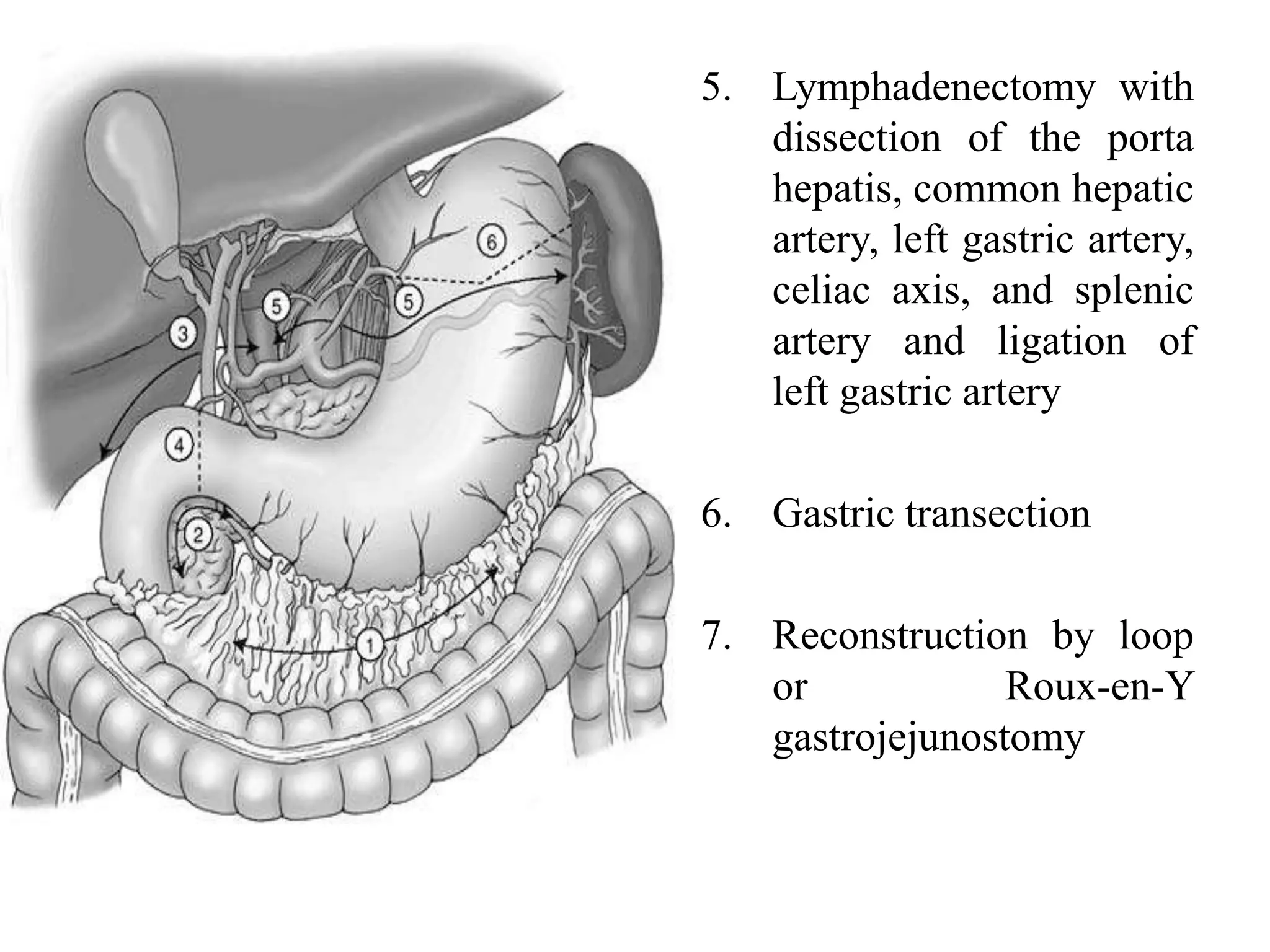
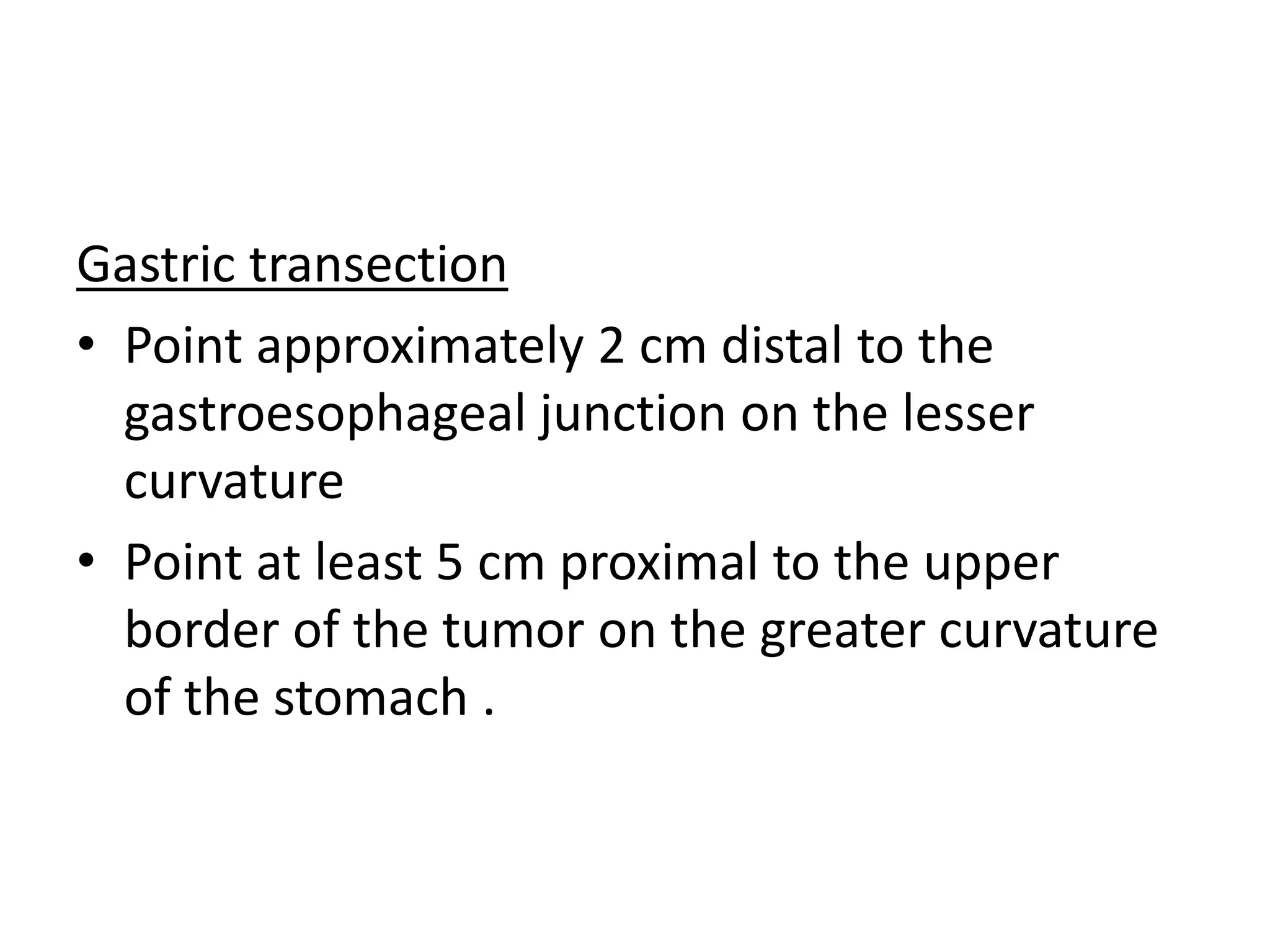
Operative surgery - Gastrectomy
Dr Suhas U
Anatomy
• Asymmetrical, pearshaped, most proximal
abdominal organ of the digestive tract
• Fixed at upper cardioesophageal and lower
pyloric sphinter
• The organs abutting the stomach - liver, colon, spleen,
pancreas, and occasionally the kidney.
• The left lateral segment of the liver usually covers a
large part of the anterior stomach.
• Inferiorly, the stomach is attached to the transverse
colon by the gastrocolic omentum.
• The lesser curvature is tethered to the liver by the
hepatogastric ligament, also referred to as the lesser
omentum or pars flaccida.
• Posterior to the stomach is the lesser omental bursa
and the pancreas.
Blood supply
Nerve supply
History
• Earliest recorded operations on the stomach were
performed for penetrating injuries
• In 1881, Rydygier performed the 1st successful
pylorectomy, and in 1884 he performed the 1st
gastroenterostomy(peptic ulcer)
• In 1881, Billroth performed the 1st successful
pylorectomy for malignancy(duodenum and lesser
curvature anastomosis)
• In 1885, Billroth performed a resection of a large
pyloric carcinoma, using an anterior
gastrojejunostomy for the reconstruction
• Common indications for gastric resections include peptic ulcer
disease and tumors of the stomach
• Safe performance of surgery involves understanding
1. The physiology of vagal innervation and gastric emptying;
2. The surface and vascular anatomy of the stomach
3. The principles of reconstruction following resection, specifically
the Billroth I (B-I) gastroduodenostomy, the Billroth II (B-II)
gastrojejunostomy, and the Roux-en-Y configuration
4. The principles of surgical stapling techniques as well as hand-sewn
suturing techniques
5. The specific early and late postoperative complications that arise
from different gastric resections and different forms of
reconstruction
Degrees of resection
• Wedge resections and closure of gastric wall for
ulcers, polyps, or tumors derived from
neuroendocrine or submucosal tissues
• Distal gastric resection, focusing on antrectomy or
hemigastrectomy (with or without vagotomy) for
peptic ulcer disease and when the major decision
involves the choice of B-I or B-II reconstruction
• Management of gastric carcinoma, focusing on
proximal, subtotal, or total resection, and the
techniques of regional node dissection
Wedge resection
Principles
– Minimum 2cm distance away from GE junction/
pylorus
– 2cm margin of resection
Indications
– Peptic ulcers
– Polyps
– Tumors derived from neuroendocrine or
submucosal tissues
• Midline laparotomy
• Location of lesion
• Omental adhesions of
tumour left in contact
• Omentum away are
ligated
• 2cm scoring of serosa
from base of
tumour(cautery)
• Deepened through
muscularis
• Submucosal vessels
pop out(control)
• Excision of
tumour
• Closure in 2
layers
Tumour on the lesser curve
• Gastrotomy
• To stop bleeding from ulcer
• Excision from mucosal side
• Might require excising both nerves of laterjet and
hence pyloroplasty
• Alternatively
• Distal gastrectomy with B1/B2 procedure /
pauchet procedure
Distal gastrectomy
• Removal of the distal portion of the stomach
• According to disease (ulcer or carcinoma) and the
location (duodenal ulcer, gastriculcer, high-gastric
ulcer), they are classified as
Antral
Two-thirds
Four-fifths
High subtotal gastrectomy
• Distal partial gastrectomy is named according to
the type of anastomosis between the small
intestine and the gastric remnant
• Indications
Recurrent gastric ulcer after adequate antisecretory
treatment and eradication of Helicobacter pylori.
Prepyloric ulcer
Complicated ulcers(perforation, bleeding,
obstruction , intractable)
Early carcinoma and Ca antrum
• Not recommended as standard treatment
• Early and well differentiated T1/T2 N0 lesions
Technique
• Position – supine
• Anaesthesia – GA +EA
• Incision
– Midline
– Transverse epigastric rectus muscle cutting
– Chevron incision
• Self retaining retractors
• Entering lesser sac at middle of gastric curvature
– Incising the gastrocolic ligament
– Dissection made between gastric wall and gastroepiploic
vessels
• Flimsy part of lesser omentum opened with blunt dissection
and penrose drain inserted
• Dissection along greater curvature towards duodenum with
ligation of vessels
Mobilsing the duodenum
• By Kochers maneuver
• By stretching the stomach , dissection proceeds along
greater curvature to the medial duodnenal wall, then
posterior wall and lateral wall( 3 -5 cm of posterior
wall is exposed )
• Transition between first and second part – entry of
gastroduodenal artery
Dissection along lesser curvature
• Ligation of right gastric artery
• Selective vagotomy of gastric remnant
• Dissection proceeds along lesser curvature depending
on the location of ulcer
• Left gastric artery(ascending branch ) should remain
intact
Resection of specimen
• Cutting the duodenum after applying stay sutures
(just distal to pyloric ring)
• Stomach resection
– Stapler (TA90/TL90)
– Angle of 45degrees to lesser curvature
– Clamp fitted at right angle to greater curvature
– The aboral end of greater curvature is cut after stay sutures
• Anastomosis is performed without clamps
Anastomosis
• Two layered technique
– Outer seromuscular (3-0 silk )
– Inner full thickness suture (3-0 vicryl)
• Jammerecke (angle of sorrow)
– Junction of suture anastomosis and the staple line
– Site of leakage
– Triple seromuscular sutures at this angle
• Patency checked with fingers
• NG tube crosses the anastomosis
Alternative methods
• End to side gastroduosenostomy
• Stapler anastomosis (circular stapler)
• Laparoscopic B1 Gastrectomy
B2 gastrectomy
Initial surgical steps
• Dissection of lesser and greater curvature
• Mobilising duodenum and circumferential
dissection
• Ligating right epiploic vessels and right gastric
artery
• Duodenum divided 2cm distal to pyloric ring
with linear stapler
Gastric resection
• Transverse apllication of linear stapler
• Removal of specimen
• Selective vagotomy of gastric remnant
Mobising the jejunum
• Tension free and retrocolonic fashion
• Placed oppsite to greater curvature
• Braun Jejunojejunostomy between ascending and
descending loop
Anastomosis
• 4-5cm stapler line at greater curvature is incised
• Anastomosis done in 2 layers
• Before anterior layer closure , NG tube is passed into
the descending jejunal segment
Roux en Y GJ
• Resection of specimen
• Mobilising the jejunum
• Identifying jejunum at ligament of Treitz
• Proximal jejunum , that reaches the stomach without
tension is selected (15- 20 cm from treitz ligament)
• Jejunum transected with linear stapler
• Mobilsing the distal jejunal segment
• End to side gastrojejunal anastomosis in two layers
and isoperistaltic
• Proximal jejunal segment anastomosed to the jejunum
around 45cm away from GJ as end to side
anastomosis
Total gastrectomy
• Complete operative resection remains the
only potentially curative modality for gastric
adenocarcinoma
• Indicated when 4 to 6 cm of negative margins
cannot be obtained from the primary tumor
• Supine, with consideration given to the
possibility of left or right thoracic approach
• Sandbag placed beneath left costal margin
• A split-lumen endotracheal tube is placed in
all patients in whom thoracotomy is likely
• Incisions
• Midline / bilateral subcostal / left
thoracoabdominal incision
• Fixed retractors are placed.
• Dissection of the greater omentum from the colon and entering
into the anterior leaf of the mesocolon
• The dissection is continued back to the inferior border of the
pancreas, and the pancreatic capsule is dissected upward
• Branches to the right gastroepiploic vessels are divided just at the
inferior border of the pancreas, and venous tributaries ligated.
• The dissection continued laterally, along the superior aspect of the
pancreas, skeletonizing the splenic artery
• Division of short gastric vessels close to the spleen
• Dissecting the lesser omentum from the undersurface of the liver,
extending back to the right crus and mobilizing the right aspect of
the gastroesophageal junction.
• Division of the duodenum
– two straight Kocher clamps /GIA stapler
– Invaginating the closure.
• Divided duodenum
– Elevated upward and forward
– Gaining easy access to the dissection of the node-
bearing areas
• Dissection in the porta
– Begin from above
– Isolating the bifurcation of the hepatic artery
– Bringing the node-bearing tissue inferiorly
• Dissecting the portal vein, to the left of the left hepatic
artery and in the area between the common hepatic and
the superior border of the pancreas
• This dissection moves towards celiac axis
– Picking up the dissection of the superior border of the pancreas
at the junction of the splenic artery with the celiac.
• The left gastric artery is then divided at its origin
• Dissection from above along the right crus
allows the aortic junction of the celiac axis to
be identified and cleared.
• The extent of the dissection of the splenic
hilum depends on the extent of disease
present.
• The short gastric vessels are identified and
ligated close to the spleen
• The left crus dissection downwards and all tissue
reflected from it.
• The left adrenal gland should be clearly identified and
preserved from harm.
• At this point, the entire stomach mobilized
• The left gastric artery can be divided at its origin
• Entire stomach lifted forward
• Paracardial lymph nodes are reflected inferiorly
• The gastroesophageal junction is mobilized and
satinsky atraumatic vascular clamp applied
Anastomosis
• Roux-en-Y method end to side esophagojejunal
anastomosis using 2 layered technique
Subtotal gastrectomy
Indication
• When 4-6cm margin from the tumour is
present
• Incision , position and anaesthesia same
• Steps
1. Mobilization of the
greater curvature with
omentectomy and
division of the left
gastroepiploic artery
2. Infrapyloric
mobilization with
ligation of the right
gastroepiploic artery
and vein as it enters
the gastrocolic trunk
3. Suprapyloric
mobilization with
ligation of the right
gastric artery
5. Lymphadenectomy with
dissection of the porta
hepatis, common hepatic
artery, left gastric artery,
celiac axis, and splenic
artery and ligation of
left gastric artery
6. Gastric transection
7. Reconstruction by loop
or Roux-en-Y
gastrojejunostomy
Gastric transection
• Point approximately 2 cm distal to the
gastroesophageal junction on the lesser
curvature
• Point at least 5 cm proximal to the upper
border of the tumor on the greater curvature
of the stomach .

gastrectomy.pptx

  • 1.
    Operative surgery -Gastrectomy Dr Suhas U
  • 2.
    Anatomy • Asymmetrical, pearshaped,most proximal abdominal organ of the digestive tract • Fixed at upper cardioesophageal and lower pyloric sphinter
  • 4.
    • The organsabutting the stomach - liver, colon, spleen, pancreas, and occasionally the kidney. • The left lateral segment of the liver usually covers a large part of the anterior stomach. • Inferiorly, the stomach is attached to the transverse colon by the gastrocolic omentum. • The lesser curvature is tethered to the liver by the hepatogastric ligament, also referred to as the lesser omentum or pars flaccida. • Posterior to the stomach is the lesser omental bursa and the pancreas.
  • 5.
  • 7.
  • 8.
    History • Earliest recordedoperations on the stomach were performed for penetrating injuries • In 1881, Rydygier performed the 1st successful pylorectomy, and in 1884 he performed the 1st gastroenterostomy(peptic ulcer) • In 1881, Billroth performed the 1st successful pylorectomy for malignancy(duodenum and lesser curvature anastomosis) • In 1885, Billroth performed a resection of a large pyloric carcinoma, using an anterior gastrojejunostomy for the reconstruction
  • 9.
    • Common indicationsfor gastric resections include peptic ulcer disease and tumors of the stomach • Safe performance of surgery involves understanding 1. The physiology of vagal innervation and gastric emptying; 2. The surface and vascular anatomy of the stomach 3. The principles of reconstruction following resection, specifically the Billroth I (B-I) gastroduodenostomy, the Billroth II (B-II) gastrojejunostomy, and the Roux-en-Y configuration 4. The principles of surgical stapling techniques as well as hand-sewn suturing techniques 5. The specific early and late postoperative complications that arise from different gastric resections and different forms of reconstruction
  • 10.
    Degrees of resection •Wedge resections and closure of gastric wall for ulcers, polyps, or tumors derived from neuroendocrine or submucosal tissues • Distal gastric resection, focusing on antrectomy or hemigastrectomy (with or without vagotomy) for peptic ulcer disease and when the major decision involves the choice of B-I or B-II reconstruction • Management of gastric carcinoma, focusing on proximal, subtotal, or total resection, and the techniques of regional node dissection
  • 11.
    Wedge resection Principles – Minimum2cm distance away from GE junction/ pylorus – 2cm margin of resection Indications – Peptic ulcers – Polyps – Tumors derived from neuroendocrine or submucosal tissues
  • 12.
    • Midline laparotomy •Location of lesion • Omental adhesions of tumour left in contact • Omentum away are ligated • 2cm scoring of serosa from base of tumour(cautery) • Deepened through muscularis • Submucosal vessels pop out(control)
  • 13.
    • Excision of tumour •Closure in 2 layers
  • 14.
    Tumour on thelesser curve • Gastrotomy • To stop bleeding from ulcer • Excision from mucosal side • Might require excising both nerves of laterjet and hence pyloroplasty • Alternatively • Distal gastrectomy with B1/B2 procedure / pauchet procedure
  • 15.
    Distal gastrectomy • Removalof the distal portion of the stomach • According to disease (ulcer or carcinoma) and the location (duodenal ulcer, gastriculcer, high-gastric ulcer), they are classified as Antral Two-thirds Four-fifths High subtotal gastrectomy • Distal partial gastrectomy is named according to the type of anastomosis between the small intestine and the gastric remnant
  • 16.
    • Indications Recurrent gastriculcer after adequate antisecretory treatment and eradication of Helicobacter pylori. Prepyloric ulcer Complicated ulcers(perforation, bleeding, obstruction , intractable) Early carcinoma and Ca antrum • Not recommended as standard treatment • Early and well differentiated T1/T2 N0 lesions
  • 17.
    Technique • Position –supine • Anaesthesia – GA +EA • Incision – Midline – Transverse epigastric rectus muscle cutting – Chevron incision • Self retaining retractors
  • 18.
    • Entering lessersac at middle of gastric curvature – Incising the gastrocolic ligament – Dissection made between gastric wall and gastroepiploic vessels • Flimsy part of lesser omentum opened with blunt dissection and penrose drain inserted • Dissection along greater curvature towards duodenum with ligation of vessels
  • 19.
    Mobilsing the duodenum •By Kochers maneuver • By stretching the stomach , dissection proceeds along greater curvature to the medial duodnenal wall, then posterior wall and lateral wall( 3 -5 cm of posterior wall is exposed ) • Transition between first and second part – entry of gastroduodenal artery
  • 20.
    Dissection along lessercurvature • Ligation of right gastric artery • Selective vagotomy of gastric remnant • Dissection proceeds along lesser curvature depending on the location of ulcer • Left gastric artery(ascending branch ) should remain intact
  • 21.
    Resection of specimen •Cutting the duodenum after applying stay sutures (just distal to pyloric ring) • Stomach resection – Stapler (TA90/TL90) – Angle of 45degrees to lesser curvature – Clamp fitted at right angle to greater curvature – The aboral end of greater curvature is cut after stay sutures • Anastomosis is performed without clamps
  • 22.
    Anastomosis • Two layeredtechnique – Outer seromuscular (3-0 silk ) – Inner full thickness suture (3-0 vicryl) • Jammerecke (angle of sorrow) – Junction of suture anastomosis and the staple line – Site of leakage – Triple seromuscular sutures at this angle • Patency checked with fingers • NG tube crosses the anastomosis
  • 23.
    Alternative methods • Endto side gastroduosenostomy • Stapler anastomosis (circular stapler) • Laparoscopic B1 Gastrectomy
  • 24.
    B2 gastrectomy Initial surgicalsteps • Dissection of lesser and greater curvature • Mobilising duodenum and circumferential dissection • Ligating right epiploic vessels and right gastric artery • Duodenum divided 2cm distal to pyloric ring with linear stapler
  • 25.
    Gastric resection • Transverseapllication of linear stapler • Removal of specimen • Selective vagotomy of gastric remnant Mobising the jejunum • Tension free and retrocolonic fashion • Placed oppsite to greater curvature • Braun Jejunojejunostomy between ascending and descending loop
  • 26.
    Anastomosis • 4-5cm staplerline at greater curvature is incised • Anastomosis done in 2 layers • Before anterior layer closure , NG tube is passed into the descending jejunal segment
  • 27.
    Roux en YGJ • Resection of specimen • Mobilising the jejunum • Identifying jejunum at ligament of Treitz • Proximal jejunum , that reaches the stomach without tension is selected (15- 20 cm from treitz ligament) • Jejunum transected with linear stapler
  • 28.
    • Mobilsing thedistal jejunal segment • End to side gastrojejunal anastomosis in two layers and isoperistaltic • Proximal jejunal segment anastomosed to the jejunum around 45cm away from GJ as end to side anastomosis
  • 29.
    Total gastrectomy • Completeoperative resection remains the only potentially curative modality for gastric adenocarcinoma • Indicated when 4 to 6 cm of negative margins cannot be obtained from the primary tumor
  • 30.
    • Supine, withconsideration given to the possibility of left or right thoracic approach • Sandbag placed beneath left costal margin • A split-lumen endotracheal tube is placed in all patients in whom thoracotomy is likely • Incisions • Midline / bilateral subcostal / left thoracoabdominal incision
  • 31.
    • Fixed retractorsare placed. • Dissection of the greater omentum from the colon and entering into the anterior leaf of the mesocolon • The dissection is continued back to the inferior border of the pancreas, and the pancreatic capsule is dissected upward • Branches to the right gastroepiploic vessels are divided just at the inferior border of the pancreas, and venous tributaries ligated. • The dissection continued laterally, along the superior aspect of the pancreas, skeletonizing the splenic artery • Division of short gastric vessels close to the spleen • Dissecting the lesser omentum from the undersurface of the liver, extending back to the right crus and mobilizing the right aspect of the gastroesophageal junction.
  • 32.
    • Division ofthe duodenum – two straight Kocher clamps /GIA stapler – Invaginating the closure. • Divided duodenum – Elevated upward and forward – Gaining easy access to the dissection of the node- bearing areas
  • 33.
    • Dissection inthe porta – Begin from above – Isolating the bifurcation of the hepatic artery – Bringing the node-bearing tissue inferiorly • Dissecting the portal vein, to the left of the left hepatic artery and in the area between the common hepatic and the superior border of the pancreas • This dissection moves towards celiac axis – Picking up the dissection of the superior border of the pancreas at the junction of the splenic artery with the celiac. • The left gastric artery is then divided at its origin
  • 34.
    • Dissection fromabove along the right crus allows the aortic junction of the celiac axis to be identified and cleared. • The extent of the dissection of the splenic hilum depends on the extent of disease present. • The short gastric vessels are identified and ligated close to the spleen
  • 35.
    • The leftcrus dissection downwards and all tissue reflected from it. • The left adrenal gland should be clearly identified and preserved from harm. • At this point, the entire stomach mobilized • The left gastric artery can be divided at its origin • Entire stomach lifted forward • Paracardial lymph nodes are reflected inferiorly • The gastroesophageal junction is mobilized and satinsky atraumatic vascular clamp applied
  • 36.
    Anastomosis • Roux-en-Y methodend to side esophagojejunal anastomosis using 2 layered technique
  • 37.
    Subtotal gastrectomy Indication • When4-6cm margin from the tumour is present • Incision , position and anaesthesia same
  • 38.
    • Steps 1. Mobilizationof the greater curvature with omentectomy and division of the left gastroepiploic artery 2. Infrapyloric mobilization with ligation of the right gastroepiploic artery and vein as it enters the gastrocolic trunk 3. Suprapyloric mobilization with ligation of the right gastric artery
  • 39.
    5. Lymphadenectomy with dissectionof the porta hepatis, common hepatic artery, left gastric artery, celiac axis, and splenic artery and ligation of left gastric artery 6. Gastric transection 7. Reconstruction by loop or Roux-en-Y gastrojejunostomy
  • 40.
    Gastric transection • Pointapproximately 2 cm distal to the gastroesophageal junction on the lesser curvature • Point at least 5 cm proximal to the upper border of the tumor on the greater curvature of the stomach .