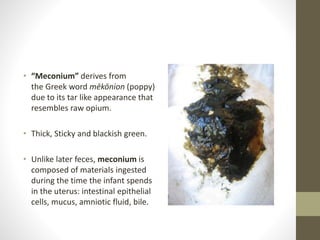
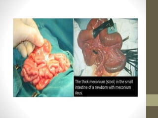
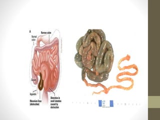
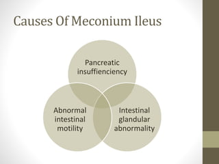
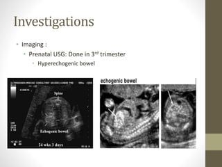
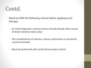
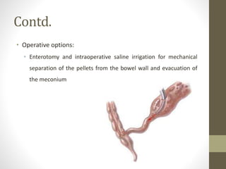
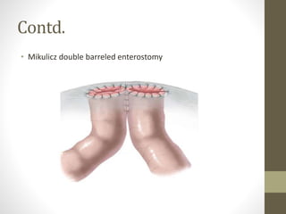
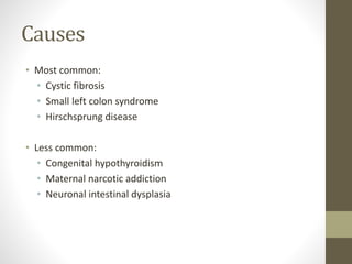
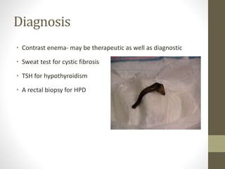
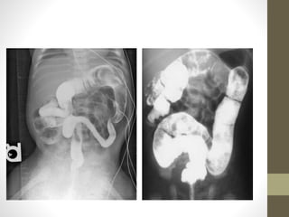
Meconium Disease
Dr. Shirish Silwal
Dept. Of Paediatric Surgery, BSMMU
Contents
• What is meconium?
• Common meconium diseases
1. Meconium ileus
2. Meconium plug syndrome
Neonatal intestinal obstruction
1. Intestinal atresia
2. Meconium ileus
3.Hirschsprung's disease (HD)
4.Malrotation of the gut with or without volvulus
Meconium
Meconium is the First Stool Passed by New-Born
• “Meconium” derives from
the Greek word mēkōnion (poppy)
due to its tar like appearance that
resembles raw opium.
• Thick, Sticky and blackish green.
• Unlike later feces, meconium is
composed of materials ingested
during the time the infant spends
in the uterus: intestinal epithelial
cells, mucus, amniotic fluid, bile.
NormalPassageOf Meconium
Healthy full term infants:
99% pass meconium within 24 hrs of birth
100% pass meconium within 48 hrs of birth
Preterm infants:
37% pass within 24 hrs
99% pass within 9th day of birth
CommonDifferentialsof MeconiumDisease
Meconium ileus
Meconium plug syndrome
First sign or symptom that we suspect its a
meconium ileus ??
Neonate born with abdominal distention before
neonate swallows air.
Meconium Ileus
Common cause of neonatal intestinal
obstruction
Characterized by extremely viscid, protein-rich,
inspissated meconium in mid ileum causing
obstruction
Earliest manifestation of cystic fibrosis(CF)
Incidence
• 9 – 33% of all neonatal intestinal obstruction
• Predominant in white countries where the rate of CF is higher
(1 in 1200 to 1 in 2700 live birth)
• Nearly absent in some Asian people and black Africans
• No difference between sexes
Causes Of Meconium Ileus
Pancreatic
insuffienciency
Intestinal
glandular
abnormality
Abnormal
intestinal
motility
Composition
• Meconium is thick, viscid, inspissated – due to
• Water: 65% (normal – 75%)
• Lower sucrase & lactase level
• Increased albumin, nitrogen
• Decreased pancreatic enzyme & carbohydrate
• Reduced Na, K, Mg, heavy metals
• Presence of abnormal mucoprotein
Clinical features
• Uncomplicated/simple (58%):
• Abdominal distension with palpable doughy
bowel loops (putty sign present)
• Bilious vomiting
• Failure to pass stool
• On DRE, narrowing of anus and rectum, with
only a dense rubberlike gray meconium
sticking to the anal wall
Contd.
• Complicated (42%):
• Presents symptoms immediately after birth
due to in utero perforation
• meconium peritonitis
• Signs of peritonitis (abdominal distension,
edema & erythema of abdominal wall,
abdominal tenderness, sepsis)
• NG tube insertion : >20ml bile stained gastric
fluid
Investigations
• Imaging :
• Prenatal USG: Done in 3rd trimester
• Hyperechogenic bowel
Contd.
• Plain x-ray abdomen:
• Disparity in the size of the
intestinal loops
• No or few air fluid levels on
erect film
• Granular soap bubble
(Neuhauser sign)
Contd.
Water soluble contrast
enema:
• Unused or microcolon
often containing small,
inspissated rabbit pellets of
meconium.
• If reflux into the terminal
ileum pellets of meconium
can be seen
Contd.
• Laboratory testing:
• Sweat chloride test:
• Sweat collected from forearm, leg or back
• >60mEq/L of chloride diagnostic of CF
• Amniocentesis with fetal DNA restriction
fragment length polymorphism analysis (if
family history of CF)
• Albumin concentration in meconium:
• Tetra bromophenolethylester blue detects
albumin >20mg/gm of stool
• Normally meconium contains <5mg/gm
albumin
• In CF: albumin >80mg/gm of stool
Contd.
• Stool trypsin & chymotrypsin analysis:
• Trypsin <80mg/gm of stool
• Histopathology:
• Goblet cell hyperplasia
• Accumulation of secretion within crypts or lumen
Management
• Non operative
• Operative
Non Operative Management
• Neonates should be managed as neonatal intestinal obstruction:
Nothing per oral
Aggressive fluid resuscitation
Nasogastric tube to suction
Mechanical respiratory support
Correction of coagulation disorder
Empirical broad spectrum antibiotic coverage
• Hyperosmolar contrast enema:
• Gastrograffin (standard)
Contd.
• Need to fulfill the following criteria before applying such
therapy:
• An initial diagnostic contrast enema should exclude other causes
of distal intestinal obstruction
• The complications of volvulus, atresia, perforation or peritonitis
must be excluded
• Must be performed with careful fluoroscopic control
Contd.
• I/V antibiotics should be administered
• Should be attended by a pediatric surgeon
• The patient should have a full fluid resuscitation with fluid given
aggressively during the procedure
• The patient should be prepared for imminent operation should
complication develop
Operative Management
• Simple meconium ileus:
• Indications of surgery:
• Inadequate meconium evacuation or a complication from the
contrast enema
• Failure of non-operative treatment with the contrast enema
• An unsuspected associated intestinal atresia
• If the enema failed to promote passage of meconium within 24 to
48 hours
• Two attempts at washout are unsuccessful
Contd.
• Operative options:
• Enterotomy and intraoperative saline irrigation for mechanical
separation of the pellets from the bowel wall and evacuation of
the meconium
Contd.
• Mikulicz double barreled enterostomy
Contd.
• The Bishop-Koop procedure
Contd.
• The Santulli procedure
Contd.
Complicated meconium ileus:
• Surgery is indicated in complicated MI.
• Surgical management includes:
• Early diagnosis
• Debridement of necrotic material
• Pseudocyst resection
• Diverting stoma’s
• Antibiotics
• Meticulous post operative care
Post Operative Management
• Initial management- ongoing resuscitation
• Instillation of 2% or 4% N-acetylcysteine will help solubilize
residual meconium
• Nutritional management- breast milk/infant formula along
with supplemental pancreatic enzyme(2000-4000 unit
lipase/120ml) & vitamins.
• Prophylactic pulmonary care with chest physiotherapy
Prognosis
• Current 5 years survival is approaching 100%
• 72% survival at 10 years
• Current operative mortality is reported to be 10-20%
• Current life expectancy for patients with cystic fibrosis is 35
years
Meconium Plug Syndrome
• Usually, the terminal 2 cm of neonatal meconium is firm in
texture, forming a whitish cap that is passed before, during or
shortly after delivery.
• 1 in 500 newborns will have a longer, more tenacious
obstructive plug
• Failure to pass this plug results in meconium plug syndrome
• Hypothesis- either colonic motility abnormality or the
character of the meconium was altered
• Most of the infants are found to be healthy
Causes
• Most common:
• Cystic fibrosis
• Small left colon syndrome
• Hirschsprung disease
• Less common:
• Congenital hypothyroidism
• Maternal narcotic addiction
• Neuronal intestinal dysplasia
Clinical Features
• Failure to pass meconium
• Bilious vomiting
• Abdominal distention
• Obstructive pattern on plain abdominal radiograph
• The meconium plug may become dislodged after digital
stimulation of the anus and rectum
Diagnosis
• Contrast enema- may be therapeutic as well as diagnostic
• Sweat test for cystic fibrosis
• TSH for hypothyroidism
• A rectal biopsy for HPD
Prognosis
• Excellent outcome after relief of the obstruction, and no
further intervention is required
ManagementofMeconiumIleus:5years'experienceatDhaka
Shishu(Children)Hospital(2011-2016)
KMNFerdous,MSHasan,KAKabir,MKIslam,TBanu
Take Home Message
• Timely passage of the first stool is a hallmark of the well-being
of the newborn infant.
• Early diagnosis is of paramount importance.
• Clinical history and physical examination combined with plain
abdominal radiographs, contrast enema radiographic
examination and rectal examination eventually yield the
diagnosis
THANK YOU

Meconium disease

  • 1.
    Meconium Disease Dr. ShirishSilwal Dept. Of Paediatric Surgery, BSMMU
  • 2.
    Contents • What ismeconium? • Common meconium diseases 1. Meconium ileus 2. Meconium plug syndrome
  • 3.
    Neonatal intestinal obstruction 1.Intestinal atresia 2. Meconium ileus 3.Hirschsprung's disease (HD) 4.Malrotation of the gut with or without volvulus
  • 4.
    Meconium Meconium is theFirst Stool Passed by New-Born
  • 5.
    • “Meconium” derivesfrom the Greek word mēkōnion (poppy) due to its tar like appearance that resembles raw opium. • Thick, Sticky and blackish green. • Unlike later feces, meconium is composed of materials ingested during the time the infant spends in the uterus: intestinal epithelial cells, mucus, amniotic fluid, bile.
  • 6.
    NormalPassageOf Meconium Healthy fullterm infants: 99% pass meconium within 24 hrs of birth 100% pass meconium within 48 hrs of birth Preterm infants: 37% pass within 24 hrs 99% pass within 9th day of birth
  • 7.
  • 8.
    First sign orsymptom that we suspect its a meconium ileus ?? Neonate born with abdominal distention before neonate swallows air.
  • 9.
    Meconium Ileus Common causeof neonatal intestinal obstruction Characterized by extremely viscid, protein-rich, inspissated meconium in mid ileum causing obstruction Earliest manifestation of cystic fibrosis(CF)
  • 12.
    Incidence • 9 –33% of all neonatal intestinal obstruction • Predominant in white countries where the rate of CF is higher (1 in 1200 to 1 in 2700 live birth) • Nearly absent in some Asian people and black Africans • No difference between sexes
  • 13.
    Causes Of MeconiumIleus Pancreatic insuffienciency Intestinal glandular abnormality Abnormal intestinal motility
  • 14.
    Composition • Meconium isthick, viscid, inspissated – due to • Water: 65% (normal – 75%) • Lower sucrase & lactase level • Increased albumin, nitrogen • Decreased pancreatic enzyme & carbohydrate • Reduced Na, K, Mg, heavy metals • Presence of abnormal mucoprotein
  • 15.
    Clinical features • Uncomplicated/simple(58%): • Abdominal distension with palpable doughy bowel loops (putty sign present) • Bilious vomiting • Failure to pass stool • On DRE, narrowing of anus and rectum, with only a dense rubberlike gray meconium sticking to the anal wall
  • 16.
    Contd. • Complicated (42%): •Presents symptoms immediately after birth due to in utero perforation • meconium peritonitis • Signs of peritonitis (abdominal distension, edema & erythema of abdominal wall, abdominal tenderness, sepsis) • NG tube insertion : >20ml bile stained gastric fluid
  • 17.
    Investigations • Imaging : •Prenatal USG: Done in 3rd trimester • Hyperechogenic bowel
  • 18.
    Contd. • Plain x-rayabdomen: • Disparity in the size of the intestinal loops • No or few air fluid levels on erect film • Granular soap bubble (Neuhauser sign)
  • 19.
    Contd. Water soluble contrast enema: •Unused or microcolon often containing small, inspissated rabbit pellets of meconium. • If reflux into the terminal ileum pellets of meconium can be seen
  • 20.
    Contd. • Laboratory testing: •Sweat chloride test: • Sweat collected from forearm, leg or back • >60mEq/L of chloride diagnostic of CF • Amniocentesis with fetal DNA restriction fragment length polymorphism analysis (if family history of CF)
  • 21.
    • Albumin concentrationin meconium: • Tetra bromophenolethylester blue detects albumin >20mg/gm of stool • Normally meconium contains <5mg/gm albumin • In CF: albumin >80mg/gm of stool
  • 22.
    Contd. • Stool trypsin& chymotrypsin analysis: • Trypsin <80mg/gm of stool • Histopathology: • Goblet cell hyperplasia • Accumulation of secretion within crypts or lumen
  • 23.
  • 24.
    Non Operative Management •Neonates should be managed as neonatal intestinal obstruction: Nothing per oral Aggressive fluid resuscitation Nasogastric tube to suction Mechanical respiratory support Correction of coagulation disorder Empirical broad spectrum antibiotic coverage • Hyperosmolar contrast enema: • Gastrograffin (standard)
  • 25.
    Contd. • Need tofulfill the following criteria before applying such therapy: • An initial diagnostic contrast enema should exclude other causes of distal intestinal obstruction • The complications of volvulus, atresia, perforation or peritonitis must be excluded • Must be performed with careful fluoroscopic control
  • 26.
    Contd. • I/V antibioticsshould be administered • Should be attended by a pediatric surgeon • The patient should have a full fluid resuscitation with fluid given aggressively during the procedure • The patient should be prepared for imminent operation should complication develop
  • 27.
    Operative Management • Simplemeconium ileus: • Indications of surgery: • Inadequate meconium evacuation or a complication from the contrast enema • Failure of non-operative treatment with the contrast enema • An unsuspected associated intestinal atresia • If the enema failed to promote passage of meconium within 24 to 48 hours • Two attempts at washout are unsuccessful
  • 28.
    Contd. • Operative options: •Enterotomy and intraoperative saline irrigation for mechanical separation of the pellets from the bowel wall and evacuation of the meconium
  • 29.
    Contd. • Mikulicz doublebarreled enterostomy
  • 30.
  • 31.
  • 32.
    Contd. Complicated meconium ileus: •Surgery is indicated in complicated MI. • Surgical management includes: • Early diagnosis • Debridement of necrotic material • Pseudocyst resection • Diverting stoma’s • Antibiotics • Meticulous post operative care
  • 33.
    Post Operative Management •Initial management- ongoing resuscitation • Instillation of 2% or 4% N-acetylcysteine will help solubilize residual meconium • Nutritional management- breast milk/infant formula along with supplemental pancreatic enzyme(2000-4000 unit lipase/120ml) & vitamins. • Prophylactic pulmonary care with chest physiotherapy
  • 34.
    Prognosis • Current 5years survival is approaching 100% • 72% survival at 10 years • Current operative mortality is reported to be 10-20% • Current life expectancy for patients with cystic fibrosis is 35 years
  • 35.
    Meconium Plug Syndrome •Usually, the terminal 2 cm of neonatal meconium is firm in texture, forming a whitish cap that is passed before, during or shortly after delivery. • 1 in 500 newborns will have a longer, more tenacious obstructive plug
  • 36.
    • Failure topass this plug results in meconium plug syndrome • Hypothesis- either colonic motility abnormality or the character of the meconium was altered • Most of the infants are found to be healthy
  • 37.
    Causes • Most common: •Cystic fibrosis • Small left colon syndrome • Hirschsprung disease • Less common: • Congenital hypothyroidism • Maternal narcotic addiction • Neuronal intestinal dysplasia
  • 38.
    Clinical Features • Failureto pass meconium • Bilious vomiting • Abdominal distention • Obstructive pattern on plain abdominal radiograph • The meconium plug may become dislodged after digital stimulation of the anus and rectum
  • 39.
    Diagnosis • Contrast enema-may be therapeutic as well as diagnostic • Sweat test for cystic fibrosis • TSH for hypothyroidism • A rectal biopsy for HPD
  • 41.
    Prognosis • Excellent outcomeafter relief of the obstruction, and no further intervention is required
  • 42.
  • 43.
    Take Home Message •Timely passage of the first stool is a hallmark of the well-being of the newborn infant. • Early diagnosis is of paramount importance. • Clinical history and physical examination combined with plain abdominal radiographs, contrast enema radiographic examination and rectal examination eventually yield the diagnosis
  • 44.

Editor's Notes

  • #4 ARM-73, HD-47, meconium ileus-38, intestinal atresia-29, malrotation-13 and others-5.