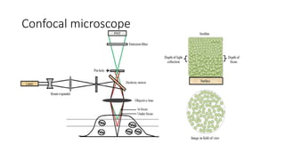
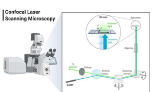
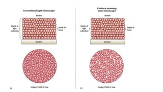
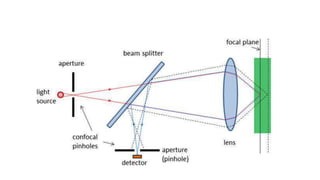
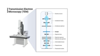
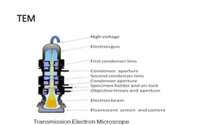
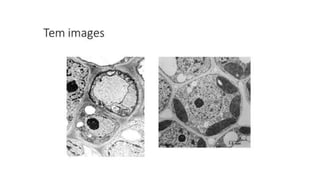
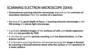
Confocal and electron
microscopy
Sem III
Confocal microscope
• The first commercial confocal microscope was developed in 1987 with
improved optics and electronics, powerful lasers with high scanning
efficiency.
• The modern confocal Microscope has all the possible integration of
technology and mechanical components including optical components,
• which perform the primary function of the configuration by use of
electronic detectors, a computer and laser systems.
• The functioning of the confocal microscope is a collective role of all its
components to produce an electronic image.
• To date, these microscopes have been used to investigate molecules,
microbial cells, and tissues.
Principle
• In wide-field or Fluorescent microscopes, the whole specimen
receives light,
• receiving complete excitement and emitting light
• which is detected by a photodetector on the microscope.
• However, with the confocal microscope, point illumination is the
principle working mechanism.
• A specimen is stained with fluorochrome is examined.
• When a beam of light is focused at a particular point of the fluoro-chromatic
specimen,
• it produces an illumination that is focused by the objective lens to a plane above
the objectives.
• The objective has an aperture on the focal plane located above it, which
primarily functions to block any stray light from reaching the specimen.
• A measure of the illumination point is about 0.25 to 0.8 um in diameter,
determined by the objective numerical aperture and 0.5 to 1.5 um deep, with
the brightest intensity.
• The specimen normally lies between the camera lens and the perfect point
Confocal microscope
Applications of the Confocal Microscope
• The Confocal Microscope is used in a wide range of fields including
Biomedical sciences,
• Cells Biology,
• genetics,
• Microbiology,
• Developmental Biology,
• Spectroscopy,
• Nanoscience (nanoimaging) and
• Quantum Optics.
Applications of the Confocal Microscope
1. In Biomedical sciences, it is used in the analysis of eye corneal
infections, by quantifying and qualitatively analyzing the endothelial cells
of the cornea.
2. Used to identify the presence of fungal elements in the corneal stroma,
during keratomycosis infection, or rapid diagnosis and quick therapeutic
response.
3. It is used in pharmaceutical industries, to ensure the maintenance of
thin-film pharmaceuticals, allowing control of the quality and uniformity
of drug distribution.
4. It is used to retrieve data from some 3D optical storage systems. This
has helped in quantifying the age of Magdalen papyrus.
ELECTRON MICROSCOPY:
• An electron microscope is a microscope that uses a beam of
accelerated electrons as a source of illumination.
• It is a special type of microscope having a high resolution of images,
able to magnify objects in nanometres,
• which are formed by controlled use of electrons in a vacuum captured
on a phosphorescent screen.
• Ernst Ruska (1906-1988), a German engineer and academic professor,
built the first Electron Microscope in 1931.
TYPES OF ELECTRON MICROSCOPY:
• TRANSMISSION ELECTRON MICROSCOPE (TEM):
• SCANNING ELECTRON MICROSCOPE (SEM):
TRANSMISSION ELECTRON MICROSCOPE (TEM):
• The transmission electron microscope is used to view thin specimens through
which electrons can pass generating a projection image.
• The TEM is analogous in many ways to the conventional (compound) light
microscope.
• TEM is used, among other things, to image the interior of cells (in thin sections),
• the structure of protein molecules (contrasted by metal shadowing),
• the organization of molecules in viruses and cytoskeletal filaments
• (prepared by the negative staining technique),
• and the arrangement of protein molecules in cell membranes (by freeze-
fracture).
• Grater magnification with high resolution
• Resolving power 10A
• 100times grater than light microscope (200nm).
• Wave length of electron is 100000 shorter than photons
• Ultrathin sections are made
• Embedding
• freezing
• Sectioning by diamond or glass knife.
• Staining with heavy metals
• Gold or palladium
• Evacuated vacuum
• Electrons are allowed to pass
• Accelerated by anode +100Ke (40 to 400Ke).
• Electrostatic and electromagnetic lenses
TEM
• Conventional scanning electron microscopy depends on the emission of
secondary electrons from the surface of a specimen.
• Because of its great depth of focus, a scanning electron microscope is the
EM analog of a stereo light microscope.
• It provides detailed images of the surfaces of cells and whole organisms
that are not possible by TEM.
• It can also be used for particle counting and size determination, and for
process control.
• It is termed a scanning electron microscope because the image is formed
by scanning a focused electron beam onto the surface of the specimen in
a raster pattern.
SCANNING ELECTRON MICROSCOPE (SEM):
• The SEM produces images by probing the specimen with a focused
electron beam that is scanned across a rectangular area of the
specimen (raster scanning).
• When the electron beam interacts with the specimen, it loses energy
by a variety of mechanisms.
• The lost energy is converted into alternative forms such as heat,
emission of low-energy secondary electrons and high-energy
backscattered electrons, light emission (cathodoluminescence) or X-
ray emission,
• all of which provide signals carrying information about the properties
of the specimen surface, such as its topography and composition.
SCANNING ELECTRON MICROSCOPE (SEM):
PRINCIPLE OF ELECTRON MICROSCOPY (SEM AND TEM):
• Electron microscopes use signals arising from the interaction of an
electron beam with the sample to obtain information about
structure, morphology, and composition.
• 1. The electron gun generates electrons.
• 2. Two sets of condenser lenses focus the electron beam on the
specimen and then into a thin tight beam.
• 3. To move electrons down the column, an accelerating voltage
(mostly between 100 kV-1000 kV) is applied between the tungsten
filament and anode.
• 4. The specimen to be examined is made extremely thin, at least 200
times thinner than those used in the optical microscope.
• Ultra-thin sections of 20-100 nm are cut which is already placed on
the specimen holder.
• 5. The electronic beam passes through the specimen and electrons
are scattered depending upon the thickness or refractive index of
different parts of the specimen.
• 6. The denser regions in the specimen scatter more electrons and
therefore appear darker in the image since fewer electrons strike
that area of the screen. In contrast, transparent regions are brighter.
• 7. The electron beam coming out of the specimen passes to the
objective lens, which has high power and forms the intermediate
magnified image.
• 8. The ocular lenses then produce the final further magnified image.
APPLICATIONS OF ELECTRON MICROSCOPY (SEM AND
TEM):
• 1. Electron microscopes are used to investigate the ultrastructure of a wide
range of biological
• and inorganic specimens including microorganisms, cells, large molecules,
biopsy samples, metals, and crystals.
• 2. Industrially, electron microscopes are often used for quality
control and failure analysis.
• 3. Modern electron microscopes produce electron micrographs using
• specialized digital cameras and frame grabbers to capture the images.
• 4. The science of microbiology owes its development to the electron
microscope.
• The study of microorganisms like bacteria, virus, and other pathogens
have made the treatment of diseases very effective
Confocal and electron microscopy.pptx

Confocal and electron microscopy.pptx

  • 1.
  • 2.
    Confocal microscope • Thefirst commercial confocal microscope was developed in 1987 with improved optics and electronics, powerful lasers with high scanning efficiency. • The modern confocal Microscope has all the possible integration of technology and mechanical components including optical components, • which perform the primary function of the configuration by use of electronic detectors, a computer and laser systems.
  • 3.
    • The functioningof the confocal microscope is a collective role of all its components to produce an electronic image. • To date, these microscopes have been used to investigate molecules, microbial cells, and tissues.
  • 4.
    Principle • In wide-fieldor Fluorescent microscopes, the whole specimen receives light, • receiving complete excitement and emitting light • which is detected by a photodetector on the microscope. • However, with the confocal microscope, point illumination is the principle working mechanism.
  • 5.
    • A specimenis stained with fluorochrome is examined. • When a beam of light is focused at a particular point of the fluoro-chromatic specimen, • it produces an illumination that is focused by the objective lens to a plane above the objectives. • The objective has an aperture on the focal plane located above it, which primarily functions to block any stray light from reaching the specimen. • A measure of the illumination point is about 0.25 to 0.8 um in diameter, determined by the objective numerical aperture and 0.5 to 1.5 um deep, with the brightest intensity. • The specimen normally lies between the camera lens and the perfect point
  • 6.
  • 10.
    Applications of theConfocal Microscope • The Confocal Microscope is used in a wide range of fields including Biomedical sciences, • Cells Biology, • genetics, • Microbiology, • Developmental Biology, • Spectroscopy, • Nanoscience (nanoimaging) and • Quantum Optics.
  • 11.
    Applications of theConfocal Microscope 1. In Biomedical sciences, it is used in the analysis of eye corneal infections, by quantifying and qualitatively analyzing the endothelial cells of the cornea. 2. Used to identify the presence of fungal elements in the corneal stroma, during keratomycosis infection, or rapid diagnosis and quick therapeutic response. 3. It is used in pharmaceutical industries, to ensure the maintenance of thin-film pharmaceuticals, allowing control of the quality and uniformity of drug distribution. 4. It is used to retrieve data from some 3D optical storage systems. This has helped in quantifying the age of Magdalen papyrus.
  • 13.
    ELECTRON MICROSCOPY: • Anelectron microscope is a microscope that uses a beam of accelerated electrons as a source of illumination. • It is a special type of microscope having a high resolution of images, able to magnify objects in nanometres, • which are formed by controlled use of electrons in a vacuum captured on a phosphorescent screen. • Ernst Ruska (1906-1988), a German engineer and academic professor, built the first Electron Microscope in 1931.
  • 14.
    TYPES OF ELECTRONMICROSCOPY: • TRANSMISSION ELECTRON MICROSCOPE (TEM): • SCANNING ELECTRON MICROSCOPE (SEM):
  • 15.
    TRANSMISSION ELECTRON MICROSCOPE(TEM): • The transmission electron microscope is used to view thin specimens through which electrons can pass generating a projection image. • The TEM is analogous in many ways to the conventional (compound) light microscope. • TEM is used, among other things, to image the interior of cells (in thin sections), • the structure of protein molecules (contrasted by metal shadowing), • the organization of molecules in viruses and cytoskeletal filaments • (prepared by the negative staining technique), • and the arrangement of protein molecules in cell membranes (by freeze- fracture).
  • 16.
    • Grater magnificationwith high resolution • Resolving power 10A • 100times grater than light microscope (200nm). • Wave length of electron is 100000 shorter than photons • Ultrathin sections are made • Embedding • freezing • Sectioning by diamond or glass knife.
  • 17.
    • Staining withheavy metals • Gold or palladium • Evacuated vacuum • Electrons are allowed to pass • Accelerated by anode +100Ke (40 to 400Ke). • Electrostatic and electromagnetic lenses
  • 19.
  • 21.
    • Conventional scanningelectron microscopy depends on the emission of secondary electrons from the surface of a specimen. • Because of its great depth of focus, a scanning electron microscope is the EM analog of a stereo light microscope. • It provides detailed images of the surfaces of cells and whole organisms that are not possible by TEM. • It can also be used for particle counting and size determination, and for process control. • It is termed a scanning electron microscope because the image is formed by scanning a focused electron beam onto the surface of the specimen in a raster pattern. SCANNING ELECTRON MICROSCOPE (SEM):
  • 22.
    • The SEMproduces images by probing the specimen with a focused electron beam that is scanned across a rectangular area of the specimen (raster scanning). • When the electron beam interacts with the specimen, it loses energy by a variety of mechanisms. • The lost energy is converted into alternative forms such as heat, emission of low-energy secondary electrons and high-energy backscattered electrons, light emission (cathodoluminescence) or X- ray emission, • all of which provide signals carrying information about the properties of the specimen surface, such as its topography and composition.
  • 23.
  • 25.
    PRINCIPLE OF ELECTRONMICROSCOPY (SEM AND TEM): • Electron microscopes use signals arising from the interaction of an electron beam with the sample to obtain information about structure, morphology, and composition. • 1. The electron gun generates electrons. • 2. Two sets of condenser lenses focus the electron beam on the specimen and then into a thin tight beam. • 3. To move electrons down the column, an accelerating voltage (mostly between 100 kV-1000 kV) is applied between the tungsten filament and anode.
  • 26.
    • 4. Thespecimen to be examined is made extremely thin, at least 200 times thinner than those used in the optical microscope. • Ultra-thin sections of 20-100 nm are cut which is already placed on the specimen holder. • 5. The electronic beam passes through the specimen and electrons are scattered depending upon the thickness or refractive index of different parts of the specimen. • 6. The denser regions in the specimen scatter more electrons and therefore appear darker in the image since fewer electrons strike that area of the screen. In contrast, transparent regions are brighter.
  • 27.
    • 7. Theelectron beam coming out of the specimen passes to the objective lens, which has high power and forms the intermediate magnified image. • 8. The ocular lenses then produce the final further magnified image.
  • 28.
    APPLICATIONS OF ELECTRONMICROSCOPY (SEM AND TEM): • 1. Electron microscopes are used to investigate the ultrastructure of a wide range of biological • and inorganic specimens including microorganisms, cells, large molecules, biopsy samples, metals, and crystals. • 2. Industrially, electron microscopes are often used for quality control and failure analysis.
  • 29.
    • 3. Modernelectron microscopes produce electron micrographs using • specialized digital cameras and frame grabbers to capture the images. • 4. The science of microbiology owes its development to the electron microscope. • The study of microorganisms like bacteria, virus, and other pathogens have made the treatment of diseases very effective