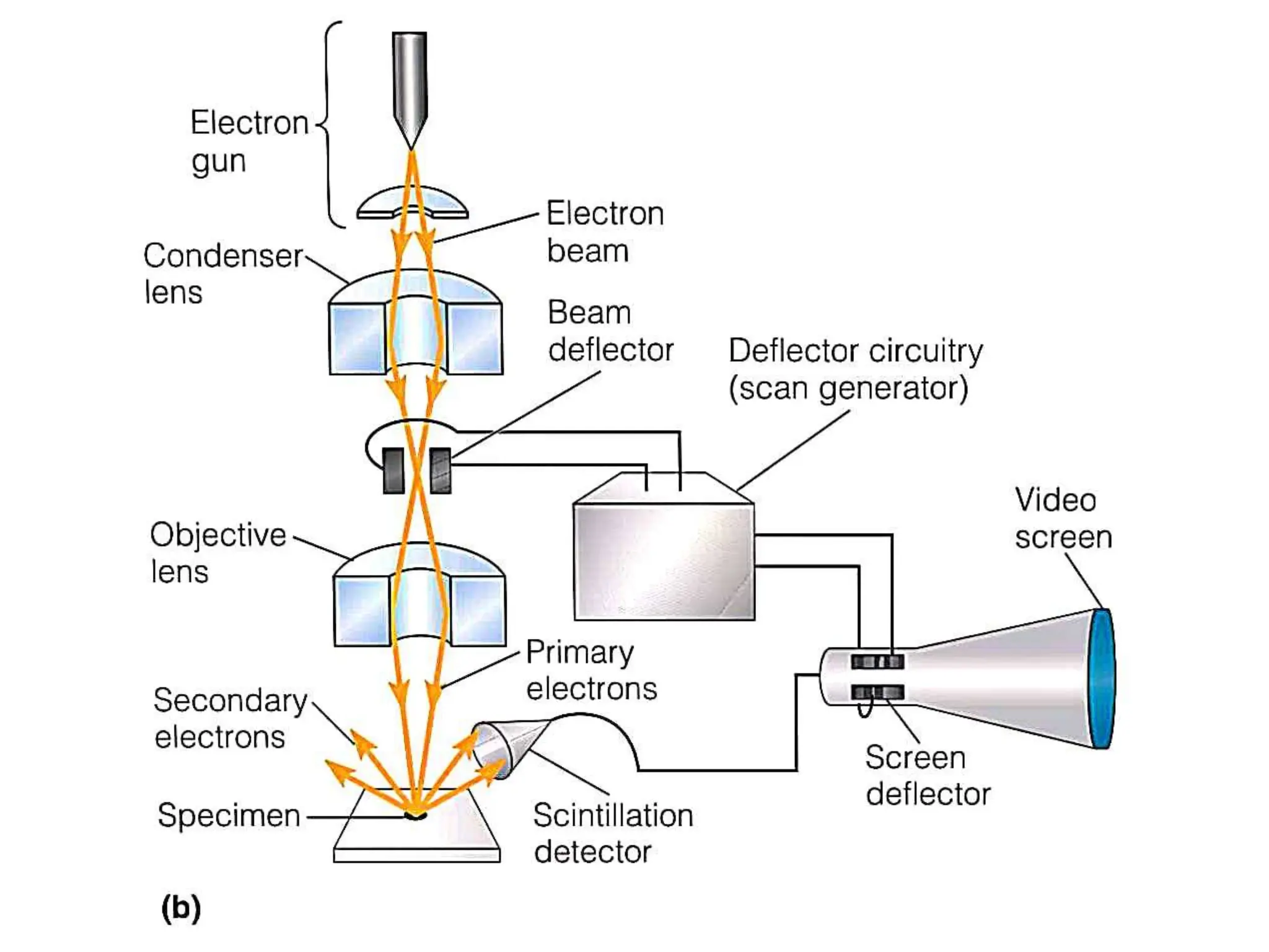
ELECTRON
MICROSCOPY-
SEM & TEM
What is Electron Microscope?
An electron microscope is a microscope that uses a beam
of accelerated electrons as a source of illumination.
It is a special type of microscope having a high
resolution of images, able to magnify objects in
nanometres, which are formed by controlled use of
electrons in a vacuum captured on a phosphorescent
screen.
In electron microscopy, resolution is much better, but
specimen preparation and instrument operation are often
more difficult.
Electron microscopes are of two basic designs: the
transmission electron microscope(TEM) and the scanning
electron microscope(SEM).
Scanning Electron Microscopy(SEM)
As the name implies, a scanning electron microscope
(SEM) generates such an image by scanning the
specimen’s surface with a beam of electrons.
Scanning electron microscopy(SEM) is a fundamentally
different type of electron microscopy that produces
images from electrons deflected from a specimen’s outer
surface (rather than electrons transmitted through the
specimen).
It is an especially spectacular technique because of the
sense of depth it gives to biological structures, thereby
allowing surface topography to be studied.
It gives 3 dimensional(3D) black and white surface image of the
specimen.
Electromagnetic lense system is mainly used.
Construction of (SEM)
Vaccum pump:
It creates vaccum in the microscope column.Metal strips
with liquid Nitrogen add in the column on which gas
molecules trap and creates vaccum.
Cathode(Tungsten lamp)
It is an electron source.It generates electrons when
heated.It is kept at 5-30 V.
Anode
It is kept at 0 V. It guides electron flow to downward
through the hole at its center.Both anode and cathode
attached in column and generates accelerating voltage.
Electromagnetic lenses
The condenser lens focuses the electron beam on the
specimen. A second condenser lens forms the electrons
into a thin tight beam.Magnets are used to create magnetic
field to attract electrons.
The electron beam coming out of the condenser lenses
passes down the second of magnetic coils called
the objective lens, which focuses the beam of electrons at
the point on the fixed specimen.
Beam Deflectors
It helps in focusing beam of electrons on sample by
moving back and forth over specimen
Scintillators
Emit photons of light when excited electrons fall on it.The
photons are used to generate an electronic signal to a
video screen.
The image develop point by point,line by line on screen
by analysis of secondary electron detection by detector
like Everhart-Thornley detector.
Rather than secondary electron pattern other signals like
back scattered electrons,transmitted electrons e.t.c. also
generated.
Transmission Electron Microscopy(SEM)
In a Transmission electron microscope, the electron beam
is transmitted through a very thin specimen or object and
forms a highly magnified and detailed image of the
sample.
This microscope uses electron beams instead of light.
The specimen used in Transmission Electron
Microscope, should be very thin, less than 100 nm thick
A Transmission Electron Microscope can create a much
higher resolution and magnified image than a light
microscope, because of the shorter wavelength of the
electron as compared to photons.
It gives 2 Dimentional sectional image of the specimen
In TEM the sample’s image is formed by the interaction
between the transmitted electron beam and sample.
TEM can tell us the structure, crystallization,
morphology, and stress of the specimen in a better way as
compared to a simple microscope.
The formed image is then magnified and visualized on
a fluorescent screen (layer of photographic film).
Construction is same as SEM except positions of lenses
and varry in detection as well as image formation pattern.
Objective lens,Intermediate lens and Projector lens
combine forms image on a viewing screen that fluorosces
when struck by elecrons.
If few electrons pass through the specimen then pattern
for that portion is dark and area where maximum electrons
have pass through the specimen it appear light
Electrons strike on the fluoroscent screen or
photographic film.
Film allows one to create photographic print called as
electron micrograph which becomes permanent
photograph of that specimen.
ELECTRON MICROSCOPY-Scanning Electron MicroscopySEM.pptx

ELECTRON MICROSCOPY-Scanning Electron MicroscopySEM.pptx

  • 1.
  • 4.
    What is ElectronMicroscope? An electron microscope is a microscope that uses a beam of accelerated electrons as a source of illumination. It is a special type of microscope having a high resolution of images, able to magnify objects in nanometres, which are formed by controlled use of electrons in a vacuum captured on a phosphorescent screen. In electron microscopy, resolution is much better, but specimen preparation and instrument operation are often more difficult. Electron microscopes are of two basic designs: the transmission electron microscope(TEM) and the scanning electron microscope(SEM).
  • 5.
    Scanning Electron Microscopy(SEM) Asthe name implies, a scanning electron microscope (SEM) generates such an image by scanning the specimen’s surface with a beam of electrons. Scanning electron microscopy(SEM) is a fundamentally different type of electron microscopy that produces images from electrons deflected from a specimen’s outer surface (rather than electrons transmitted through the specimen). It is an especially spectacular technique because of the sense of depth it gives to biological structures, thereby allowing surface topography to be studied. It gives 3 dimensional(3D) black and white surface image of the specimen. Electromagnetic lense system is mainly used.
  • 7.
    Construction of (SEM) Vaccumpump: It creates vaccum in the microscope column.Metal strips with liquid Nitrogen add in the column on which gas molecules trap and creates vaccum. Cathode(Tungsten lamp) It is an electron source.It generates electrons when heated.It is kept at 5-30 V. Anode It is kept at 0 V. It guides electron flow to downward through the hole at its center.Both anode and cathode attached in column and generates accelerating voltage.
  • 8.
    Electromagnetic lenses The condenserlens focuses the electron beam on the specimen. A second condenser lens forms the electrons into a thin tight beam.Magnets are used to create magnetic field to attract electrons. The electron beam coming out of the condenser lenses passes down the second of magnetic coils called the objective lens, which focuses the beam of electrons at the point on the fixed specimen. Beam Deflectors It helps in focusing beam of electrons on sample by moving back and forth over specimen Scintillators Emit photons of light when excited electrons fall on it.The photons are used to generate an electronic signal to a
  • 9.
    video screen. The imagedevelop point by point,line by line on screen by analysis of secondary electron detection by detector like Everhart-Thornley detector. Rather than secondary electron pattern other signals like back scattered electrons,transmitted electrons e.t.c. also generated.
  • 10.
    Transmission Electron Microscopy(SEM) Ina Transmission electron microscope, the electron beam is transmitted through a very thin specimen or object and forms a highly magnified and detailed image of the sample. This microscope uses electron beams instead of light. The specimen used in Transmission Electron Microscope, should be very thin, less than 100 nm thick A Transmission Electron Microscope can create a much higher resolution and magnified image than a light microscope, because of the shorter wavelength of the electron as compared to photons. It gives 2 Dimentional sectional image of the specimen
  • 11.
    In TEM thesample’s image is formed by the interaction between the transmitted electron beam and sample. TEM can tell us the structure, crystallization, morphology, and stress of the specimen in a better way as compared to a simple microscope. The formed image is then magnified and visualized on a fluorescent screen (layer of photographic film). Construction is same as SEM except positions of lenses and varry in detection as well as image formation pattern.
  • 13.
    Objective lens,Intermediate lensand Projector lens combine forms image on a viewing screen that fluorosces when struck by elecrons. If few electrons pass through the specimen then pattern for that portion is dark and area where maximum electrons have pass through the specimen it appear light Electrons strike on the fluoroscent screen or photographic film. Film allows one to create photographic print called as electron micrograph which becomes permanent photograph of that specimen.