Downloaded 58 times


![Update
• A Cover of Glass: First Report of Biomineralized Silicon in a Ciliate, Maryna umbrellata (Ciliophora:
Colpodea)
• Using hydrofluoric acid, scanning electron microscope-assisted X-ray microanalysis, and
energy-filtered transmission electron microscopy, we present the first definite proof of
biomineralized silicon [(SiO2)]nin a ciliophoran protist, Maryna umbrellata, a common
inhabitant of ephemeral pools. In the trophic specimen, the amorphic silicon (glass)
granules are accumulated in the anterior half of the body. When entering the dormant
stage, most glass granules are excreted to form the surface cover of the globular resting
cyst. Most likely, the silicon granules are synthesized in vesicles of the Golgi apparatus.
First, nanospheres with a size of 20–40 nm are formed in a fibrous matrix; they grow to be
spongious complexes, eventually becoming amorphous glass granules with an average size
of 819 nm × 630 nm. In the transmission electron microscope, the silicon granules show
the characteristic fracture pattern of glass known from many other silicon-bearing
organisms. A literature survey suggests that silicon is very rare in ciliates. The fine structure
and genesis of silicon granules in M. umbrellata are very similar to those of other
organisms, including vascular plants and animals, indicating a common mechanism. Light
perception and protection against mechanical stress and predators might be functions of
the silicon granules in M. umbrellata. The palaeontological significance of glass cysts in
ciliates is also discussed.](https://image.slidesharecdn.com/ciliatedprotozoa-130228073229-phpapp01/85/Ciliated-protozoa-12-320.jpg)


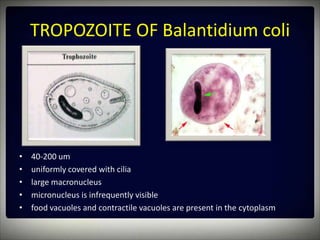
This document discusses the ciliated protozoan Balantidium coli. It provides details on the morphology and life cycle of both the trophozoite and cyst stages. It examines prepared slides under the microscope and compares the pathology caused by B. coli to Entamoeba histolytica. The reservoir host of B. coli is pigs and the laboratory methods for identification include stool examination and sigmoidoscopy. An update section discusses the first report of biomineralized silicon discovered in the ciliate Maryna umbrellata.


