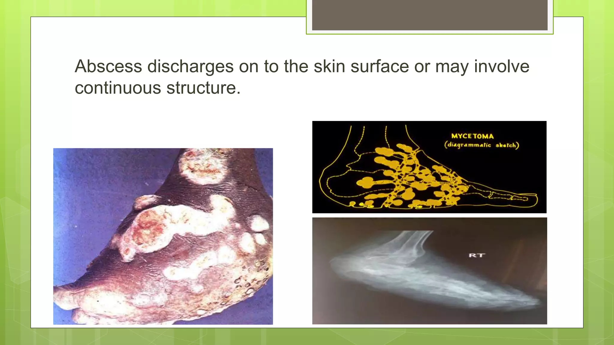
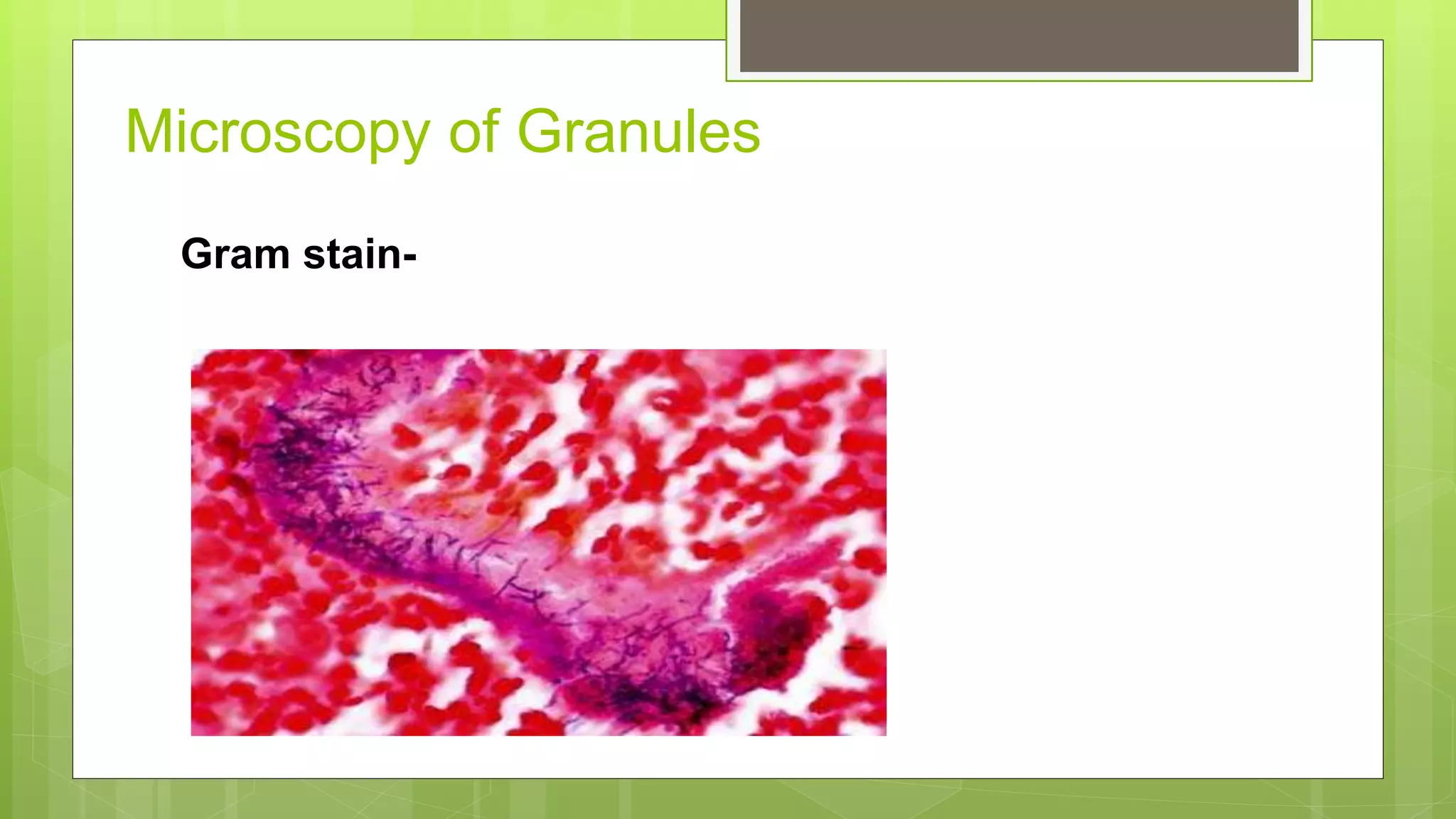
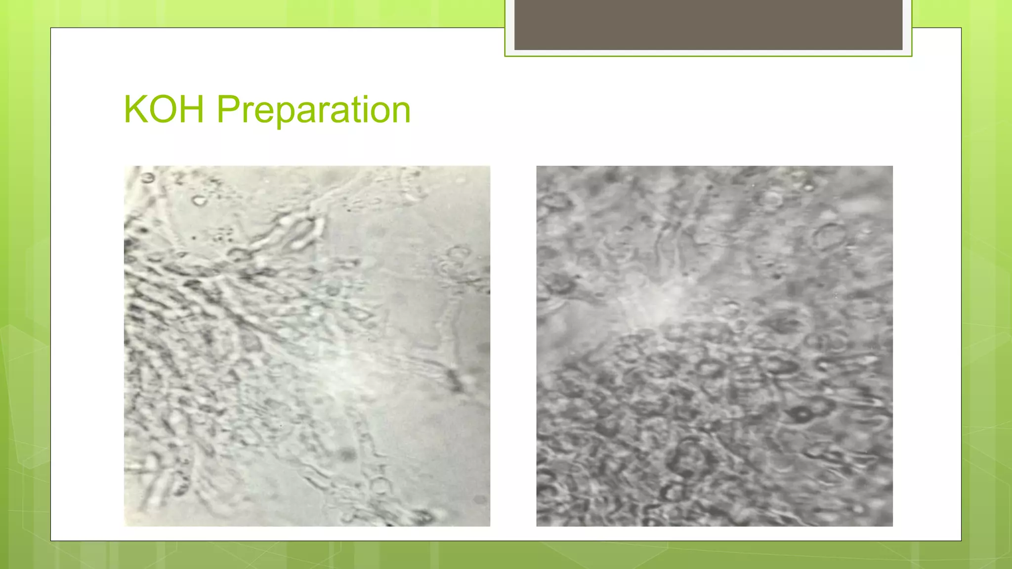
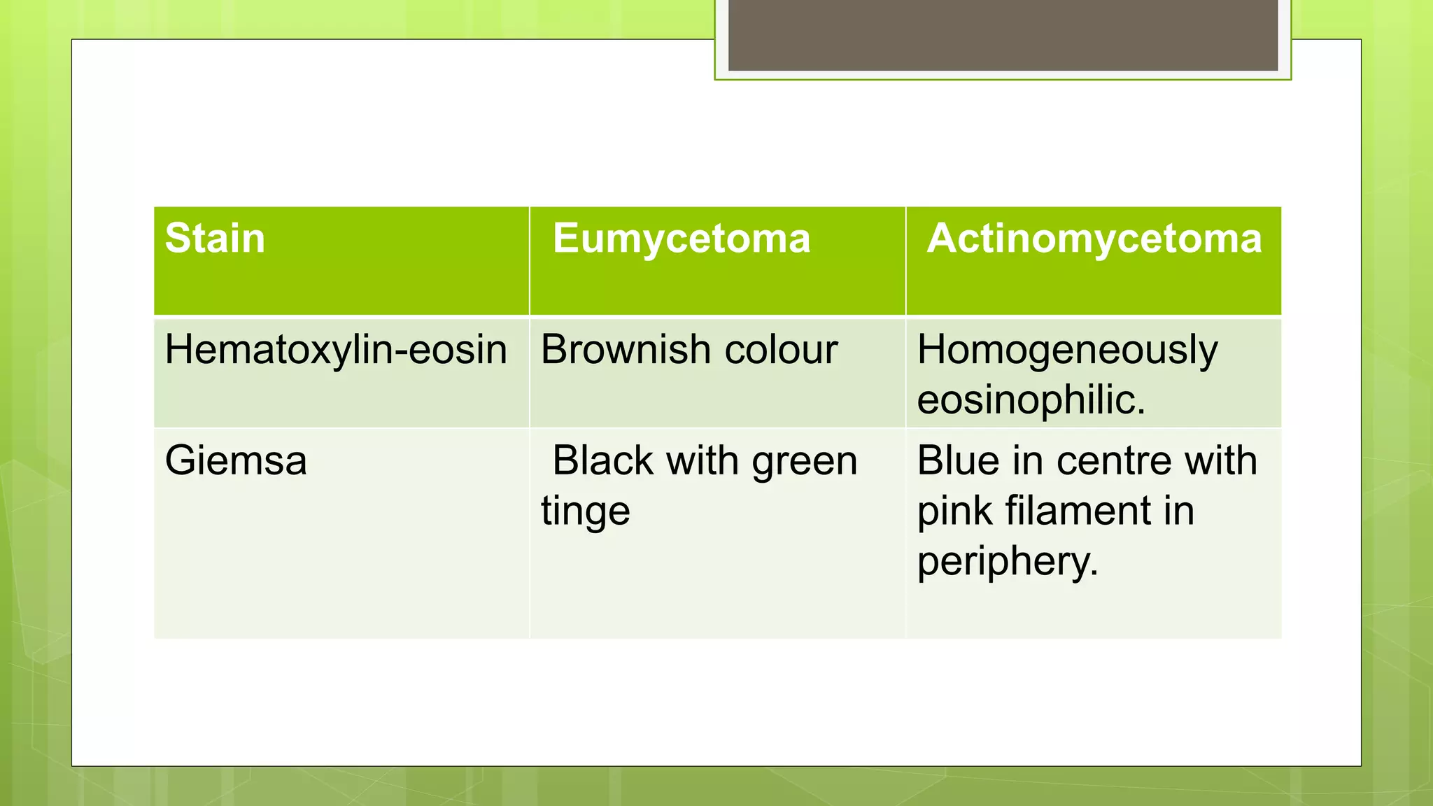
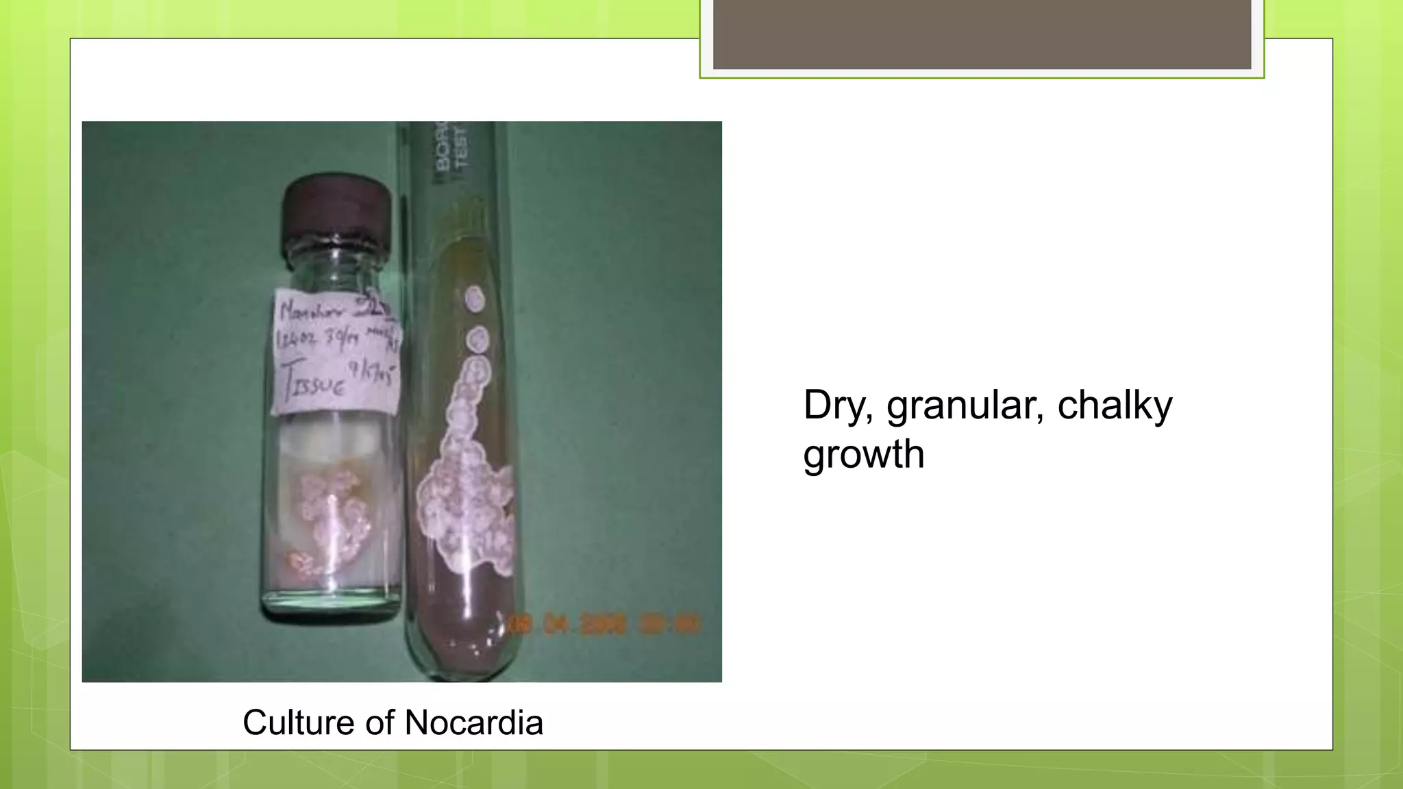
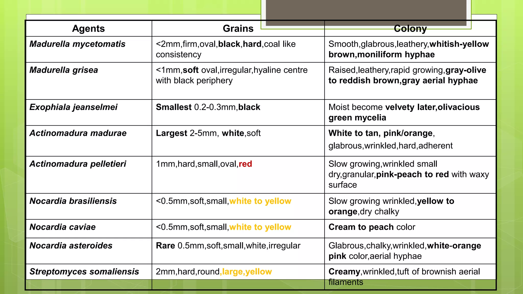
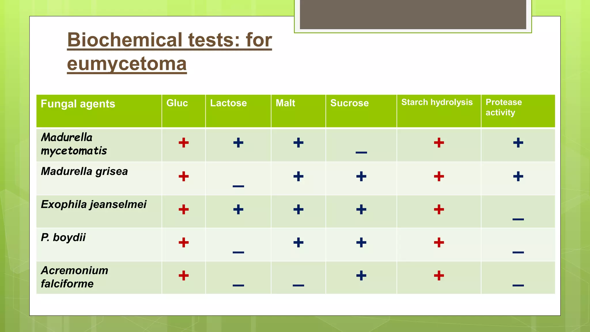
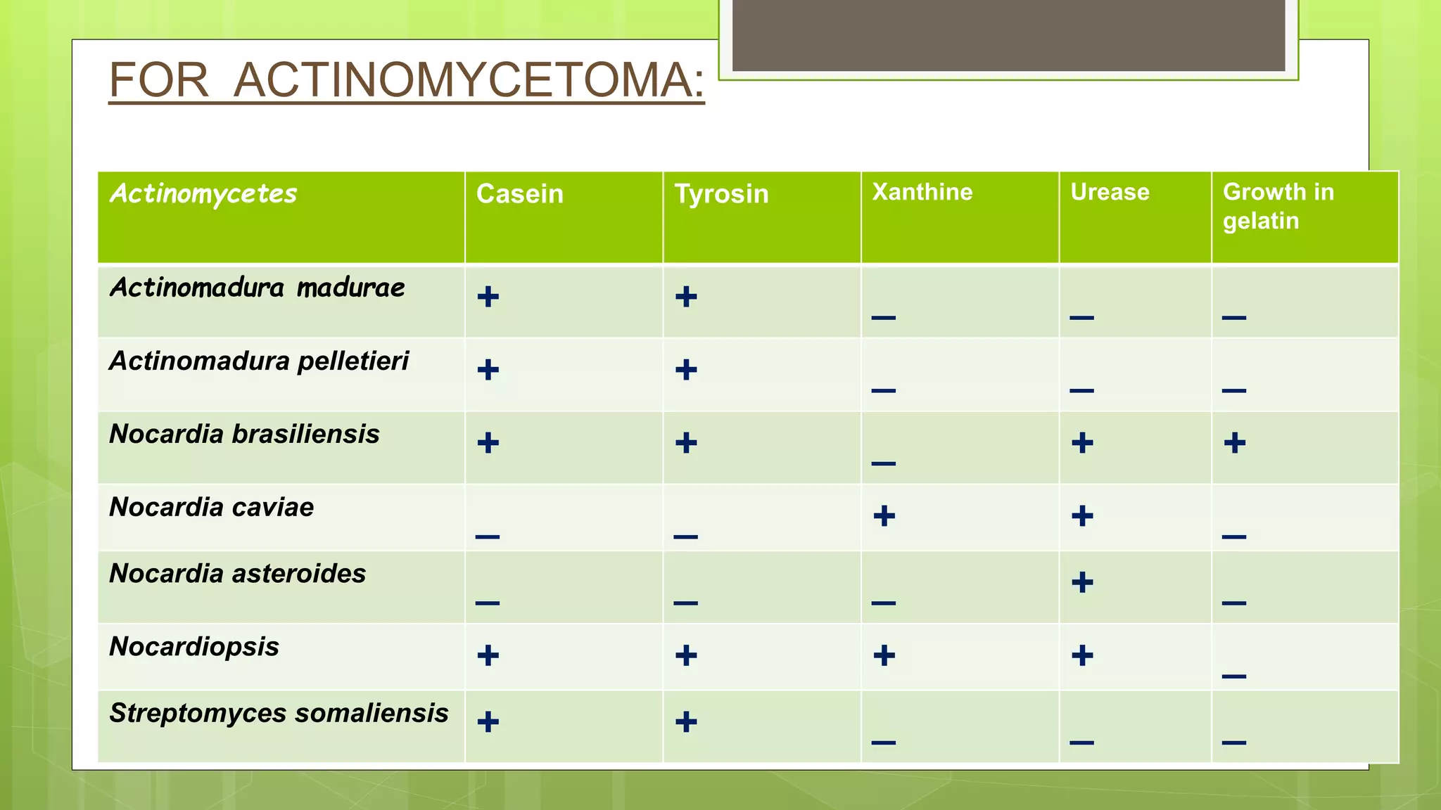
MYCETOMA
Dr. Sshrutkirti Gupta
Consultant Microbiologist
KEM Mumbai
OVERVIEW
History
Definition
Classification
Epidemiology
Etiology
Pathogenesis
Clinical manifestation
Diagnosis
Treatment
Prevention
History
 Ancient disease: roots are found in 4th Samhita of an
Atharva veda – Pada valmikam
 1694 - Kaempfer wrote in his thesis about ulcerous
hypersarcosis of foot.
A tuberous disease affecting feet with
discharging granules
 1842- John Gill describe it first time at Madurai
 1846- Colebrook describe a term “Madura Foot”
 1860- Henry Carter establish fungal etiology and coined
term Mycetoma
 1906- Brumpt first isolate Madurella mycetomi.
 1913- Pinoy separated disease in 2 groups
 2013-WHO declared it as Neglected tropical
disease.
Definition
A localised swollen lesion
Subject to a trauma
Contains granulomas and abscesses
which suppurate and drain through
sinus tracts.
Involves skin, subcutaneous tissue,
fascia and bone.
Localized
swelling
Sinus tract
Production of granules within
sinus tract
Classification
I. EUMYCETOMAS-caused by Eumycetes (fungi)
II. Schizomycetomas- caused by schizomycetes
A. ACTINOMYCETOMAS- caused by actinomycetes
B. BOTRYOMYCOSIS- bacterial pseudomycosis,
caused by true bacteria.
Epidemiology
Place : tropical and subtropical climate
Age : 20-40 yrs
M:F = 3.5-4 : 1
Seen commonly with agricultural workers, laborers.
Associated with decrease protective clothing.
World
Eumycetoma : Actinomycetoma -- 40% : 60%
SE Asia it is 35%:65%
In India ,Actinomycetoma -75% and
Eumycetoma --- more common in northern region
Etiology
I.EUMYCETES
Madurella mycetomatis
Madurella grisea
Allescheria boydii
Cephalosposporium spp.
Phialophora jeanselemei
Pyrenochaeta romeroi
Acremonium kilience
Leptosphaeria
senegalensis
Neotestudina rosatii
II.SCHIZOMYCETES
A.-ACTINOMYCETOMA
Nocardia brasilensis
Nocardia asteroides
Nocardia caviae
Actinomyces israelli
Actinomadura madurae
Actinomadura pilletieri
Streptomyces
somaliensis
B. BOTRYOMYCOSIS
Staphylococcus species
E. coli
Actinobacillus lingieresi
Proteus species
Pseudomonas aeruginosa
Streptococcus spp,
Pathogenesis
Route of Infection: Inoculation of organism in pre existing
abrasion or trauma.
Organism grow and multiply- development of grains
Three types of immune responses- to grains of
mycetoma
a) Neutrophils degranulation and adherence to grain
surface-leading to disintegration of grains.
b) Disappearance of neutrophils and arrival of
macrophages to clear grains and neutrophil debris
c) Formation of epithelioid granuloma-
macrophages, lymphocytes and plasma cells
Th2-like responses (interleukin (IL)-10 and IL-4) were
found in primary lesions and in draining lymph nodes
in S. somaliensis
 Factors related to Pathogen- Cell wall thickening and
melanin production
Immune system attempts
to engulf organism but
fails resulting in abscess
containing granules with
granulomatous
inflammation and fibrosis
Abscess discharges on to the skin surface or may involve
continuous structure.
Clinical manifestation
Lower Limb :Upper Limb = 3:1
Single area is involved,
Exposed region is more involved
Can affect neck, head and face
Lesion can appear after long time
Spontaneous healing of older sinus and simultaneous
spread of disease is also seen
Tendon & nerves are usually spared from direct
invasion
Morbidity increased due to secondary bacterial
infection.
Clinical features Actinomycetoma Eumycetoma
1. Organism Aerobic Actinomycetes Deuteromycetes or
ascomycetes.
2. Tumour mass Multiple,diffuse with ill defined
margins
Usually single,well defined
margins.
3.Sinuses Appear early, more in number Appear late, less in number
4.Sinus opening Raised, inflammed and flared
up
Flat opening, nor flared up
5.Flap of opening Easily removed Not easy to remove
6.Discharge Usually purulent Serous or sero-sanguinous
7.Grains White mostly Black or white
8.Extent of Involvement More extensive,obliterative with
hypertrophic, lytic lesion.
Less extensive,only
osteosclerotic lesion of bone.
DIFF OF CLINICAL FEATURES:
Differential diagnosis
Chronic bacterial osteomyelitis
Tubercular osteomyelitis
Chromoblastomycosis
Soft tissue tumor
Diagnosis
Localized
swelling
Sinus tract
Production of granules
within sinus tract
Lab diagnosis
To identify the organism and give appropriate treatment.
Gross examination of lesion and granules
Microscopy of granules
Culture
Serodiagnosis
Histopathology
Molecular methods
Sample collection
From an unruptured pustule.
 A large deep biopsy after removing mouth of an sinus
tract.
Collection of grains
Grains frequently found underneath the pus, collected
from the removed bandages
Collected by lifting the crust at the opening of a sinus
Aspirated from undrained sinuses
Collection of biopsy
Tissue is collected from center and edge of the lesion
Specimen placed between two sterile gauze pads,
sterile petridish or tube which contains 2-3 ml of
sterile NS or brain heart infusion broth
Gross examination
Size
Shape
Texture
Color
Eumycotic mycetoma
Dark grains
Madurella spp.
Leptosphaeria spp.
Exophiala spp.
Phaeoacremonium spp.
Phialophora spp.
Pale grains(white or
yellow)
Pseudallescheria boydii
Acremonium spp.
Neotestudina spp.
Actinomycetoma
Pale grains (white or yellow)
Actinomadura madurae
Nocardia brasiliensis
Yellow to brown grains
Streptomyces somalinensis
Red to brown grains
Actinomadurae pelletieri
LARGE SIZED PALE GRAINS PRODUCED IN
ACTINOMYCETOMA CAUSED BY Actinomadure madurae
Microscopy of Granules
Gram stain-
Gram stain-Actinomycetoma
Gram stain-
Gram positive
branching
filamentous bacteria
embedded in grain
material
(actinomycetoma).
10% KOH preparation -Grains are mounted KOH
and allowed to stand for 1–2 hours.
Eumycetoma grains:-
Seen with 2-6 µm (upto 15 µm ) wide interwoven
hyphae with or without large chlamydospores.
Actinomycotic grains:-
Have filaments with diameter of 0.5 to 1 µm, as well
as coccoid and bacillary form.
KOH Preparation
Modified ZN stain:
Nocardia species are red-pink acid fast filamentous
bacteria.
Others are non acid fast
Further confirmed by fungal stains.
Stain Eumycetoma Actinomycetoma
Hematoxylin-eosin Brownish colour Homogeneously
eosinophilic.
Giemsa Black with green
tinge
Blue in centre with
pink filament in
periphery.
Centrally, the organisms are
filamentous and basophilic;
Specular, red deposits have formed
at the periphery of the colonies
Splendore-Hoeppli phenomena
CULTURE
Done on different sets of media- for fungi and bacteria.
The rate of growth is highly variable and therefore cultures
should be kept for at least 6-8 weeks before they are discarded
as negative.
If actinomycetoma suspected on microscopy
Grains are washed several times with normal saline(NS)
without antibiotics,
then inoculated on- SDA without antibiotics,blood agar, LJ
media and brain heart infusion agar.
If eumycetoma suspected on microscopy:
Grains are washed several times in NS with antibiotics
(streptomycin, penicillin),
Then inoculated on SDA with antibiotic (chloramphenicol &
gentamycin).
Inoculate as many as grains as possible on several
plates .
Culture is incubated at 25°c, 37°c and 44°c.
(A) White to grey,
heaped, radially folded
colonies of Madurella
mycetomatis on
sabouraud dextrose
agar.
(B) Reverse of the agar
showing brown black
diffusible pigment
Culture of Nocardia
Dry, granular, chalky
growth
Paraffin Baiting Technique,
a) KOH mount
showing mycelial
clumps with septate
hyphae.
b) Lactophenol cotton blue
mount revealing septate
hyphae with
chlamydospores
M.mycetomatis SHOWING PHIALIDES
Agents Grains Colony
Madurella mycetomatis <2mm,firm,oval,black,hard,coal like
consistency
Smooth,glabrous,leathery,whitish-yellow
brown,moniliform hyphae
Madurella grisea <1mm,soft oval,irregular,hyaline centre
with black periphery
Raised,leathery,rapid growing,gray-olive
to reddish brown,gray aerial hyphae
Exophiala jeanselmei Smallest 0.2-0.3mm,black Moist become velvety later,olivacious
green mycelia
Actinomadura madurae Largest 2-5mm, white,soft White to tan, pink/orange,
glabrous,wrinkled,hard,adherent
Actinomadura pelletieri 1mm,hard,small,oval,red Slow growing,wrinkled small
dry,granular,pink-peach to red with waxy
surface
Nocardia brasiliensis <0.5mm,soft,small,white to yellow Slow growing wrinkled,yellow to
orange,dry chalky
Nocardia caviae <0.5mm,soft,small,white to yellow Cream to peach color
Nocardia asteroides Rare 0.5mm,soft,small,white,irregular Glabrous,chalky,wrinkled,white-orange
pink color,aerial hyphae
Streptomyces somaliensis 2mm,hard,round,large,yellow Creamy,wrinkled,tuft of brownish aerial
filaments
Fungal agents Gluc Lactose Malt Sucrose Starch hydrolysis Protease
activity
Madurella
mycetomatis + + + _ + +
Madurella grisea
+ _ + + + +
Exophila jeanselmei
+ + + + + _
P. boydii
+ _ + + + _
Acremonium
falciforme + _ _ + + _
Biochemical tests: for
eumycetoma
Actinomycetes Casein Tyrosin Xanthine Urease Growth in
gelatin
Actinomadura madurae
+ + _ _ _
Actinomadura pelletieri
+ + _ _ _
Nocardia brasiliensis
+ + _ + +
Nocardia caviae
_ _ + + _
Nocardia asteroides
_ _ _ + _
Nocardiopsis
+ + + + _
Streptomyces somaliensis
+ + _ _ _
FOR ACTINOMYCETOMA:
DST
Pen
Tobra
Ery Amk
Gen
Resistance of Drugs if
Gentamicin---- 15 mm
Tobramycin --- < 20 mm
Amikacin---- 20 mm
Erythromycin--- < 30 mm
Ciprofloxacin--- < 35 mm
Cefotaxime-----< 20 mm
CTX
SERODIAGNOSIS
No established serological study
Methods such as - Complement fixation test
Immunodiffusion
Counter-immuno-electrophoresis
Enzyme linked immunosorbent assays
Western blot
Skin test not diagnostically useful
Cell extracts rather than exoantigens are used in most of the tests
• 26s rRNA ITS sequencing
(Internal Transcribed Spacer (ITS) sequences)
• Multilocus sequence analysis- large subunit, small
subunit 18s nrDNA, B-tubulin and chitin synthase 1
regions.
MOLECULAR METHODS
RADIO-DIAGNOSIS
USG: soft tissue and bone swelling
X-Ray:
– Soft tissue swelling
– Endosteal cavitations
– Small cystic lesion
– Periosteal bone erosion
– Hyper osteosis
MRI: Anatomical distribution
TREATMENT
Treatment of eumycetomas with chemotherapy is difficult
and their management often involves conservative
approaches involving observation, a trial of chemotherapy,
and surgery.
 Conservative management:
 Eumycetomas are seldom life-threatening and are often asymptomatic.
 Relief of pain and dressings of sinuses may be required from time to
time.
 Chemotherapy:
 In some cases, responses have been seen with amphotericin B, either
in conventional formulation or in combination with lipids or liposomes;
ketoconazole may also be effective.
 Eumycetoma- ketoconazole 200 mg daily for 3 months.
 Actinomycetoma- antibiotics with antifungals.
 Griseofulvin-slows the progress of mycetomas in some pateints, an
active inhibitor of leukocyte chemotaxis and suppresses the
accumulation of neutrophils in lesions.
Pretreatment Post treatment
• Amenable to medical treatment
• First line: Streptomycin + dapsone
 If no response: replace dapsone with cotrimoxazole
Amikacin with cotrimoxazole (Welsh regimen)
• Second line: Rifampicin, Sulfadoxine + pyrimethamine ,
sulphonamides.
• Mean duration: 1 year
• Cure rate: 60-90%
ACTINOMYCETOMA:RX
Pretreatment Post treatment
The ultimate method of eradicating a fungal mycetoma is surgical
excision.
 It has to be sufficiently radical to ensure an adequate margin of
excision, otherwise relapse is inevitable.
The definitive curative process is adequate amputation, again
ensuring that the margins are well clear of infected bone or
subcutaneous tissue.
SURGERY
PREVENTION
No preventive Vaccine
Reduce traumatic inoculation of organism
Protective clothing, footwears
Early identification & treatment of lesion
THANK YOU..

Mycetoma.pptx

  • 1.
  • 2.
  • 3.
    History  Ancient disease:roots are found in 4th Samhita of an Atharva veda – Pada valmikam  1694 - Kaempfer wrote in his thesis about ulcerous hypersarcosis of foot. A tuberous disease affecting feet with discharging granules
  • 4.
     1842- JohnGill describe it first time at Madurai  1846- Colebrook describe a term “Madura Foot”  1860- Henry Carter establish fungal etiology and coined term Mycetoma  1906- Brumpt first isolate Madurella mycetomi.  1913- Pinoy separated disease in 2 groups
  • 5.
     2013-WHO declaredit as Neglected tropical disease.
  • 6.
    Definition A localised swollenlesion Subject to a trauma Contains granulomas and abscesses which suppurate and drain through sinus tracts. Involves skin, subcutaneous tissue, fascia and bone.
  • 7.
  • 8.
    Classification I. EUMYCETOMAS-caused byEumycetes (fungi) II. Schizomycetomas- caused by schizomycetes A. ACTINOMYCETOMAS- caused by actinomycetes B. BOTRYOMYCOSIS- bacterial pseudomycosis, caused by true bacteria.
  • 9.
    Epidemiology Place : tropicaland subtropical climate Age : 20-40 yrs M:F = 3.5-4 : 1 Seen commonly with agricultural workers, laborers. Associated with decrease protective clothing.
  • 10.
    World Eumycetoma : Actinomycetoma-- 40% : 60% SE Asia it is 35%:65% In India ,Actinomycetoma -75% and Eumycetoma --- more common in northern region
  • 11.
    Etiology I.EUMYCETES Madurella mycetomatis Madurella grisea Allescheriaboydii Cephalosposporium spp. Phialophora jeanselemei Pyrenochaeta romeroi Acremonium kilience Leptosphaeria senegalensis Neotestudina rosatii II.SCHIZOMYCETES A.-ACTINOMYCETOMA Nocardia brasilensis Nocardia asteroides Nocardia caviae Actinomyces israelli Actinomadura madurae Actinomadura pilletieri Streptomyces somaliensis
  • 12.
    B. BOTRYOMYCOSIS Staphylococcus species E.coli Actinobacillus lingieresi Proteus species Pseudomonas aeruginosa Streptococcus spp,
  • 13.
    Pathogenesis Route of Infection:Inoculation of organism in pre existing abrasion or trauma. Organism grow and multiply- development of grains Three types of immune responses- to grains of mycetoma a) Neutrophils degranulation and adherence to grain surface-leading to disintegration of grains.
  • 14.
    b) Disappearance ofneutrophils and arrival of macrophages to clear grains and neutrophil debris c) Formation of epithelioid granuloma- macrophages, lymphocytes and plasma cells Th2-like responses (interleukin (IL)-10 and IL-4) were found in primary lesions and in draining lymph nodes in S. somaliensis  Factors related to Pathogen- Cell wall thickening and melanin production
  • 15.
    Immune system attempts toengulf organism but fails resulting in abscess containing granules with granulomatous inflammation and fibrosis
  • 16.
    Abscess discharges onto the skin surface or may involve continuous structure.
  • 17.
    Clinical manifestation Lower Limb:Upper Limb = 3:1 Single area is involved, Exposed region is more involved Can affect neck, head and face Lesion can appear after long time
  • 18.
    Spontaneous healing ofolder sinus and simultaneous spread of disease is also seen Tendon & nerves are usually spared from direct invasion Morbidity increased due to secondary bacterial infection.
  • 19.
    Clinical features ActinomycetomaEumycetoma 1. Organism Aerobic Actinomycetes Deuteromycetes or ascomycetes. 2. Tumour mass Multiple,diffuse with ill defined margins Usually single,well defined margins. 3.Sinuses Appear early, more in number Appear late, less in number 4.Sinus opening Raised, inflammed and flared up Flat opening, nor flared up 5.Flap of opening Easily removed Not easy to remove 6.Discharge Usually purulent Serous or sero-sanguinous 7.Grains White mostly Black or white 8.Extent of Involvement More extensive,obliterative with hypertrophic, lytic lesion. Less extensive,only osteosclerotic lesion of bone. DIFF OF CLINICAL FEATURES:
  • 20.
    Differential diagnosis Chronic bacterialosteomyelitis Tubercular osteomyelitis Chromoblastomycosis Soft tissue tumor
  • 21.
  • 22.
  • 23.
    Lab diagnosis To identifythe organism and give appropriate treatment. Gross examination of lesion and granules Microscopy of granules Culture Serodiagnosis Histopathology Molecular methods
  • 24.
    Sample collection From anunruptured pustule.  A large deep biopsy after removing mouth of an sinus tract.
  • 25.
    Collection of grains Grainsfrequently found underneath the pus, collected from the removed bandages Collected by lifting the crust at the opening of a sinus Aspirated from undrained sinuses
  • 26.
    Collection of biopsy Tissueis collected from center and edge of the lesion Specimen placed between two sterile gauze pads, sterile petridish or tube which contains 2-3 ml of sterile NS or brain heart infusion broth
  • 27.
  • 28.
    Eumycotic mycetoma Dark grains Madurellaspp. Leptosphaeria spp. Exophiala spp. Phaeoacremonium spp. Phialophora spp. Pale grains(white or yellow) Pseudallescheria boydii Acremonium spp. Neotestudina spp.
  • 29.
    Actinomycetoma Pale grains (whiteor yellow) Actinomadura madurae Nocardia brasiliensis Yellow to brown grains Streptomyces somalinensis Red to brown grains Actinomadurae pelletieri
  • 30.
    LARGE SIZED PALEGRAINS PRODUCED IN ACTINOMYCETOMA CAUSED BY Actinomadure madurae
  • 31.
  • 32.
    Gram stain-Actinomycetoma Gram stain- Grampositive branching filamentous bacteria embedded in grain material (actinomycetoma).
  • 33.
    10% KOH preparation-Grains are mounted KOH and allowed to stand for 1–2 hours. Eumycetoma grains:- Seen with 2-6 µm (upto 15 µm ) wide interwoven hyphae with or without large chlamydospores. Actinomycotic grains:- Have filaments with diameter of 0.5 to 1 µm, as well as coccoid and bacillary form.
  • 34.
  • 35.
    Modified ZN stain: Nocardiaspecies are red-pink acid fast filamentous bacteria. Others are non acid fast Further confirmed by fungal stains.
  • 36.
    Stain Eumycetoma Actinomycetoma Hematoxylin-eosinBrownish colour Homogeneously eosinophilic. Giemsa Black with green tinge Blue in centre with pink filament in periphery.
  • 37.
    Centrally, the organismsare filamentous and basophilic; Specular, red deposits have formed at the periphery of the colonies Splendore-Hoeppli phenomena
  • 38.
    CULTURE Done on differentsets of media- for fungi and bacteria. The rate of growth is highly variable and therefore cultures should be kept for at least 6-8 weeks before they are discarded as negative.
  • 39.
    If actinomycetoma suspectedon microscopy Grains are washed several times with normal saline(NS) without antibiotics, then inoculated on- SDA without antibiotics,blood agar, LJ media and brain heart infusion agar. If eumycetoma suspected on microscopy: Grains are washed several times in NS with antibiotics (streptomycin, penicillin), Then inoculated on SDA with antibiotic (chloramphenicol & gentamycin).
  • 40.
    Inoculate as manyas grains as possible on several plates . Culture is incubated at 25°c, 37°c and 44°c.
  • 41.
    (A) White togrey, heaped, radially folded colonies of Madurella mycetomatis on sabouraud dextrose agar. (B) Reverse of the agar showing brown black diffusible pigment
  • 42.
    Culture of Nocardia Dry,granular, chalky growth
  • 43.
  • 44.
    a) KOH mount showingmycelial clumps with septate hyphae. b) Lactophenol cotton blue mount revealing septate hyphae with chlamydospores
  • 45.
  • 46.
    Agents Grains Colony Madurellamycetomatis <2mm,firm,oval,black,hard,coal like consistency Smooth,glabrous,leathery,whitish-yellow brown,moniliform hyphae Madurella grisea <1mm,soft oval,irregular,hyaline centre with black periphery Raised,leathery,rapid growing,gray-olive to reddish brown,gray aerial hyphae Exophiala jeanselmei Smallest 0.2-0.3mm,black Moist become velvety later,olivacious green mycelia Actinomadura madurae Largest 2-5mm, white,soft White to tan, pink/orange, glabrous,wrinkled,hard,adherent Actinomadura pelletieri 1mm,hard,small,oval,red Slow growing,wrinkled small dry,granular,pink-peach to red with waxy surface Nocardia brasiliensis <0.5mm,soft,small,white to yellow Slow growing wrinkled,yellow to orange,dry chalky Nocardia caviae <0.5mm,soft,small,white to yellow Cream to peach color Nocardia asteroides Rare 0.5mm,soft,small,white,irregular Glabrous,chalky,wrinkled,white-orange pink color,aerial hyphae Streptomyces somaliensis 2mm,hard,round,large,yellow Creamy,wrinkled,tuft of brownish aerial filaments
  • 47.
    Fungal agents GlucLactose Malt Sucrose Starch hydrolysis Protease activity Madurella mycetomatis + + + _ + + Madurella grisea + _ + + + + Exophila jeanselmei + + + + + _ P. boydii + _ + + + _ Acremonium falciforme + _ _ + + _ Biochemical tests: for eumycetoma
  • 48.
    Actinomycetes Casein TyrosinXanthine Urease Growth in gelatin Actinomadura madurae + + _ _ _ Actinomadura pelletieri + + _ _ _ Nocardia brasiliensis + + _ + + Nocardia caviae _ _ + + _ Nocardia asteroides _ _ _ + _ Nocardiopsis + + + + _ Streptomyces somaliensis + + _ _ _ FOR ACTINOMYCETOMA:
  • 49.
    DST Pen Tobra Ery Amk Gen Resistance ofDrugs if Gentamicin---- 15 mm Tobramycin --- < 20 mm Amikacin---- 20 mm Erythromycin--- < 30 mm Ciprofloxacin--- < 35 mm Cefotaxime-----< 20 mm CTX
  • 50.
    SERODIAGNOSIS No established serologicalstudy Methods such as - Complement fixation test Immunodiffusion Counter-immuno-electrophoresis Enzyme linked immunosorbent assays Western blot Skin test not diagnostically useful Cell extracts rather than exoantigens are used in most of the tests
  • 51.
    • 26s rRNAITS sequencing (Internal Transcribed Spacer (ITS) sequences) • Multilocus sequence analysis- large subunit, small subunit 18s nrDNA, B-tubulin and chitin synthase 1 regions. MOLECULAR METHODS
  • 52.
    RADIO-DIAGNOSIS USG: soft tissueand bone swelling X-Ray: – Soft tissue swelling – Endosteal cavitations – Small cystic lesion – Periosteal bone erosion – Hyper osteosis MRI: Anatomical distribution
  • 53.
    TREATMENT Treatment of eumycetomaswith chemotherapy is difficult and their management often involves conservative approaches involving observation, a trial of chemotherapy, and surgery.
  • 54.
     Conservative management: Eumycetomas are seldom life-threatening and are often asymptomatic.  Relief of pain and dressings of sinuses may be required from time to time.  Chemotherapy:  In some cases, responses have been seen with amphotericin B, either in conventional formulation or in combination with lipids or liposomes; ketoconazole may also be effective.  Eumycetoma- ketoconazole 200 mg daily for 3 months.  Actinomycetoma- antibiotics with antifungals.  Griseofulvin-slows the progress of mycetomas in some pateints, an active inhibitor of leukocyte chemotaxis and suppresses the accumulation of neutrophils in lesions.
  • 55.
  • 56.
    • Amenable tomedical treatment • First line: Streptomycin + dapsone  If no response: replace dapsone with cotrimoxazole Amikacin with cotrimoxazole (Welsh regimen) • Second line: Rifampicin, Sulfadoxine + pyrimethamine , sulphonamides. • Mean duration: 1 year • Cure rate: 60-90% ACTINOMYCETOMA:RX
  • 57.
  • 58.
    The ultimate methodof eradicating a fungal mycetoma is surgical excision.  It has to be sufficiently radical to ensure an adequate margin of excision, otherwise relapse is inevitable. The definitive curative process is adequate amputation, again ensuring that the margins are well clear of infected bone or subcutaneous tissue. SURGERY
  • 59.
    PREVENTION No preventive Vaccine Reducetraumatic inoculation of organism Protective clothing, footwears Early identification & treatment of lesion
  • 60.