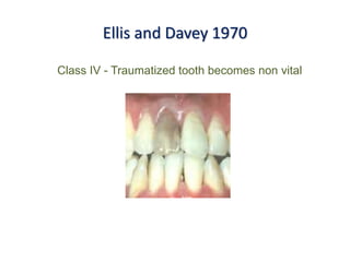
Introduction
• Trauma: Trauma refers to injury;
damage; impairment; external
violence producing injury or
degeneration.
• Traumatic injury: It may be defined as
a damage to a part of the body tissue.
Etiology
Falls or Collision
fall from baby carriage, child begins to crawl, stand or walk.
Child abuse – inflicted, non accidental injuries
Sports – teenagers, contact type of sports.
Automobile, bicycle accidents
Fights
Grouped in to
• Intentional (Abuse)
• Unintentional (Sporting
activity, falls)
• Direct trauma
• Indirect trauma: Blow to
chin – sudden closure of
mandibular teeth with their
opponents
Predisposing Factors
Angle’s class II type 1 malocclusion - Increased overjet with
protrusion of upper incisors and insufficient lip closure
Epileptic patients
Children with cerebral palsy
o Abnormal muscle tone
o Poor skeletal and muscle co- ordination
Dentinogenesis Imperfecta
Deleterious oral habits
Epidemiology
• Boys affected almost
twice as often as girls.
• Peak incidence of dental
injuries at 2 to 4 and 8 to
10 years of age.
Classification
Ellis and Davey 1970
Class I - Simple fracture of crown involving little or no dentin
Ellis and Davey 1970
Class II - Extensive fracture of crown involving dentin but
no pulp
Ellis and Davey 1970
Class III - Extensive involvement of crown with pulp exposure
Ellis and Davey 1970
Class IV - Traumatized tooth becomes non vital
Ellis and Davey 1970
Class V - Tooth lost as a result of trauma
Ellis and Davey 1970
Class VI – Root fracture with or without loss of crown
structure
Ellis and Davey 1970
Class VII - Displacement of tooth without fracture of
crown or root
Ellis and Davey 1970
Class VIII - Fracture of crown en masse and its replacement
Ellis and Davey 1970
Class IX - Traumatic injuries to primary teeth
WHO CLASSIFICATION -1992
• Injuries to the Hard Dental Tissues and Pulp
• Injuries to periodontal tissues
• Injuries of the Supporting Bone
• Injuries to Gingiva or Oral Mucosa
Injuries to the Hard Dental Tissues and Pulp
Crown infraction - N 502.50
Injuries to the Hard Dental Tissues and Pulp
Enamel fracture - N 502.50
Injuries to the Hard Dental Tissues and Pulp
Crown fracture without pulpal involvement – N 502.51
Injuries to the Hard Dental Tissues and Pulp
Crown fracture with pulpal involvement – N 502.52
Injuries to the Hard Dental Tissues and Pulp
Root fracture – N 502.53
Injuries to the Hard Dental Tissues and Pulp
Crown root fracture without pulpal involvement – N 502.54
&
Crown root fracture with pulpal involvement – N 502.54
Uncomplicated Complicated
Injuries to the Periodontal Tissues
Concussion – N 503.20
Injury to the tooth supporting structures
without abnormal loosening or
displacement of teeth. Blood supply to the
pulp is rarely affected
Injuries to the Periodontal Tissues
Subluxation – N 503.20
Slight increase in mobility, but without
malposition of teeth. The blood supply
to the pulp may be affected.
Injuries to the Periodontal Tissues
Extrusive Luxation – N 503.20
Injuries to the Periodontal Tissues
Lateral Luxation – N 503.20
Injuries to the Periodontal Tissues
Intrusive Luxation – N 503.21
Injuries to the Periodontal Tissues
Avulsion – N 503.22
Injuries to Gingiva or Oral mucosa
Abrasion S 00.50
Superficial wound in which epithelial
tissue is rubbed or scratched
Injuries to Gingiva or Oral mucosa
Hemorrhage of subcutaneous tissue
without laceration of epithelial tissue.
Etiology: Blunt object hitting the tissue
Contusion S 00.50
Injuries to Gingiva or Oral mucosa
Tearing of tissues caused by
sharp object
Laceration S 01.50
Injuries of the supporting bone
Comminution of alveolar socket
(Mandible N802.20, Maxilla 802.40)
Crushing and compression of the
alveolar socket. Intrusion and lateral
luxation.
Injuries of the supporting bone
Fracture of the alveolar socket wall
(Mandible N802.20, Maxilla N802.40)
A fracture contained to the facial or
lingual socket wall.
Injuries of the supporting bone
Fracture of the alveolar process
(Mandible N802.20, Maxilla N802.40)
A fracture of the alveolar process
which may or may not involve the
alveolar socket.
Injuries of the supporting bone
Fracture of the Mandible and maxilla
(Mandible N802.21, Maxilla N802.42)
• A fracture involving the base of the mandible or maxilla and
often the alveolar process (jaw fracture).
Examination & Diagnosis
Trauma Triad
• When the injury occur? - will imply a time factor.
** Time interval between injury and treatment – affects Prognosis of
pulp exposure, displacement and avulsion.
• Where did the injury occur? - indicate the possibility of
contamination of wounds. Tetanus prophylaxis
• How did the injury occur? - indicate the location of possible injury
zones
• History of previous dental injuries?
• Was there a period of unconsciousness? Episodes of headache?
Amnesia? Nausea? Vomiting? - Signs of cerebral involvement and
require medical attention.
• General Health
• Is there spontaneous pain from the teeth? - indicates damage to
PDL(hyperemia), Pulp (crown fracture and crown root fracture)
• Is there any reaction in the teeth to cold and/or heat? - Exposure of
dentin or pulp.
• Is there any disturbance in the bite? - Tooth luxation, alveolar
fracture, jaw fracture.
• Recording of extraoral wounds
Wound (Chin) – premolar & molar regions, condylar fracture
• Recording of injuries to oral mucosa
Wounds penetrating the entire thickness of lip (Two parallel wounds
– possibility of tooth fragment burried between the lacerations)
• Examination of the crowns of teeth - Presence and extent of
fractures, pulpal exposure, or changes in color.
• Recording of displacement of teeth.
- Visual examination.
- Direction of the dislocation as well as extent (in mm).
• Disturbances in occlusion
• Abnormal mobility of teeth or alveolar fragments
- Horizontally and axially (Mobility of teeth).
- Typical sign of alveolar fracture is movement of adjacent teeth
when the mobility of a single tooth is tested.
• Tenderness of teeth to percussion.
• Percussion tone
Hard Metallic - Tooth is locked into bone (intrusion).
Dull sound - Subluxation or Extrusive luxation.
Pulp vitality
• Positive/Negative response – Pulp alive/death
• Following trauma – tooth in a state of shock – false response
• Recommended pulp testing: immediately, 2 weeks, 1,2,6,12 months
and then at yearly interval for next 3 years.
Various methods of pulp vitality testing
• Thermal stimulation – Heated GP, CO2snow, ice
• Electrical pup testing
• Laser doppler flowmetry
• Pulse oximetry
Radiographic examination
• Reveals the stage of root formation
• Discloses injuries affecting the root
portion of the tooth and Pdl structures.
• Widening of the periodontal space,
Blurred periodontal space.
• Dislocated tooth fragments within a lip
laceration
ENAMEL INFRACTIONS
 Crazing within the enamel substance which
do not cross the DEJ.
 Horizontal, vertical or diverging.
 Transillumination.
TREATMENT
 Multiple infraction lines – seal with unfilled
resin and acid etch technique.
UNCOMPLICATED CROWN FRACTURE
Uncomplicated
crown fracture
Enamel fractures
Small Fracture
Selective
grinding
Large fracture
Composite
resin
Crown
fragment
Reattachment
Enamel-Dentin Fractures
Dentin coverage
Ca(OH)2
Composite
resin
Crown
fragment
Reattachment
Selective Grinding
Composite Resin Restoration
Dentin Coverage
Fragment Reattachment
• Improved esthetics.
• Incisal edge of the tooth fragment wears at a similar rate to
the adjacent teeth.
• More economical.
Uncomplicated crown fracture
Broken fragment
Fragment mounted (sticky wax)
Dentin coverage
Acid etching
Removal of etchant
Dentin conditioning
Fragment Bonding Curing
Groove preparation
Final restoration
Finishing
Fragment Reattachment Technique
COMPLICATED CROWN FRACTURE
Four factors contribute the management of Complicated
crown fracture:
1. The Length Of Time Elapsed Since The Injury Occur.
2. The Size Of The Pulp Exposure.
3. The Condition Of The Pulp (Vital Or Non Vital).
4. Stage Of Root Development
COMPLICATED CROWN FRACTURE
Complicated Crown
fracture
Vital Pulp
Tooth with
open apex
Smaller pulp
exposure
Direct pulp
capping
Larger pulp
exposure
Pulpotomy
Tooth with
closed apex
Small
exposure
Direct pulp
capping
Larger
exposure
Root canal
treatment
Non-vital
pulp
Tooth with
open apex
Apexification
Tooth with
closed apex
Root canal
treatment
Direct Pulp capping (DPC)
Pulpotomy
1. Fractured Crown With Pulp Exposure.
2. Pulpotomy Using MTA For Pulpal Protection.
3. One-year Follow-up; Note Continued Root Formation.
4. Two-year Recall Showing Further Root Development.
Pulpotomy
Root canal Treatment
• Removal of the infected soft tissue within the tooth and
its replacement by an artificial inert ‘filling’ material
Apexification
CROWN ROOT FRACTURE
Crown Root fracture
Uncomplicated (a, b)
1.Position of fracture
- near crestal bony margin
2. No or slight bleeding
3. Coronal Fragment Mobile.
4. Pulp vitality: +ve for apical fragment
5. Radiographically, Apical extension of
fracture - not visible
Complicated (C)
1. Position of fracture
- infrabony margin
2. Bleeding from Pulp & PDL
3. Coronal Fragment Mobile.
4. Pulp vitality: +ve for apical fragment
5. Apical extension of fracture - not
visible
Treatment
Emergency
Temporary stabilization of
a loose segment to
adjacent teeth (Splint)
Definitive
1 - Fragment removal & Supragingival restoration
2 - Fragment removal & Gingivectomy
3 - Orthodontic extrusion
4 - Surgical extrusion
Fragment Removal
 Removal of superficial coronal crown-root fragment and
subsequent restoration of exposed dentin.
 Indicated in uncomplicated crown root fracture.
 Easy to perform, Restoration can be completed soon after injury.
Fragment removal Clean with water
Wait for 2- 3 weeks for
gingival healing (Temporary
restoration)
Disinfect with sodium
hypochloride
Apply GIC to exposed
dentine
Restore with composite
Fragment Removal
Fragment Removal With Gingivectomy
 Removal of segment of the fractured fragment with subsequent
endodontic treatment and restored with crown.
 Preceded by Gingivectomy and sometimes Osteotomy.
 Indicated in where denudation of the fracture site does not
compromise esthetics.
 Easy to perform.
Fragment removal
Amputate the coronal
pulp
GIC application
Gingivectomy and ostectomy. Restore with crown.
Orthodontic Extrusion of Apical Fragment
 To orthodontically move the fracture to a supragingival position.
 Indicated in all types of fractures, assuming that reasonable root
length can be achieved after extrusion.
 Stable position of the restored tooth. Optimal gingival health.
 Time consuming procedure.
Complicated crown root fracture Endodontic treatment
Orthodontic extrusion Postoperative
Surgical Extrusion
 To surgically move the fracture to a supragingival position
 Indicated in all types of fractures (except crown-root fractures in
teeth with open apices)
 Rapid procedure, Stable position of the tooth.
 Risk for root resorption and marginal breakdown of the
periodontium.
Extract & reposition apical
fragment with forceps
Perform pulp extirpation &
seal root canal
Apply resin splint for 4
weeks.
RCT initiated 3-4 weeks later.
After another 1-2 months, the tooth can be restored with a post-retained crown.
ROOT FRACTURE
• Maxillary central incisor region
in the age group of 11 to 20
years.
• Clinically, slightly extruded
tooth.
Root fracture
Horizontal
Apical 3rd Middle 3rd Coronal 3rd
Vertical
Extraction
• RCT till the possible working length
• Apical surgery to remove the apical fragment
Apical 3rd
(C)
• RCT involving both fragments and obturated
with silver points (splint)
Middle 3rd
(B)
• Orthodontic or surgical extrusion
• Immobilization and later crown fabrication
Coronal 3rd
(A)
Clinical findings Radiographic
findings
Treatment
• Tooth is tender to percussion
• Not displaced
• No mobility
• No radiographic
abnormalities
• No treatment is
needed.
• Slight adjustment of
opposing tooth to
relieve occlusion
• Soft diet for 10 – 14
days.
Concussion
Subluxation
Clinical findings Radiographic
findings
Treatment
• Tooth is tender to percussion
• Increased mobility
• Not displaced.
• Bleeding from gingival crevice.
• No Radiographic
abnormalities
• Splinting for 10 days
• Soft diet for 10 – 14
days.
Extrusive luxation
Clinical findings Radiographic
findings
Treatment
Tooth is mobile.
Clinically crown appears longer
Bleeding from gingival crevice.
Tooth is tender to Percussion
The width of the
periodontal ligament
space is increased
Administer L.A if forceful
positioning is anticipated
Reposition the tooth.
Splint the tooth for 2 -3 weeks.
Advice soft diet
Follow up period of 1 year
Lateral luxation
• Treatment
• Treatment
Clinical findings Radiographic findings Treatment
• Tooth is displaced in palatal
or labial direction.
• Fracture of vestibular part of
the socket wall.
• Immobile
• Percussion - metallic.
• The widened PDL
space
• Administer L.A
• Reposition the tooth
• Splint the tooth for 6 – 8
weeks
• Advice soft diet
• Follow up period of 1 year
Forceful displacement of the root tip
through the facial alveolar wall.
To dislodge the root tip from its bony lock, firm
digital pressure is applied (incisal direction)
Then pushed apically into its correct position
After tooth repositioning, the labial &palatal bone
plates - compressed
Lacerated gingiva re-adapted to the neck of the
tooth and sutured.
The tooth should be splinted in its normal position
Intrusive luxation
Clinical findings Radiographic
findings
Treatment
• Tooth is displaced axially
into the alveolar bone.
• Clinically crown appears
shorter
• Immobile
• Percussion - metallic.
• The PDL space
may be absent
from all or part
of the root .
Teeth with open apex:
• Allow spontaneous
repositioning to take place.
• If no movement is noted within
3 weeks, recommend rapid
orthodontic repositioning.
Teeth with closed apex:
• Repositioned either
orthodontically or surgically as
soon as possible and initiate
RCT with in 3 weeks .
Avulsion
Clinical findings Radiographic findings Treatment
• Bleeding socket with
missing tooth.
• Empty socket
• Associated bone
fractures.
• Reimplantation with
subsequent endodontic
therapy.
8- to 10-year-olds
Permanent maxillary central incisor.
Replantation
• Prognosis depends on the amount of time the tooth was extraorally
and the medium used for the storage.
• To minimize the PDL tissue damage and pulpal infection.
Instructions on telephone
Locate the tooth
Rinse gently under tap water to clean the debris
No scrubbing or scraping of the root
Should be handled by holding the crown
Insert the tooth into socket and gently occlude with guaze and should
visit the dental office as soon as possible.
If it cannot be replanted, the tooth should be placed in suitable medium.
Visit the dentist as
soon as possible
Storage media
• HBSS (Hanks balanced salt solution)
• Milk
• Isotonic saline
• Buccal vestibule or under the tongue
• Contact lens solution
• Saliva
• Physiological pH range (7.2 - 7.6)
• Provides cells with a source of water
and essential inorganic ions, and a
carbohydrate as an energy source.
• Save-A-Tooth®
- Emergency Tooth Preserving System
Hanks’ balanced salt solution (HBSS)
Soak in 1 % doxy
soln - 5 mts
Category 2
Immediately
replanted at
the accident
site
Extraoral time < 20 mts
& Transported - HBSS
or milk (20 mts – 6 hrs)
Tooth kept moist (In water,
saliva or non physiologic
media) - 20 - 60 mts
Extraoral
dry time
< 60 mts
Extraoral
dry time
> 60 mts
Category 1 Category 3
Change transport to
HBSS or cold milk
Debride - soft pumice
prophylaxis or 3%
citric acid for 3 mts &
rinse well or gentle
scaling & root planing
Place in NAF
for 5 mts
Obtain radiographs to verify position
Rx: Doxycycline or Pencillin VK for 7 days
Assess tetanus vaccination
Provide postoperative instructions
Follow up: 1 week, 1, 3, 6, 12 months and annualy for 5 years
Category 2
Immediately
replanted at
the accident
site
Extraoral time < 20mts
& transported - HBSS
or milk (20 mts – 6 hrs)
Tooth - kept moist (In water,
saliva or other non physiologic
media) - 20 - 60 mts
> 60 mts
extraoral
dry time
Category 1 Category 3
Change transport to
HBSS, cold milk
Debride - soft pumice
prophylaxis or 3%
citric acid for 3 mts &
rinse well or gentle
scaling & root planing
Place in NAF
for 5 mts
Obtain radiographs to verify position
Rx: Doxycycline or Pencillin VK for 7 days
Assess tetanus vaccination
Category 4
 Pulp extirpation/debridement as soon as possible
 Place Ca(OH)2 slurry & change every 3 months
 Complete root canal therapy
Follow up: 1 week, 1, 3, 6, 12 months and annualy for 5 years
Healing of replanted teeth
• Surface resorption
• Replacement resorption (Ankylosis)
• Inflammatory resorption
Surface resorption
• Characterized by superficial resorption lacunae repaired by new
cementum
• Clinically tooth is normal and normal percussion tone
Replacement resorption (Ankylosis)
• Etiology: Absence of vital PDL cover on the root surface
• Bone precursor cells occupies the damaged root surface
• Osteoclasts – resorbs dentin, osteoblasts – lays the bone.
• Clinically: infraocclusion in the developing dentition,
Inflammatory resorption
Injury to PDL tissue
during trauma
Bacterial
contamination
Small resorption
cavities on root
surface
Cavities expose
dentinal tubules
Toxins from the
infected necrotic pulp
penetrate along these
tubules to PDL tissue
Provokes
inflammatory
response
In turn will intensify
the resorption and
within few months the
entire root is resorbed
• Radiographically, radiolucent bowl shaped cavitations along root
surface with corresponding excavations in adjacent bone
• Clinically the replanted tooth is loose, extruded and sensitive to
percussion with dull tone
SPLINTING
• Stabilization of repositioned or replanted teeth following trauma.
• Removable or fixed
: Sved splint
Ideal requirements
• Should be quick and easy to fabricate
• should not traumatize the teeth and gingiva
• should stabilize the injured tooth
• should not interfere with occlusion
• should have access to endodontic therapy if needed
• should allow a normal physiologic tooth mobility
• should be easily removed.
INSTRUCTION TO PARENTS AFTER
TRAUMATIC INJURY
• Give the child soft diet for few days
• Maintain the child’s fluid intake to avoid dehydration
• Maintain good oral hygiene
• Use of CHX mouthwash twice a day for 1 week
• Inform the possible complications that would arise in the injured
teeth
TRAUMATIC  INJURIES.pptx

TRAUMATIC INJURIES.pptx

  • 2.
    Introduction • Trauma: Traumarefers to injury; damage; impairment; external violence producing injury or degeneration. • Traumatic injury: It may be defined as a damage to a part of the body tissue.
  • 3.
    Etiology Falls or Collision fallfrom baby carriage, child begins to crawl, stand or walk.
  • 4.
    Child abuse –inflicted, non accidental injuries
  • 5.
    Sports – teenagers,contact type of sports.
  • 6.
  • 7.
  • 8.
    Grouped in to •Intentional (Abuse) • Unintentional (Sporting activity, falls) • Direct trauma • Indirect trauma: Blow to chin – sudden closure of mandibular teeth with their opponents
  • 9.
    Predisposing Factors Angle’s classII type 1 malocclusion - Increased overjet with protrusion of upper incisors and insufficient lip closure
  • 10.
  • 11.
    Children with cerebralpalsy o Abnormal muscle tone o Poor skeletal and muscle co- ordination
  • 12.
  • 13.
  • 14.
    Epidemiology • Boys affectedalmost twice as often as girls. • Peak incidence of dental injuries at 2 to 4 and 8 to 10 years of age.
  • 15.
  • 16.
    Ellis and Davey1970 Class I - Simple fracture of crown involving little or no dentin
  • 17.
    Ellis and Davey1970 Class II - Extensive fracture of crown involving dentin but no pulp
  • 18.
    Ellis and Davey1970 Class III - Extensive involvement of crown with pulp exposure
  • 19.
    Ellis and Davey1970 Class IV - Traumatized tooth becomes non vital
  • 20.
    Ellis and Davey1970 Class V - Tooth lost as a result of trauma
  • 21.
    Ellis and Davey1970 Class VI – Root fracture with or without loss of crown structure
  • 22.
    Ellis and Davey1970 Class VII - Displacement of tooth without fracture of crown or root
  • 23.
    Ellis and Davey1970 Class VIII - Fracture of crown en masse and its replacement
  • 24.
    Ellis and Davey1970 Class IX - Traumatic injuries to primary teeth
  • 25.
    WHO CLASSIFICATION -1992 •Injuries to the Hard Dental Tissues and Pulp • Injuries to periodontal tissues • Injuries of the Supporting Bone • Injuries to Gingiva or Oral Mucosa
  • 26.
    Injuries to theHard Dental Tissues and Pulp Crown infraction - N 502.50
  • 27.
    Injuries to theHard Dental Tissues and Pulp Enamel fracture - N 502.50
  • 28.
    Injuries to theHard Dental Tissues and Pulp Crown fracture without pulpal involvement – N 502.51
  • 29.
    Injuries to theHard Dental Tissues and Pulp Crown fracture with pulpal involvement – N 502.52
  • 30.
    Injuries to theHard Dental Tissues and Pulp Root fracture – N 502.53
  • 31.
    Injuries to theHard Dental Tissues and Pulp Crown root fracture without pulpal involvement – N 502.54 & Crown root fracture with pulpal involvement – N 502.54 Uncomplicated Complicated
  • 32.
    Injuries to thePeriodontal Tissues Concussion – N 503.20 Injury to the tooth supporting structures without abnormal loosening or displacement of teeth. Blood supply to the pulp is rarely affected
  • 33.
    Injuries to thePeriodontal Tissues Subluxation – N 503.20 Slight increase in mobility, but without malposition of teeth. The blood supply to the pulp may be affected.
  • 34.
    Injuries to thePeriodontal Tissues Extrusive Luxation – N 503.20
  • 35.
    Injuries to thePeriodontal Tissues Lateral Luxation – N 503.20
  • 36.
    Injuries to thePeriodontal Tissues Intrusive Luxation – N 503.21
  • 37.
    Injuries to thePeriodontal Tissues Avulsion – N 503.22
  • 38.
    Injuries to Gingivaor Oral mucosa Abrasion S 00.50 Superficial wound in which epithelial tissue is rubbed or scratched
  • 39.
    Injuries to Gingivaor Oral mucosa Hemorrhage of subcutaneous tissue without laceration of epithelial tissue. Etiology: Blunt object hitting the tissue Contusion S 00.50
  • 40.
    Injuries to Gingivaor Oral mucosa Tearing of tissues caused by sharp object Laceration S 01.50
  • 41.
    Injuries of thesupporting bone Comminution of alveolar socket (Mandible N802.20, Maxilla 802.40) Crushing and compression of the alveolar socket. Intrusion and lateral luxation.
  • 42.
    Injuries of thesupporting bone Fracture of the alveolar socket wall (Mandible N802.20, Maxilla N802.40) A fracture contained to the facial or lingual socket wall.
  • 43.
    Injuries of thesupporting bone Fracture of the alveolar process (Mandible N802.20, Maxilla N802.40) A fracture of the alveolar process which may or may not involve the alveolar socket.
  • 44.
    Injuries of thesupporting bone Fracture of the Mandible and maxilla (Mandible N802.21, Maxilla N802.42) • A fracture involving the base of the mandible or maxilla and often the alveolar process (jaw fracture).
  • 45.
  • 46.
  • 47.
    • When theinjury occur? - will imply a time factor. ** Time interval between injury and treatment – affects Prognosis of pulp exposure, displacement and avulsion. • Where did the injury occur? - indicate the possibility of contamination of wounds. Tetanus prophylaxis • How did the injury occur? - indicate the location of possible injury zones
  • 48.
    • History ofprevious dental injuries? • Was there a period of unconsciousness? Episodes of headache? Amnesia? Nausea? Vomiting? - Signs of cerebral involvement and require medical attention. • General Health
  • 49.
    • Is therespontaneous pain from the teeth? - indicates damage to PDL(hyperemia), Pulp (crown fracture and crown root fracture) • Is there any reaction in the teeth to cold and/or heat? - Exposure of dentin or pulp. • Is there any disturbance in the bite? - Tooth luxation, alveolar fracture, jaw fracture.
  • 50.
    • Recording ofextraoral wounds Wound (Chin) – premolar & molar regions, condylar fracture • Recording of injuries to oral mucosa Wounds penetrating the entire thickness of lip (Two parallel wounds – possibility of tooth fragment burried between the lacerations)
  • 51.
    • Examination ofthe crowns of teeth - Presence and extent of fractures, pulpal exposure, or changes in color. • Recording of displacement of teeth. - Visual examination. - Direction of the dislocation as well as extent (in mm).
  • 52.
    • Disturbances inocclusion • Abnormal mobility of teeth or alveolar fragments - Horizontally and axially (Mobility of teeth). - Typical sign of alveolar fracture is movement of adjacent teeth when the mobility of a single tooth is tested.
  • 53.
    • Tenderness ofteeth to percussion. • Percussion tone Hard Metallic - Tooth is locked into bone (intrusion). Dull sound - Subluxation or Extrusive luxation.
  • 54.
    Pulp vitality • Positive/Negativeresponse – Pulp alive/death • Following trauma – tooth in a state of shock – false response • Recommended pulp testing: immediately, 2 weeks, 1,2,6,12 months and then at yearly interval for next 3 years.
  • 55.
    Various methods ofpulp vitality testing • Thermal stimulation – Heated GP, CO2snow, ice • Electrical pup testing • Laser doppler flowmetry • Pulse oximetry
  • 56.
    Radiographic examination • Revealsthe stage of root formation • Discloses injuries affecting the root portion of the tooth and Pdl structures. • Widening of the periodontal space, Blurred periodontal space. • Dislocated tooth fragments within a lip laceration
  • 57.
    ENAMEL INFRACTIONS  Crazingwithin the enamel substance which do not cross the DEJ.  Horizontal, vertical or diverging.  Transillumination. TREATMENT  Multiple infraction lines – seal with unfilled resin and acid etch technique.
  • 58.
    UNCOMPLICATED CROWN FRACTURE Uncomplicated crownfracture Enamel fractures Small Fracture Selective grinding Large fracture Composite resin Crown fragment Reattachment Enamel-Dentin Fractures Dentin coverage Ca(OH)2 Composite resin Crown fragment Reattachment
  • 59.
  • 60.
  • 61.
    Fragment Reattachment • Improvedesthetics. • Incisal edge of the tooth fragment wears at a similar rate to the adjacent teeth. • More economical.
  • 62.
    Uncomplicated crown fracture Brokenfragment Fragment mounted (sticky wax) Dentin coverage Acid etching Removal of etchant Dentin conditioning Fragment Bonding Curing Groove preparation Final restoration Finishing Fragment Reattachment Technique
  • 63.
    COMPLICATED CROWN FRACTURE Fourfactors contribute the management of Complicated crown fracture: 1. The Length Of Time Elapsed Since The Injury Occur. 2. The Size Of The Pulp Exposure. 3. The Condition Of The Pulp (Vital Or Non Vital). 4. Stage Of Root Development
  • 64.
    COMPLICATED CROWN FRACTURE ComplicatedCrown fracture Vital Pulp Tooth with open apex Smaller pulp exposure Direct pulp capping Larger pulp exposure Pulpotomy Tooth with closed apex Small exposure Direct pulp capping Larger exposure Root canal treatment Non-vital pulp Tooth with open apex Apexification Tooth with closed apex Root canal treatment
  • 65.
  • 66.
  • 67.
    1. Fractured CrownWith Pulp Exposure. 2. Pulpotomy Using MTA For Pulpal Protection. 3. One-year Follow-up; Note Continued Root Formation. 4. Two-year Recall Showing Further Root Development. Pulpotomy
  • 68.
    Root canal Treatment •Removal of the infected soft tissue within the tooth and its replacement by an artificial inert ‘filling’ material
  • 69.
  • 70.
    CROWN ROOT FRACTURE CrownRoot fracture Uncomplicated (a, b) 1.Position of fracture - near crestal bony margin 2. No or slight bleeding 3. Coronal Fragment Mobile. 4. Pulp vitality: +ve for apical fragment 5. Radiographically, Apical extension of fracture - not visible Complicated (C) 1. Position of fracture - infrabony margin 2. Bleeding from Pulp & PDL 3. Coronal Fragment Mobile. 4. Pulp vitality: +ve for apical fragment 5. Apical extension of fracture - not visible
  • 71.
    Treatment Emergency Temporary stabilization of aloose segment to adjacent teeth (Splint) Definitive 1 - Fragment removal & Supragingival restoration 2 - Fragment removal & Gingivectomy 3 - Orthodontic extrusion 4 - Surgical extrusion
  • 72.
    Fragment Removal  Removalof superficial coronal crown-root fragment and subsequent restoration of exposed dentin.  Indicated in uncomplicated crown root fracture.  Easy to perform, Restoration can be completed soon after injury.
  • 73.
    Fragment removal Cleanwith water Wait for 2- 3 weeks for gingival healing (Temporary restoration) Disinfect with sodium hypochloride Apply GIC to exposed dentine Restore with composite Fragment Removal
  • 74.
    Fragment Removal WithGingivectomy  Removal of segment of the fractured fragment with subsequent endodontic treatment and restored with crown.  Preceded by Gingivectomy and sometimes Osteotomy.  Indicated in where denudation of the fracture site does not compromise esthetics.  Easy to perform.
  • 75.
    Fragment removal Amputate thecoronal pulp GIC application Gingivectomy and ostectomy. Restore with crown.
  • 76.
    Orthodontic Extrusion ofApical Fragment  To orthodontically move the fracture to a supragingival position.  Indicated in all types of fractures, assuming that reasonable root length can be achieved after extrusion.  Stable position of the restored tooth. Optimal gingival health.  Time consuming procedure.
  • 77.
    Complicated crown rootfracture Endodontic treatment Orthodontic extrusion Postoperative
  • 78.
    Surgical Extrusion  Tosurgically move the fracture to a supragingival position  Indicated in all types of fractures (except crown-root fractures in teeth with open apices)  Rapid procedure, Stable position of the tooth.  Risk for root resorption and marginal breakdown of the periodontium.
  • 79.
    Extract & repositionapical fragment with forceps Perform pulp extirpation & seal root canal Apply resin splint for 4 weeks. RCT initiated 3-4 weeks later. After another 1-2 months, the tooth can be restored with a post-retained crown.
  • 80.
    ROOT FRACTURE • Maxillarycentral incisor region in the age group of 11 to 20 years. • Clinically, slightly extruded tooth. Root fracture Horizontal Apical 3rd Middle 3rd Coronal 3rd Vertical Extraction
  • 81.
    • RCT tillthe possible working length • Apical surgery to remove the apical fragment Apical 3rd (C) • RCT involving both fragments and obturated with silver points (splint) Middle 3rd (B) • Orthodontic or surgical extrusion • Immobilization and later crown fabrication Coronal 3rd (A)
  • 83.
    Clinical findings Radiographic findings Treatment •Tooth is tender to percussion • Not displaced • No mobility • No radiographic abnormalities • No treatment is needed. • Slight adjustment of opposing tooth to relieve occlusion • Soft diet for 10 – 14 days. Concussion
  • 84.
    Subluxation Clinical findings Radiographic findings Treatment •Tooth is tender to percussion • Increased mobility • Not displaced. • Bleeding from gingival crevice. • No Radiographic abnormalities • Splinting for 10 days • Soft diet for 10 – 14 days.
  • 85.
    Extrusive luxation Clinical findingsRadiographic findings Treatment Tooth is mobile. Clinically crown appears longer Bleeding from gingival crevice. Tooth is tender to Percussion The width of the periodontal ligament space is increased Administer L.A if forceful positioning is anticipated Reposition the tooth. Splint the tooth for 2 -3 weeks. Advice soft diet Follow up period of 1 year
  • 87.
    Lateral luxation • Treatment •Treatment Clinical findings Radiographic findings Treatment • Tooth is displaced in palatal or labial direction. • Fracture of vestibular part of the socket wall. • Immobile • Percussion - metallic. • The widened PDL space • Administer L.A • Reposition the tooth • Splint the tooth for 6 – 8 weeks • Advice soft diet • Follow up period of 1 year
  • 88.
    Forceful displacement ofthe root tip through the facial alveolar wall. To dislodge the root tip from its bony lock, firm digital pressure is applied (incisal direction) Then pushed apically into its correct position After tooth repositioning, the labial &palatal bone plates - compressed Lacerated gingiva re-adapted to the neck of the tooth and sutured. The tooth should be splinted in its normal position
  • 89.
    Intrusive luxation Clinical findingsRadiographic findings Treatment • Tooth is displaced axially into the alveolar bone. • Clinically crown appears shorter • Immobile • Percussion - metallic. • The PDL space may be absent from all or part of the root . Teeth with open apex: • Allow spontaneous repositioning to take place. • If no movement is noted within 3 weeks, recommend rapid orthodontic repositioning. Teeth with closed apex: • Repositioned either orthodontically or surgically as soon as possible and initiate RCT with in 3 weeks .
  • 91.
    Avulsion Clinical findings Radiographicfindings Treatment • Bleeding socket with missing tooth. • Empty socket • Associated bone fractures. • Reimplantation with subsequent endodontic therapy. 8- to 10-year-olds Permanent maxillary central incisor.
  • 92.
    Replantation • Prognosis dependson the amount of time the tooth was extraorally and the medium used for the storage. • To minimize the PDL tissue damage and pulpal infection.
  • 93.
    Instructions on telephone Locatethe tooth Rinse gently under tap water to clean the debris No scrubbing or scraping of the root Should be handled by holding the crown Insert the tooth into socket and gently occlude with guaze and should visit the dental office as soon as possible. If it cannot be replanted, the tooth should be placed in suitable medium.
  • 94.
    Visit the dentistas soon as possible
  • 95.
    Storage media • HBSS(Hanks balanced salt solution) • Milk • Isotonic saline • Buccal vestibule or under the tongue • Contact lens solution • Saliva
  • 96.
    • Physiological pHrange (7.2 - 7.6) • Provides cells with a source of water and essential inorganic ions, and a carbohydrate as an energy source. • Save-A-Tooth® - Emergency Tooth Preserving System Hanks’ balanced salt solution (HBSS)
  • 97.
    Soak in 1% doxy soln - 5 mts Category 2 Immediately replanted at the accident site Extraoral time < 20 mts & Transported - HBSS or milk (20 mts – 6 hrs) Tooth kept moist (In water, saliva or non physiologic media) - 20 - 60 mts Extraoral dry time < 60 mts Extraoral dry time > 60 mts Category 1 Category 3 Change transport to HBSS or cold milk Debride - soft pumice prophylaxis or 3% citric acid for 3 mts & rinse well or gentle scaling & root planing Place in NAF for 5 mts Obtain radiographs to verify position Rx: Doxycycline or Pencillin VK for 7 days Assess tetanus vaccination Provide postoperative instructions Follow up: 1 week, 1, 3, 6, 12 months and annualy for 5 years
  • 98.
    Category 2 Immediately replanted at theaccident site Extraoral time < 20mts & transported - HBSS or milk (20 mts – 6 hrs) Tooth - kept moist (In water, saliva or other non physiologic media) - 20 - 60 mts > 60 mts extraoral dry time Category 1 Category 3 Change transport to HBSS, cold milk Debride - soft pumice prophylaxis or 3% citric acid for 3 mts & rinse well or gentle scaling & root planing Place in NAF for 5 mts Obtain radiographs to verify position Rx: Doxycycline or Pencillin VK for 7 days Assess tetanus vaccination Category 4  Pulp extirpation/debridement as soon as possible  Place Ca(OH)2 slurry & change every 3 months  Complete root canal therapy Follow up: 1 week, 1, 3, 6, 12 months and annualy for 5 years
  • 99.
    Healing of replantedteeth • Surface resorption • Replacement resorption (Ankylosis) • Inflammatory resorption
  • 100.
    Surface resorption • Characterizedby superficial resorption lacunae repaired by new cementum • Clinically tooth is normal and normal percussion tone
  • 101.
    Replacement resorption (Ankylosis) •Etiology: Absence of vital PDL cover on the root surface • Bone precursor cells occupies the damaged root surface • Osteoclasts – resorbs dentin, osteoblasts – lays the bone. • Clinically: infraocclusion in the developing dentition,
  • 102.
    Inflammatory resorption Injury toPDL tissue during trauma Bacterial contamination Small resorption cavities on root surface Cavities expose dentinal tubules Toxins from the infected necrotic pulp penetrate along these tubules to PDL tissue Provokes inflammatory response In turn will intensify the resorption and within few months the entire root is resorbed
  • 103.
    • Radiographically, radiolucentbowl shaped cavitations along root surface with corresponding excavations in adjacent bone • Clinically the replanted tooth is loose, extruded and sensitive to percussion with dull tone
  • 104.
    SPLINTING • Stabilization ofrepositioned or replanted teeth following trauma. • Removable or fixed : Sved splint
  • 106.
    Ideal requirements • Shouldbe quick and easy to fabricate • should not traumatize the teeth and gingiva • should stabilize the injured tooth • should not interfere with occlusion • should have access to endodontic therapy if needed • should allow a normal physiologic tooth mobility • should be easily removed.
  • 107.
    INSTRUCTION TO PARENTSAFTER TRAUMATIC INJURY • Give the child soft diet for few days • Maintain the child’s fluid intake to avoid dehydration • Maintain good oral hygiene • Use of CHX mouthwash twice a day for 1 week • Inform the possible complications that would arise in the injured teeth