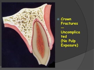
CONTENTS:
 INTRODUCTION
 CLASSIFICATION
 ETIOLOGY & EPIDEMIOLOGY
 ENAMEL FRACTURES
 CROWN FRACTURES – UNCOMPLICATED
 CROWN FRACTURES - COMPLICATED
 CROWN –ROOT FRACTURES
 ROOT FRACTURES
 LUXATION
 SUBLUXATION
 LATERAL INTRUSION
 AVULSION
INTRODUCTION
 Trauma to the tooth may result either in injury to the pulp , with or
without damage to the crown or root , or in displacement of the
tooth from its socket .
 Sudden impact involving the head or face may result in trauma to
the teeth & supporting structures.
 The most frequent cause being while running, traffic accidents,
acts of violence and sports.
 They are most commonly seen between 8 & 12 years, when
children are most active.
 Teeth most vulnerable is maxillary central incisor, which sustains
80% of dental injuries… followed by max. lateral incisor &
mandibular central and lateral.
 Management of traumatic injuries … both demanding and
challenging.
 Apart from physical injury, they are generally accompanied by
emotional factors… specially if there is hemorrhage.
 Favorable healing after traumatic injury requires quick emergency
intervention followed by evaluation & possible treatment at decisive
times during healing phase.
 Recent advances in the understanding of wound healing related to
dental trauma, tooth transplantation & implantation have opened
up new treatment avenues that have made it possible to fully
restore even the most severely traumatized teeth.
CLASSIFICATION
ELLIS CLASSIFICATION
 Class I –Simple crown fracture with little or no dentine affected.
 Class II – Extensive crown fracture with considerable loss of dentin, but
with the pulp not affected.
 Class III – Extensive crown fracture with considerable loss of dentin
and pulp exposure.
 Class IV – A tooth devitalized by trauma with or without loss of tooth
 structure.
 Class V – Tooth lost as a result of trauma.
 Class VI – Root fracture with or without the loss of crown fracture.
 Class VII – Displacement of the tooth with neither root nor crown
fracture.
 Class VIII – Complete crown fracture and its replacement.
 Class IX – Traumatic injuries of primary teeth.
W.H.O. CLASSIFICATION
 The World Health Organization adopted the following
classification in 1978 with a code number corresponding to the
international classification of disease:
 873.60  Enamel fracture.
 873.61  Crown fracture involving enamel / dentin without pulp
exposure.
 873.62  Crown fracture with pulp exposure.
 873.63  Root fracture.
 873.64  Crown root fracture.
 873.66  Luxation.
 873.67  Intrusion or extrusion.
 873.68  Avulsion.
 873.69  Other injuries such as soft tissues.
BASRANI CLASSIFICATION
 Crown fractures.
 Fracture of enamel.
 Fracture of enamel and dentin.
○ Without pulp exposure.
○ With pulp exposure.
 Root fractures.
 Crown root fractures.
ANDREASEN’S CLASSIFICATION MODIFIED: traumatic
injuries to teeth
1. Enamel infarction
2. Enamel fracture
3. Enamel – dentin fracture (uncomplicated)
4. Complicated crown fracture
5. Uncomplicated crown-root fracture
6. Complicated crown-root fracture
7. Root fracture
8. Luxation injuries
 Concussion
 Subluxation (loosening)
 Extrusive luxation ( partial avulsion)
 Lateral luxation
 Intrusive luxation
 Avulsion (exarticulation)
ANDREASEN’S CLASSIFICATION OF SOFT TISSUE
INJURY:
 Laceration Of Gingiva
 Contusion of gingiva
 Abrasion of gingiva or oral mucosa
 Fracture of mandibular or maxillary alveolar socket wall
 Fracture of mandibular or maxillary alveolar process
 Fracture of mandible or maxilla
DENTOFACIAL INJURIES:
I. SOFT TISSUE INJURIES
- Lacerations
- Contusions
- Abrasions
II. TOOTH FRACTURES
- enamel fractures
- Crown fractures-uncomplicated
- Crown fractures – complicated
- Crown – root fractures
- Root fractures
III. LUXATION INJURIES
- Tooth concussion
- Extrusive luxation
- Lateral luxation
- Intrusive luxation
- Avulsion
IV. FACIAL SKELETAL INJURIES
- Alveolar process- maxilla / mandible
- Body of the maxillary / mandibular bone
- Temporomandibular joint
ETIOLOGY
 Sudden impact – head /face
 Frequent causes – falling while running, traffic
accidents, acts of violence, & sports
 20 to 60% traffic accidents– facial injuries
 Involve teeth – avulsions/ intrusions are common
 Common in school athletes who do not use mouth
guards
INCIDENCE
 Dental injuries commonly occur --- first two decades of life
 Most accident prone period--- 8 to 12 years
 Dental injuries also occur in age range of 2 to 3 years
 Boys : girls ratio --- 2:1 to 3:1
 Most commonly seen trauma --- facture of enamel, or enamel and
dentin, but without pulp involvement
 Also seen in --- mentally retarded patients, drug addicts, epileptic
patients, dentinogenesis imperfecta (root fracture due to reduced
hardness of dentin)
 Child abuse or “battered child” syndrome --- more apparent for
dental injuries
MEDICAL EMERGENCY
CONSIDERATIONS
 B.P , pulse rate , respiration --- preliminary examination
 Cold, pale skin , perspiration, hypotension, tachycardia, ---
shock.
 Severe hemorrahage secondary to facial injury rare --- but
could be life threatening
 Coughing, cynosis, dyspnea--- signs of foreign body
aspiration
 Chest and abdominal radiographs to confirm
 Glassgow coma scale --- 3 to 15 scale --- lower the no. more
severe the injury
 Inability to open eyes --- neurological problem
 Diplopia – zygomatic / maxillary fracture --- blow out orbital
fracture
 Mononuclear diplopia--- injury to eye globe
 Localozed area of parasthesia on face ---- damage to
trigeminal nerve with alveolar fracture
 Inability to protrude tongue ---- damage to hypoglossal nerve
HISTORY:
 WHERE
 WHEN
 HOW
 WHERE : location will provide information of degree of bacterial
contamination
Clean wounds --- booster tetanus--- 10 yrs
Contaminated – booster tetanus --- 5 yrs
 WHEN – necessary to determine treatment and asses prognosis
 HOW --- pattern of injury depend on site, direction& resilience of
pdl status
PREDISPOSING
FACTORS
 Increased overjet with protrussion of upper
incisors and insufficient lip closure.
MECHANISMS OF DENTAL INJURY
INDIRECT
INJURIES
DIRECT
Occurs when tooth
itself is struck
e.g: play groung
Equipment run in
anterior region
Lower anterior fully
closed
Against upper teeth
Crown/ crown – root
fractures
In PM & M
DIAGNOSIS OF TOOT FRACTURE
 Symptoms depend on --- pulp exposed or not, damage to
pulp, and age of patient
 Common symptoms --- pain on chewing, pain on exposure to
cold & in some cases sweet & sour food
 Pulp involved – bacterial infection or periodontal ligament is
reached
 Diffuse protracted pain
 Visual examination – transillumination, dyes like mythelene
blue
 Use of microscope along with dye – fracture quickly visible
 Removal of any restoration if present --- restoration hamper
the detection of fracture
 BITING TEST : Biting on burlow wheel, rubber wheel, cotton tip
applicator, tooth sloth & Farc Finder can elicit pain.
 Used to differentiate between pain from restoration from
microleakage & pain from tooth infarction.
 A singnificant response to biting ---- release of biting pressure.
 Referred to as “rebound pain” or “referred pain”.
 EPT / electric pulp test: defferentiate between vital pulp and
necrotic pulp.
 Cold test in form of CO2 / ice: reliable but not quatifiable …..
More useful in differentiating between reversible pulpitis &
irreversible pulpitis.
 Heat test: limited use in pulp testing tramatized tooth.
Soft tissue injuries:
 DESCRIPTION: injuries to oral soft tissues like lacerations,
contusions, or abrasions of the epithelial layer or combination of
injuries.
 Treatment --- controlling bleeding, repositioning displaced tissue&
suturing.
 Oral soft tissues heal rather easily.
TOOTH FRACTURE:
 Includes --- fracture from enamel infarctions to
complicated crown-root fractures.
 They are the most common reported types of
dental injuries ---- incidence of 4 to 5% of all
population
 Accounting for over 1/3 rd of all dental trauma.
ENAMEL INFARCTION:
 DISCRIPTION: includes chips or cracks confined to the
enamel & not crossing enamel-dentin border.
 Diagnosed by --- under indirect light or transillumination or by
using dyes.
 Anterior teeth --- enamel chips involve either distal corners or
the central lobe of incisal edge.
 Treatment – minor smoothening of rough edges/ composite
restoration.
 Long term response--- difficult to predict.
 Pulp vitality tests --- performed immediately after injury and
again in 6 to 8 wks.
 Hoewvwr the prognosis for enamel fractures is very good.
 SEQUELE FOR ENAMEL FRACTURES:
 Pulp necrosis
 Internal resorption
 Calcification
 Trauma to primary teeth – malformation of
permanent teeth.
 Crown
Fractures
—
Uncomplica
ted
(No Pulp
Exposure)
 DESCRIPTION: crown fractures involving enamel and dentin
without involving pulp are called uncomplicated crown fractures by
Andreasen and class II fractures by Ellis.
 Include incisal proximal corners
 Anterior teeth ( more common)
Site : incisal proximal corners
incisal edges
lingual chisel type fractures
 Posterior teeth :
Incidence – 1/3 rd of dental injuries
 Crown fractures – expose dentinal tubules --- harmful bacteria ---
pathway to pulp.
 Outcome --- irritational dentin / pulp necrosis.
 DIAGNOSIS :
 Along with extent and degree of fracture– pulp vitality
 Electric pulp test --- more reliable then cold / heat test
 Non-vital --- endodontic therapy
 Percussion test --- check for tenderness
 Mobility test --- periodontal status
 Treatment – emergency / immediate follow – up
OBJECTIVE OF TREATING TOOTH WITHOUT PULP EXPOSURE
 Elimination of discomfort
 Preservation of vital pulp
 Restoration of fractured crown
 EMERGENCY :
Primary goal of treatment --- protect the pulp
1. Placement of material over exposed dentin ---
allow pulp to form a protective barrier . E.g:
Ca(OH)2.
2. fracture site covered with composite restoration.
ADVANTAGES:
 No additional tooth structure removed
 Protects the dressing material
 Provides for function and esthetics
 PRECAUTIONS:
- Do not cut tooth structure
- Tooth should be relieved from occlusion
PERMANENT TEETH:
 Treatment begins at 6 to 8 weeks after injury
 Acid – etch composite --- treatment of choice
 Esthetic reason --- crown may be required
 If the fractured crown fragment is available, it is often
advantageous to use it to restore the tooth. The
technique for Reattachment
 Expected outcome is usually good, although
resistance to refracture is about 50% less than an
intact tooth’s resistance
Crown
Fractures
—
Complicat
ed
(With
Pulp
Exposure)
 DESCRIPTION: crown fractures involving enamel,
dentin & pulp are called ‘complicated crown”
fractures by Andreasen & class 3 by Ellis.
 Degree of pulp exposure--- pinpoint exposure to
total unroofing of coronal pulp.
 Treatment compilcated --- pulp exposure
 Initial reaction --- hemorrhage --- inflammatory
response --- destructive (necrotic) / proliferative
(polyp)
INCIDENCE:
 2 to 3 % of dental injuries
DIAGNOSIS:
 Made by observation also determine the condition
of pulp.
DEPENDS ON FOUR FACTORS:
1. Length of time pulp has exposed
2. Maturity of tooth – apex formed/ not
3. Age of the patient
4. Extent of crown fracture --- pulp treatment along
with maturity.
TREATMENT:
 Depends on maturity of the pulp……
 Preservation of the pulp by vital pulp therapy--- pulp
capping / pulpotomy
Pulp Capping Modified pulpotomy technique (“Cvek
type”)
 PULP CAPPING: time honoured procedure
modified pulpotomy / Cvek pulpotomy / shallow pulpotomy
rubberdam isolation and anesthesia
removal of granulation tissue & pulp tissue upt0 1 to 2mm
plenty of irrigation --- prevent damage to remaining pulp
irrigation with saline to stop bleeding , cover with Ca(OH)2
cover with intermediate base --- zinc phosphate / GIC / composite
replace the liner / base --- prevent miocroleakage (6-12 months )
 Alternative to Ca(OH)2 --- MTA --- effective in vital pulp therapy
TOOTH anesthetized & isolated with rubberdam
tooth, fractures surface & wound disinfected with sodium hypochlorite
shallow pulpotomy --- space for MTA ( atleast 2mm)
bleeding allowed to stop before placement of MTA
presence of SMALL AMOUNT BLOOD – not a contraindication
small amount MTA placed on wound surface
after 24 hrs, restore the tooth with definitive restorative material
DIFFERENCES IN VITAL PULP THERAPY TECHNIQUE WHEN
MTA IS USED IN PLACE OF Ca(OH)2:
1. Bleeding need not to be completely stopped --- moisture is
necessary for curing of MTA.
2. Two appointment procedure --- first shallow pulpotomy , second
complete restoratoration
3. No need to re-enter the pulpotomy site as recommended for
Ca(OH)2.
FOLLOW – UP AND PROGNOSIS:
Acceptable results of evaluation following pulpotomy
should be all of the following:
1. No clinical signs or symptoms
2. No evidence of periradicular pathologies
3. No evidence of resorption – internal / external
4. Evidence of continued root formationin developing
tooth.
 Crown-
Root
Fractures
 Enamel, dentin, and cementum are involved.
 If the pulp is also involved, the case is considered
more complicated.
Description.
 Usually caused by direct trauma.
 Results in a chisel-type fracture, with the apical
extent of the fracture below the lingual gingiva.
 Fragments may be single or multiple, leaving the
fragment or fragments loose and attached only by
periodontal ligament fibers.
 In posterior teeth causes of crown-root fractures are
attributed to
 large-size restorations
 high-speed instrumentation
 pin placement
 direct trauma, such as accidental blows to the face and
jaws.
 Incidence.
• Andreasen and Andreasen reported a 5%
incidence of total dental injuries.
• Incidence may be higher when one
includes the so-called cracked tooth
syndrome and vertical fractures of
endodontically treated teeth .
Diagnosis.
 Complaint of pain, particularly when the loose fragment or
fragments are manipulated.
 The fragments are generally easy to move, and bleeding from
the periodontal ligament or pulp often fills the fracture line.
 Radiographs of anterior crown-root fractures are often
difficult to interpret.
 Important to take more than one angulation to assess the
extent of fractures. Angulations of films should include both
additional horizontal and vertical angulations.
Treatment.
 Uncomplicated # -- fragment reattached by bonding.
 Pulp exposure -- shallow pulpotomy procedure (if the tooth is still
developing) or root canal treatment (fully developed teeth) done prior
to any rebonding or crown restoration.
 Crown-root fractures extending well below the alveolar crest may
require surgical repositioning of the tissues to expose the level of
fracture.
 Extrusion either surgical or orthodontic can also be done to allow better
restoration of the fractured tooth.
Surgical Reposition
Procedure
Clinical and radiographic appearance of
a complicated crown root fracture.
Exposing the fracture site : The coronal
fragment is removed. A combined gingivectomy
and osteotomy expose the fracture surface.
Constructing a post-retained crown:
After taking an impression, a post-retained
fuIl crown is fabricated.
Finished restoration : The clinical and
radiographic condition 2 months after
insertion of the crown
Surgical Extrusion
Loose fragment is stabilized immediately.
Incision of the PDL Using a specially
contoured surgical blade, after
administration of a local anesthetic.
Luxation of the root
The root is extracted and inspected for
additional fractures
Replanting the apical fragment: The root is
tried in different positions in order to establish
where the fracture is optimally exposed yet
with minimal extrusion.
Stabilization of the apical fragment during
healing: The root is splinted to adjacent teeth.
The pulp is extirpated and the access cavity to
the root canal closed.
The root canal is obturated with gutta percha
and sealer as far apically as possible 1 month
after surgical extruslon.
Orthodontic Extrusion
Root Fracture
 DESCRIPTION: frcture involves the root only; cementum , dentin,
and pulp.
 INCIDENCE: account 2% of all dental injuries
CLASSIFICATION:
I. BASED ON ORIENTATION
- vertical/ split root / oblique
- Horizontal / transverse ---- 5 to 7%
II. BASED ON LEVEL
- Coronal 1/3 rd…. More common
- Middle 1/3 rd
- Apical 1/3 rd
III. BASED ON FRAGMENTS
- Single
- Communited
Root fractures produce new sections of root – fragments
- Coronal fragment
space between them “diastasis”
- Apical fragment
 DIAGNOSIS :
 Missed in conventional radiographs
 With conventional radiograph… 90 degree
angulation ….if fracture diagonal … missed
 Additional film angulation of 45 degree + 90
degree ---- reveal root fracture.
MANAGEMENT:
SPLINTING:
 Repositioning the coronal fragment
 Fragments with close proximity --- splinting does not
make difference
 Semirigid / no splinting --- favour healing
 splinting after4 wks– does not make difference
 Delaying treatment for 24 hrs --- does not make
difference
 TREATMENT OF CORONAL FRAGMENT:
 most simplistic.
 Fracture site located more coronally on the root --- “new apex”
which is wide and open--- open apex
 Establishment of working length ---- radiographically
 Conventional apexification
- Calcium hydroxide ---- 3 to 12 months for barrier formation
- MTA --- immediate restoration
TREATNG CORONAL FRAGMENT & REMOVAL OF
APICAL FRAGMENT:
 signs and symptoms of non-healing after coronal RCT.
 SWELLING / radiolucent area at the site / apically.
 Assuming coronal fragment stable--- surgical removal of apical
fragment.
 68% success rate
 Notable mobility of coronal fragment --- implant through coronal
fragment & into bone = endosseous implant.
TREATING CORONAL & APICAL FRAGMENTS AT THE
SAME TIME:
 It is difficult to get apical seal when endodontically treating only
coronal fragment.
 To achieve this seal --- treatment of both coronal & apical fragment
simultaneously which is almost impossible.
 low success rate
 Favorable approximation of fragments --- intra radicular splint ---
rigid type of post (cobalt – chromium alloy [vitallium])
REMOVING THE CORONAL FRAGMENT & TREATING THE
APICAL SEGMENT
A) CROWN ROOT FRACTURE:
- Coronal fragment attached only by gingiva
- Acceptable crown – to – root ratio---- 1:1
- Endodontic treatment of apical segment .
- Followed by crown lengthening --- orthodontic/ periodontal
- Placement of appropriate crown.
B) CORONAL ROOT FRACTURES:
- Excessive mobility of coronal fragment ---- only option is removal
- Endodontic treatment of apical fragment
- Laser / electrosurgery / peridontal surgery/ orthodontic forced
eruption ---- crown lengthening
- Followed by crown
SEQUELE OF ROOT FRACTURE:
1. Healing with calcified tissue : radiographically fracture line
discernable, but fragments in close contact.
2. Healing with interproximal connective tissue : radiographically
fragment appear separated with fractured edges rounded.
3. Healing with interproximal bone & connective tissue: fragments are
separated by a distant bony bridge.
4. interproximal inflammatory tissue without healing: widening of
fracture line & developing radiolucency near fracture line.
- First three types --- successful healing--- asymptomatic.
PROGNOSIS:
-poor prognosis --- middle / coronal third
Slight discoloration --- frequently seen
Occasionally --- loss of response to EPT
Endodontic treatment --- resorption/ periradicular radiolucency
Teeth treated with endodontic treatment --- good prognosis
PRIMARY TEETH:
 Root factures infrequent
 When they occur --- coronal fragment removed
 No manipultion of segment --- harm to the
succedaneous tooth
LUXATION INJURIES
 Includes impact of trauma --- minor crushing
of PDL to more tooth displacement
(avulsion).
 INCIDENCE: 30 to 40 % of all dental injuries
 They can be :
- partial --- partially displaced in socket
- Total ---- completely avulsed from socket
WHO CLASSIFICATION OF LUXATION:
A) 873.66
- Concussion
- Subluxation
- Luxation
B) 873.67
- Intrusion
- Extrusion
DIAGNOSIS:
- Luxated teeth --- loosened or slightly
displaced --- sensitive to biting & chewing
- Sensitive to pressure & palpation of
alveolus , mobility, dislocation & bleeding
from PDL.
- Radiographs – not much helpful
- Negative response to EPT --- necrosis /
calcification
TREATMENT :
 INITIAL treatment --- doing nothing , avoid the
use of tooth.
 More serious luxations --- occlusal adjustment
to repositioning (reduction) &
splinting(stabalization) for 2 to 6 weeks.
 If crown fracture with pulp exposure ---
endodontic treatment
 Concussion
 Mildest form of luxation injury
 Sensitivity to percussion
 no displacement present
 No moblilty
 Treatment --- asymptomatic --- allow the tooth to
rest as much as possible to promote recovery
 Monitor pulpal status by EPT , watch clinically for
discoloration & radiographically for resorption
 Prognosis is good.
 Subluxation
 Tooth sensitive to percussion & increased
mobility--- referred as subluxation.
 EPT --- positive / negative
 Treatment --- stabilization of tooth for a short
period ( 2 to 3 weeks) to promote periodontal
healing & reduction in mobility.
 EVALUATED LONG ENOUGH --- CHECK
FOR PULPAL HEALING
 Definitive treatment – root canal treatment ---
fully developed teeth.
 Extrusive
Luxation
 Displacement of tooth axially in coronal
direction results in partial avulsion.
 Tooth – mobile & continually traumatized by
contact with opposing tooth --- premature
occlusion
 Radiographically --- “empty” radiolucent space
 Immediate treatment – repositioning the tooth &
stabilizing by functional splint for 4 to 8 wks.
 Long stabilization --- realignment of periodontal
ligament fibres
 Definitive treatment –RCT --- NECROSIS/
INFLAMATORY ROOT RESORPTION SEEN
 Lateral
Luxation
 Traumatic injury --- displacement of tooth labially ,
distally or mesially --- lateral luxation.
 Very painfull --- premature occlusion e.g : max.incisor
pushed palatally.
 Crown makes contact long before centric occlusion.
 Treatment plan depends on ---presence or absence of
apical displacement at the time of injury.
LATERAL LUXATION WITHOUT APICAL
DISPLACEMENT:
 Teeth pushed only in facial / lingual direction with
apical root remaining in its original position within the
socket.
 Teeth loose enough – slight digital pressure to
reposition the tooth.
 Some sulcular bleeding --- typically seen
 If there is no widening of PDL space as confirmed by
radiographs --- good prognosis --- endodontic
treatment may not be necessary.
 Tooth is initially unresponsive to EPT and cold test.
LATERAL LUXATION WITH APICAL DISPLACEMENT:
 Tooth is frequently pushed palatally / lingually and firmly
located in its new position.
 Tooth will elicit dull metallic sound on percussion.
 Palpating alveolar bone --- reveal new location of apex.
 Radiographically – PDL space widened around the midportion
and coronal portion of root.
 If apex moved out of its original position --- damage to
neurovascular bundle.
 Negative response to cold and EPT .
 Closed apex cases --- advice endodontic treatment
REPOSITIONING THE TOOTH IN ITS SOCKET
SPLINTING --- 3 TO 4 WEEKS
PROGNOSIS GOOD --- if endodontic treatment done when
indicated.
 Intrusion
 An intruded tooth appear as if it is not fully erupted,
such that a portion or whole crown is submerged
subgingivally.
 Treatment depends on stage of development of tooth
--- immature tooth… little / no treatment , mature tooth
…. Aggressive initial treatment.
 TREATMENT:
- Fully developed tooth --- reposition surgically /
orthodontically
- If allowed to remain in intruded position --- ankylosis
- Pulp should be prophylactically extirpated – followed
by endodontic treatment after PDL healing.
- Exception for endodontic treatment --- spontaneous
eruption in young developing tooth.
PROGNOSIS AND FOLLOW – UP :
Complications after luxation injuries are common:
- Pulp necrosis ---20 to 12%
- Apical periodontitis --- long term follow – up
- Crown discoloration and calcification --- yellow color ---
calcification, gray color --- pulp necrosis.
- Resorption ( external / internal) --- 5 to 15% cases.
 Evaluation --- after 4 to 6 wks , then 12 wks , 6 months and yearly.
 Treatment for resorption --- RCT
 Treatment for immature tooth----
- APEXIFICATION --- best is combination therapy --- 2 wks calcium
hydroxide placement to assist disinfection followed by MTA
placement to get apical seal --- then completion of RCT.
- PROMOTE REVASCULARIZATION
PRIMARY TEETH TREATMENT:
 Injury to primary teeth --- results in damage to supporting tissues
more frequently than crown-root fractures.
 Initial observation --- gray discoloration changing to yellow
indicating calcification in contrast to permanent teeth --- grayish
discoloration indicates pulp necrosis .
 Most common sequel in primary teeth --- calcification ( internal /
external resorption , apical lesions & discoloration can also occur) .
 Endodontic treatment ---- pulp necrosis.
 Majority of intruded primary teeth re-erupt within 6 months ---
follow-up necessary.
 Tooth
Avulsion/
Exarticulation
 Avulsed tooth is completely displaced out of its socket .
 Incidence – 3% of all dental injuries
 True dental emergency – timely attention to replantation could save
the tooth.
 Sports and automobile accidents --- frequent causes.
 Examine --- tooth is replanted before coming to dental office.
 See for any debris / contaminants.
 Record the time of avulsion.
 Length of extraalveolar time --- determines treatment plan and
prognosis.
 Tooth left dry for less than 1 hr / kept in milk for no more than 4 to 6
hrs --- immediate replantation
 More than 1 hr of dry time --- delayed replantation.
 TREATMENT :
IMMEDIATE REPLANTATION:
- Sooner the avulsed tooth replanted --- better is the prognosis
- Manner of tooth transportation is important.
- Ideal ways to transport the tooth --- HBSS (Hank’s balanced salt
solution) then milk, water, patient’s vestibule / saliva.
- Place tooth in saline
- Radiographs --- fracture of alveolar bone
- Socket --- foreign bodies and debris --- scrape gently from bony
walls.
- Blood clot – gently suctioned and irrigated with saline
- Avulsed tooth --- debris gently rinsed off
- Do not hold the in hands --- always use some twizer / forceps to
hold the tooth
- Gently and slowly insert the into socket --- aneasthesia may not be
necessary.
 Check the alignment --- no hyperocclusion .
 Splinting not necessary if tooth fits firmly in socket.
 Mobility --- splinting recommended
 Orthodontic wire ( 0.3 mm) attached with composite on the labial
surface of tooth.
 Splinting should left for 1 to 2 wks
 Initial antibiotic course, tetanus prevention & RCT ( after 10 to 14
days).
 Calcium hydroxide is recommended as an intracanal medicament
--- 2 wks , during RCT.
 DELAYED REPLANTATION:
- Extra-alveolar time more than 1 h
- Examine for any debris on tooth --- after 1 h vitality of PDL fibres
not expected --- remove the pieces of soft tissue attached to the
root surface.
- Perform RCT in vitro by holding the tooth by crown & endodontic
treatment through apical approach.
- Cut off 2 to 3mm root apex , extirpate the pulp, ten fill the canal with
gutta-percha and sealer ( adv – convenience and crown left intact).
- Soak the tooth in 2.4 % fluoride solution acidulated at ph 5.5 for 20
min.
- Rinse the tooth with saline , insert in socket --- splint for 6 wks.
- Ankylosis is the expected outcome --- radiographically, no pdl
space , clinically, loss of normal mobility and metallic percussion
sound.
 AVULSED TOOTH WITH OPEN APICES:
- IMMEDIATE ENDODONTIC TREATMENT IS NOT DONE
- Monitor the tooth for revascularization ( continued root formation
without sign s of resorption or ankylosis)
- Soak the tooth in a sol.of doxycycline (1mg/20 ml) prior to
replantation.
- Replantation in primary teeth – done only if there is no trauma to
the succedaneous tooth.
PROGNOSIS AND FOLLOW-UP: external resorption frequent
1. Surface
2. Inflammatory
3. Replacement ( ankylotic)
CONCLUSION
 Bicycle and automobile accidents, home and play
ground injuries have all taken their toll in fractured
crowns and roots non-vital pulps and avulsed or
dislocated teeth.
 These dental injuries meant a life of discomfort and
disfigurement as no replacement can equal function
and esthetics of intact dental structures.
 Considering the multiplicity of etiologic factors, one
can easily understand why preventive measures are
difficult to institute.
 However, certain accident-prone individuals can be
protected. For example mouth guards have proven
effective in the prevention of dental injuries due to
contact sports and during anesthetic procedures.

TRAUMATIC INJURIES.pptx

  • 2.
    CONTENTS:  INTRODUCTION  CLASSIFICATION ETIOLOGY & EPIDEMIOLOGY  ENAMEL FRACTURES  CROWN FRACTURES – UNCOMPLICATED  CROWN FRACTURES - COMPLICATED  CROWN –ROOT FRACTURES  ROOT FRACTURES  LUXATION  SUBLUXATION  LATERAL INTRUSION  AVULSION
  • 3.
    INTRODUCTION  Trauma tothe tooth may result either in injury to the pulp , with or without damage to the crown or root , or in displacement of the tooth from its socket .  Sudden impact involving the head or face may result in trauma to the teeth & supporting structures.  The most frequent cause being while running, traffic accidents, acts of violence and sports.  They are most commonly seen between 8 & 12 years, when children are most active.  Teeth most vulnerable is maxillary central incisor, which sustains 80% of dental injuries… followed by max. lateral incisor & mandibular central and lateral.  Management of traumatic injuries … both demanding and challenging.  Apart from physical injury, they are generally accompanied by emotional factors… specially if there is hemorrhage.
  • 4.
     Favorable healingafter traumatic injury requires quick emergency intervention followed by evaluation & possible treatment at decisive times during healing phase.  Recent advances in the understanding of wound healing related to dental trauma, tooth transplantation & implantation have opened up new treatment avenues that have made it possible to fully restore even the most severely traumatized teeth.
  • 5.
    CLASSIFICATION ELLIS CLASSIFICATION  ClassI –Simple crown fracture with little or no dentine affected.  Class II – Extensive crown fracture with considerable loss of dentin, but with the pulp not affected.  Class III – Extensive crown fracture with considerable loss of dentin and pulp exposure.  Class IV – A tooth devitalized by trauma with or without loss of tooth  structure.  Class V – Tooth lost as a result of trauma.  Class VI – Root fracture with or without the loss of crown fracture.  Class VII – Displacement of the tooth with neither root nor crown fracture.  Class VIII – Complete crown fracture and its replacement.  Class IX – Traumatic injuries of primary teeth.
  • 6.
    W.H.O. CLASSIFICATION  TheWorld Health Organization adopted the following classification in 1978 with a code number corresponding to the international classification of disease:  873.60  Enamel fracture.  873.61  Crown fracture involving enamel / dentin without pulp exposure.  873.62  Crown fracture with pulp exposure.  873.63  Root fracture.  873.64  Crown root fracture.  873.66  Luxation.  873.67  Intrusion or extrusion.  873.68  Avulsion.  873.69  Other injuries such as soft tissues.
  • 7.
    BASRANI CLASSIFICATION  Crownfractures.  Fracture of enamel.  Fracture of enamel and dentin. ○ Without pulp exposure. ○ With pulp exposure.  Root fractures.  Crown root fractures.
  • 8.
    ANDREASEN’S CLASSIFICATION MODIFIED:traumatic injuries to teeth 1. Enamel infarction 2. Enamel fracture 3. Enamel – dentin fracture (uncomplicated) 4. Complicated crown fracture 5. Uncomplicated crown-root fracture 6. Complicated crown-root fracture 7. Root fracture 8. Luxation injuries  Concussion  Subluxation (loosening)  Extrusive luxation ( partial avulsion)  Lateral luxation  Intrusive luxation  Avulsion (exarticulation)
  • 9.
    ANDREASEN’S CLASSIFICATION OFSOFT TISSUE INJURY:  Laceration Of Gingiva  Contusion of gingiva  Abrasion of gingiva or oral mucosa  Fracture of mandibular or maxillary alveolar socket wall  Fracture of mandibular or maxillary alveolar process  Fracture of mandible or maxilla
  • 10.
    DENTOFACIAL INJURIES: I. SOFTTISSUE INJURIES - Lacerations - Contusions - Abrasions II. TOOTH FRACTURES - enamel fractures - Crown fractures-uncomplicated - Crown fractures – complicated - Crown – root fractures - Root fractures III. LUXATION INJURIES - Tooth concussion - Extrusive luxation - Lateral luxation - Intrusive luxation - Avulsion
  • 11.
    IV. FACIAL SKELETALINJURIES - Alveolar process- maxilla / mandible - Body of the maxillary / mandibular bone - Temporomandibular joint
  • 12.
    ETIOLOGY  Sudden impact– head /face  Frequent causes – falling while running, traffic accidents, acts of violence, & sports  20 to 60% traffic accidents– facial injuries  Involve teeth – avulsions/ intrusions are common  Common in school athletes who do not use mouth guards
  • 13.
    INCIDENCE  Dental injuriescommonly occur --- first two decades of life  Most accident prone period--- 8 to 12 years  Dental injuries also occur in age range of 2 to 3 years  Boys : girls ratio --- 2:1 to 3:1  Most commonly seen trauma --- facture of enamel, or enamel and dentin, but without pulp involvement  Also seen in --- mentally retarded patients, drug addicts, epileptic patients, dentinogenesis imperfecta (root fracture due to reduced hardness of dentin)  Child abuse or “battered child” syndrome --- more apparent for dental injuries
  • 14.
    MEDICAL EMERGENCY CONSIDERATIONS  B.P, pulse rate , respiration --- preliminary examination  Cold, pale skin , perspiration, hypotension, tachycardia, --- shock.  Severe hemorrahage secondary to facial injury rare --- but could be life threatening  Coughing, cynosis, dyspnea--- signs of foreign body aspiration  Chest and abdominal radiographs to confirm  Glassgow coma scale --- 3 to 15 scale --- lower the no. more severe the injury  Inability to open eyes --- neurological problem  Diplopia – zygomatic / maxillary fracture --- blow out orbital fracture
  • 15.
     Mononuclear diplopia---injury to eye globe  Localozed area of parasthesia on face ---- damage to trigeminal nerve with alveolar fracture  Inability to protrude tongue ---- damage to hypoglossal nerve
  • 16.
  • 17.
     WHERE :location will provide information of degree of bacterial contamination Clean wounds --- booster tetanus--- 10 yrs Contaminated – booster tetanus --- 5 yrs  WHEN – necessary to determine treatment and asses prognosis  HOW --- pattern of injury depend on site, direction& resilience of pdl status
  • 18.
    PREDISPOSING FACTORS  Increased overjetwith protrussion of upper incisors and insufficient lip closure.
  • 19.
    MECHANISMS OF DENTALINJURY INDIRECT INJURIES DIRECT Occurs when tooth itself is struck e.g: play groung Equipment run in anterior region Lower anterior fully closed Against upper teeth Crown/ crown – root fractures In PM & M
  • 20.
    DIAGNOSIS OF TOOTFRACTURE  Symptoms depend on --- pulp exposed or not, damage to pulp, and age of patient  Common symptoms --- pain on chewing, pain on exposure to cold & in some cases sweet & sour food  Pulp involved – bacterial infection or periodontal ligament is reached  Diffuse protracted pain  Visual examination – transillumination, dyes like mythelene blue  Use of microscope along with dye – fracture quickly visible  Removal of any restoration if present --- restoration hamper the detection of fracture
  • 21.
     BITING TEST: Biting on burlow wheel, rubber wheel, cotton tip applicator, tooth sloth & Farc Finder can elicit pain.  Used to differentiate between pain from restoration from microleakage & pain from tooth infarction.  A singnificant response to biting ---- release of biting pressure.  Referred to as “rebound pain” or “referred pain”.  EPT / electric pulp test: defferentiate between vital pulp and necrotic pulp.  Cold test in form of CO2 / ice: reliable but not quatifiable ….. More useful in differentiating between reversible pulpitis & irreversible pulpitis.  Heat test: limited use in pulp testing tramatized tooth.
  • 22.
    Soft tissue injuries: DESCRIPTION: injuries to oral soft tissues like lacerations, contusions, or abrasions of the epithelial layer or combination of injuries.  Treatment --- controlling bleeding, repositioning displaced tissue& suturing.  Oral soft tissues heal rather easily.
  • 23.
    TOOTH FRACTURE:  Includes--- fracture from enamel infarctions to complicated crown-root fractures.  They are the most common reported types of dental injuries ---- incidence of 4 to 5% of all population  Accounting for over 1/3 rd of all dental trauma.
  • 24.
    ENAMEL INFARCTION:  DISCRIPTION:includes chips or cracks confined to the enamel & not crossing enamel-dentin border.  Diagnosed by --- under indirect light or transillumination or by using dyes.  Anterior teeth --- enamel chips involve either distal corners or the central lobe of incisal edge.  Treatment – minor smoothening of rough edges/ composite restoration.  Long term response--- difficult to predict.  Pulp vitality tests --- performed immediately after injury and again in 6 to 8 wks.  Hoewvwr the prognosis for enamel fractures is very good.
  • 25.
     SEQUELE FORENAMEL FRACTURES:  Pulp necrosis  Internal resorption  Calcification  Trauma to primary teeth – malformation of permanent teeth.
  • 27.
  • 28.
     DESCRIPTION: crownfractures involving enamel and dentin without involving pulp are called uncomplicated crown fractures by Andreasen and class II fractures by Ellis.  Include incisal proximal corners  Anterior teeth ( more common) Site : incisal proximal corners incisal edges lingual chisel type fractures  Posterior teeth : Incidence – 1/3 rd of dental injuries  Crown fractures – expose dentinal tubules --- harmful bacteria --- pathway to pulp.  Outcome --- irritational dentin / pulp necrosis.
  • 29.
     DIAGNOSIS : Along with extent and degree of fracture– pulp vitality  Electric pulp test --- more reliable then cold / heat test  Non-vital --- endodontic therapy  Percussion test --- check for tenderness  Mobility test --- periodontal status  Treatment – emergency / immediate follow – up OBJECTIVE OF TREATING TOOTH WITHOUT PULP EXPOSURE  Elimination of discomfort  Preservation of vital pulp  Restoration of fractured crown
  • 30.
     EMERGENCY : Primarygoal of treatment --- protect the pulp 1. Placement of material over exposed dentin --- allow pulp to form a protective barrier . E.g: Ca(OH)2. 2. fracture site covered with composite restoration. ADVANTAGES:  No additional tooth structure removed  Protects the dressing material  Provides for function and esthetics
  • 31.
     PRECAUTIONS: - Donot cut tooth structure - Tooth should be relieved from occlusion PERMANENT TEETH:  Treatment begins at 6 to 8 weeks after injury  Acid – etch composite --- treatment of choice  Esthetic reason --- crown may be required
  • 33.
     If thefractured crown fragment is available, it is often advantageous to use it to restore the tooth. The technique for Reattachment  Expected outcome is usually good, although resistance to refracture is about 50% less than an intact tooth’s resistance
  • 34.
  • 35.
     DESCRIPTION: crownfractures involving enamel, dentin & pulp are called ‘complicated crown” fractures by Andreasen & class 3 by Ellis.  Degree of pulp exposure--- pinpoint exposure to total unroofing of coronal pulp.  Treatment compilcated --- pulp exposure  Initial reaction --- hemorrhage --- inflammatory response --- destructive (necrotic) / proliferative (polyp)
  • 36.
    INCIDENCE:  2 to3 % of dental injuries DIAGNOSIS:  Made by observation also determine the condition of pulp. DEPENDS ON FOUR FACTORS: 1. Length of time pulp has exposed 2. Maturity of tooth – apex formed/ not 3. Age of the patient 4. Extent of crown fracture --- pulp treatment along with maturity.
  • 37.
    TREATMENT:  Depends onmaturity of the pulp……  Preservation of the pulp by vital pulp therapy--- pulp capping / pulpotomy Pulp Capping Modified pulpotomy technique (“Cvek type”)
  • 39.
     PULP CAPPING:time honoured procedure modified pulpotomy / Cvek pulpotomy / shallow pulpotomy rubberdam isolation and anesthesia removal of granulation tissue & pulp tissue upt0 1 to 2mm plenty of irrigation --- prevent damage to remaining pulp irrigation with saline to stop bleeding , cover with Ca(OH)2 cover with intermediate base --- zinc phosphate / GIC / composite replace the liner / base --- prevent miocroleakage (6-12 months )
  • 40.
     Alternative toCa(OH)2 --- MTA --- effective in vital pulp therapy TOOTH anesthetized & isolated with rubberdam tooth, fractures surface & wound disinfected with sodium hypochlorite shallow pulpotomy --- space for MTA ( atleast 2mm) bleeding allowed to stop before placement of MTA presence of SMALL AMOUNT BLOOD – not a contraindication small amount MTA placed on wound surface after 24 hrs, restore the tooth with definitive restorative material
  • 41.
    DIFFERENCES IN VITALPULP THERAPY TECHNIQUE WHEN MTA IS USED IN PLACE OF Ca(OH)2: 1. Bleeding need not to be completely stopped --- moisture is necessary for curing of MTA. 2. Two appointment procedure --- first shallow pulpotomy , second complete restoratoration 3. No need to re-enter the pulpotomy site as recommended for Ca(OH)2.
  • 42.
    FOLLOW – UPAND PROGNOSIS: Acceptable results of evaluation following pulpotomy should be all of the following: 1. No clinical signs or symptoms 2. No evidence of periradicular pathologies 3. No evidence of resorption – internal / external 4. Evidence of continued root formationin developing tooth.
  • 43.
  • 44.
     Enamel, dentin,and cementum are involved.  If the pulp is also involved, the case is considered more complicated. Description.  Usually caused by direct trauma.  Results in a chisel-type fracture, with the apical extent of the fracture below the lingual gingiva.  Fragments may be single or multiple, leaving the fragment or fragments loose and attached only by periodontal ligament fibers.
  • 45.
     In posteriorteeth causes of crown-root fractures are attributed to  large-size restorations  high-speed instrumentation  pin placement  direct trauma, such as accidental blows to the face and jaws.
  • 46.
     Incidence. • Andreasenand Andreasen reported a 5% incidence of total dental injuries. • Incidence may be higher when one includes the so-called cracked tooth syndrome and vertical fractures of endodontically treated teeth .
  • 47.
    Diagnosis.  Complaint ofpain, particularly when the loose fragment or fragments are manipulated.  The fragments are generally easy to move, and bleeding from the periodontal ligament or pulp often fills the fracture line.  Radiographs of anterior crown-root fractures are often difficult to interpret.  Important to take more than one angulation to assess the extent of fractures. Angulations of films should include both additional horizontal and vertical angulations.
  • 48.
    Treatment.  Uncomplicated #-- fragment reattached by bonding.  Pulp exposure -- shallow pulpotomy procedure (if the tooth is still developing) or root canal treatment (fully developed teeth) done prior to any rebonding or crown restoration.  Crown-root fractures extending well below the alveolar crest may require surgical repositioning of the tissues to expose the level of fracture.  Extrusion either surgical or orthodontic can also be done to allow better restoration of the fractured tooth.
  • 49.
    Surgical Reposition Procedure Clinical andradiographic appearance of a complicated crown root fracture. Exposing the fracture site : The coronal fragment is removed. A combined gingivectomy and osteotomy expose the fracture surface. Constructing a post-retained crown: After taking an impression, a post-retained fuIl crown is fabricated. Finished restoration : The clinical and radiographic condition 2 months after insertion of the crown
  • 50.
    Surgical Extrusion Loose fragmentis stabilized immediately. Incision of the PDL Using a specially contoured surgical blade, after administration of a local anesthetic. Luxation of the root The root is extracted and inspected for additional fractures
  • 51.
    Replanting the apicalfragment: The root is tried in different positions in order to establish where the fracture is optimally exposed yet with minimal extrusion. Stabilization of the apical fragment during healing: The root is splinted to adjacent teeth. The pulp is extirpated and the access cavity to the root canal closed. The root canal is obturated with gutta percha and sealer as far apically as possible 1 month after surgical extruslon.
  • 52.
  • 53.
  • 54.
     DESCRIPTION: frctureinvolves the root only; cementum , dentin, and pulp.  INCIDENCE: account 2% of all dental injuries
  • 55.
    CLASSIFICATION: I. BASED ONORIENTATION - vertical/ split root / oblique - Horizontal / transverse ---- 5 to 7% II. BASED ON LEVEL - Coronal 1/3 rd…. More common - Middle 1/3 rd - Apical 1/3 rd III. BASED ON FRAGMENTS - Single - Communited Root fractures produce new sections of root – fragments - Coronal fragment space between them “diastasis” - Apical fragment
  • 56.
     DIAGNOSIS : Missed in conventional radiographs  With conventional radiograph… 90 degree angulation ….if fracture diagonal … missed  Additional film angulation of 45 degree + 90 degree ---- reveal root fracture.
  • 57.
    MANAGEMENT: SPLINTING:  Repositioning thecoronal fragment  Fragments with close proximity --- splinting does not make difference  Semirigid / no splinting --- favour healing  splinting after4 wks– does not make difference  Delaying treatment for 24 hrs --- does not make difference
  • 58.
     TREATMENT OFCORONAL FRAGMENT:  most simplistic.  Fracture site located more coronally on the root --- “new apex” which is wide and open--- open apex  Establishment of working length ---- radiographically  Conventional apexification - Calcium hydroxide ---- 3 to 12 months for barrier formation - MTA --- immediate restoration
  • 59.
    TREATNG CORONAL FRAGMENT& REMOVAL OF APICAL FRAGMENT:  signs and symptoms of non-healing after coronal RCT.  SWELLING / radiolucent area at the site / apically.  Assuming coronal fragment stable--- surgical removal of apical fragment.  68% success rate  Notable mobility of coronal fragment --- implant through coronal fragment & into bone = endosseous implant.
  • 60.
    TREATING CORONAL &APICAL FRAGMENTS AT THE SAME TIME:  It is difficult to get apical seal when endodontically treating only coronal fragment.  To achieve this seal --- treatment of both coronal & apical fragment simultaneously which is almost impossible.  low success rate  Favorable approximation of fragments --- intra radicular splint --- rigid type of post (cobalt – chromium alloy [vitallium])
  • 61.
    REMOVING THE CORONALFRAGMENT & TREATING THE APICAL SEGMENT A) CROWN ROOT FRACTURE: - Coronal fragment attached only by gingiva - Acceptable crown – to – root ratio---- 1:1 - Endodontic treatment of apical segment . - Followed by crown lengthening --- orthodontic/ periodontal - Placement of appropriate crown. B) CORONAL ROOT FRACTURES: - Excessive mobility of coronal fragment ---- only option is removal - Endodontic treatment of apical fragment - Laser / electrosurgery / peridontal surgery/ orthodontic forced eruption ---- crown lengthening - Followed by crown
  • 62.
    SEQUELE OF ROOTFRACTURE: 1. Healing with calcified tissue : radiographically fracture line discernable, but fragments in close contact. 2. Healing with interproximal connective tissue : radiographically fragment appear separated with fractured edges rounded. 3. Healing with interproximal bone & connective tissue: fragments are separated by a distant bony bridge. 4. interproximal inflammatory tissue without healing: widening of fracture line & developing radiolucency near fracture line. - First three types --- successful healing--- asymptomatic. PROGNOSIS: -poor prognosis --- middle / coronal third Slight discoloration --- frequently seen Occasionally --- loss of response to EPT Endodontic treatment --- resorption/ periradicular radiolucency Teeth treated with endodontic treatment --- good prognosis
  • 63.
    PRIMARY TEETH:  Rootfactures infrequent  When they occur --- coronal fragment removed  No manipultion of segment --- harm to the succedaneous tooth
  • 64.
    LUXATION INJURIES  Includesimpact of trauma --- minor crushing of PDL to more tooth displacement (avulsion).  INCIDENCE: 30 to 40 % of all dental injuries  They can be : - partial --- partially displaced in socket - Total ---- completely avulsed from socket
  • 65.
    WHO CLASSIFICATION OFLUXATION: A) 873.66 - Concussion - Subluxation - Luxation B) 873.67 - Intrusion - Extrusion
  • 66.
    DIAGNOSIS: - Luxated teeth--- loosened or slightly displaced --- sensitive to biting & chewing - Sensitive to pressure & palpation of alveolus , mobility, dislocation & bleeding from PDL. - Radiographs – not much helpful - Negative response to EPT --- necrosis / calcification
  • 67.
    TREATMENT :  INITIALtreatment --- doing nothing , avoid the use of tooth.  More serious luxations --- occlusal adjustment to repositioning (reduction) & splinting(stabalization) for 2 to 6 weeks.  If crown fracture with pulp exposure --- endodontic treatment
  • 68.
  • 69.
     Mildest formof luxation injury  Sensitivity to percussion  no displacement present  No moblilty  Treatment --- asymptomatic --- allow the tooth to rest as much as possible to promote recovery  Monitor pulpal status by EPT , watch clinically for discoloration & radiographically for resorption  Prognosis is good.
  • 70.
  • 71.
     Tooth sensitiveto percussion & increased mobility--- referred as subluxation.  EPT --- positive / negative  Treatment --- stabilization of tooth for a short period ( 2 to 3 weeks) to promote periodontal healing & reduction in mobility.  EVALUATED LONG ENOUGH --- CHECK FOR PULPAL HEALING  Definitive treatment – root canal treatment --- fully developed teeth.
  • 72.
  • 73.
     Displacement oftooth axially in coronal direction results in partial avulsion.  Tooth – mobile & continually traumatized by contact with opposing tooth --- premature occlusion  Radiographically --- “empty” radiolucent space  Immediate treatment – repositioning the tooth & stabilizing by functional splint for 4 to 8 wks.  Long stabilization --- realignment of periodontal ligament fibres  Definitive treatment –RCT --- NECROSIS/ INFLAMATORY ROOT RESORPTION SEEN
  • 74.
  • 75.
     Traumatic injury--- displacement of tooth labially , distally or mesially --- lateral luxation.  Very painfull --- premature occlusion e.g : max.incisor pushed palatally.  Crown makes contact long before centric occlusion.  Treatment plan depends on ---presence or absence of apical displacement at the time of injury.
  • 76.
    LATERAL LUXATION WITHOUTAPICAL DISPLACEMENT:  Teeth pushed only in facial / lingual direction with apical root remaining in its original position within the socket.  Teeth loose enough – slight digital pressure to reposition the tooth.  Some sulcular bleeding --- typically seen  If there is no widening of PDL space as confirmed by radiographs --- good prognosis --- endodontic treatment may not be necessary.  Tooth is initially unresponsive to EPT and cold test.
  • 77.
    LATERAL LUXATION WITHAPICAL DISPLACEMENT:  Tooth is frequently pushed palatally / lingually and firmly located in its new position.  Tooth will elicit dull metallic sound on percussion.  Palpating alveolar bone --- reveal new location of apex.  Radiographically – PDL space widened around the midportion and coronal portion of root.  If apex moved out of its original position --- damage to neurovascular bundle.  Negative response to cold and EPT .  Closed apex cases --- advice endodontic treatment REPOSITIONING THE TOOTH IN ITS SOCKET SPLINTING --- 3 TO 4 WEEKS PROGNOSIS GOOD --- if endodontic treatment done when indicated.
  • 79.
  • 80.
     An intrudedtooth appear as if it is not fully erupted, such that a portion or whole crown is submerged subgingivally.  Treatment depends on stage of development of tooth --- immature tooth… little / no treatment , mature tooth …. Aggressive initial treatment.  TREATMENT: - Fully developed tooth --- reposition surgically / orthodontically - If allowed to remain in intruded position --- ankylosis - Pulp should be prophylactically extirpated – followed by endodontic treatment after PDL healing. - Exception for endodontic treatment --- spontaneous eruption in young developing tooth.
  • 82.
    PROGNOSIS AND FOLLOW– UP : Complications after luxation injuries are common: - Pulp necrosis ---20 to 12% - Apical periodontitis --- long term follow – up - Crown discoloration and calcification --- yellow color --- calcification, gray color --- pulp necrosis. - Resorption ( external / internal) --- 5 to 15% cases.  Evaluation --- after 4 to 6 wks , then 12 wks , 6 months and yearly.  Treatment for resorption --- RCT  Treatment for immature tooth---- - APEXIFICATION --- best is combination therapy --- 2 wks calcium hydroxide placement to assist disinfection followed by MTA placement to get apical seal --- then completion of RCT. - PROMOTE REVASCULARIZATION
  • 83.
    PRIMARY TEETH TREATMENT: Injury to primary teeth --- results in damage to supporting tissues more frequently than crown-root fractures.  Initial observation --- gray discoloration changing to yellow indicating calcification in contrast to permanent teeth --- grayish discoloration indicates pulp necrosis .  Most common sequel in primary teeth --- calcification ( internal / external resorption , apical lesions & discoloration can also occur) .  Endodontic treatment ---- pulp necrosis.  Majority of intruded primary teeth re-erupt within 6 months --- follow-up necessary.
  • 84.
  • 85.
     Avulsed toothis completely displaced out of its socket .  Incidence – 3% of all dental injuries  True dental emergency – timely attention to replantation could save the tooth.  Sports and automobile accidents --- frequent causes.  Examine --- tooth is replanted before coming to dental office.  See for any debris / contaminants.  Record the time of avulsion.  Length of extraalveolar time --- determines treatment plan and prognosis.  Tooth left dry for less than 1 hr / kept in milk for no more than 4 to 6 hrs --- immediate replantation  More than 1 hr of dry time --- delayed replantation.
  • 86.
     TREATMENT : IMMEDIATEREPLANTATION: - Sooner the avulsed tooth replanted --- better is the prognosis - Manner of tooth transportation is important. - Ideal ways to transport the tooth --- HBSS (Hank’s balanced salt solution) then milk, water, patient’s vestibule / saliva. - Place tooth in saline - Radiographs --- fracture of alveolar bone - Socket --- foreign bodies and debris --- scrape gently from bony walls. - Blood clot – gently suctioned and irrigated with saline - Avulsed tooth --- debris gently rinsed off - Do not hold the in hands --- always use some twizer / forceps to hold the tooth - Gently and slowly insert the into socket --- aneasthesia may not be necessary.
  • 87.
     Check thealignment --- no hyperocclusion .  Splinting not necessary if tooth fits firmly in socket.  Mobility --- splinting recommended  Orthodontic wire ( 0.3 mm) attached with composite on the labial surface of tooth.  Splinting should left for 1 to 2 wks  Initial antibiotic course, tetanus prevention & RCT ( after 10 to 14 days).  Calcium hydroxide is recommended as an intracanal medicament --- 2 wks , during RCT.
  • 88.
     DELAYED REPLANTATION: -Extra-alveolar time more than 1 h - Examine for any debris on tooth --- after 1 h vitality of PDL fibres not expected --- remove the pieces of soft tissue attached to the root surface. - Perform RCT in vitro by holding the tooth by crown & endodontic treatment through apical approach. - Cut off 2 to 3mm root apex , extirpate the pulp, ten fill the canal with gutta-percha and sealer ( adv – convenience and crown left intact). - Soak the tooth in 2.4 % fluoride solution acidulated at ph 5.5 for 20 min. - Rinse the tooth with saline , insert in socket --- splint for 6 wks. - Ankylosis is the expected outcome --- radiographically, no pdl space , clinically, loss of normal mobility and metallic percussion sound.
  • 89.
     AVULSED TOOTHWITH OPEN APICES: - IMMEDIATE ENDODONTIC TREATMENT IS NOT DONE - Monitor the tooth for revascularization ( continued root formation without sign s of resorption or ankylosis) - Soak the tooth in a sol.of doxycycline (1mg/20 ml) prior to replantation. - Replantation in primary teeth – done only if there is no trauma to the succedaneous tooth. PROGNOSIS AND FOLLOW-UP: external resorption frequent 1. Surface 2. Inflammatory 3. Replacement ( ankylotic)
  • 90.
    CONCLUSION  Bicycle andautomobile accidents, home and play ground injuries have all taken their toll in fractured crowns and roots non-vital pulps and avulsed or dislocated teeth.  These dental injuries meant a life of discomfort and disfigurement as no replacement can equal function and esthetics of intact dental structures.  Considering the multiplicity of etiologic factors, one can easily understand why preventive measures are difficult to institute.  However, certain accident-prone individuals can be protected. For example mouth guards have proven effective in the prevention of dental injuries due to contact sports and during anesthetic procedures.