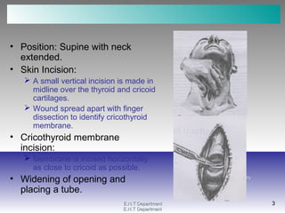
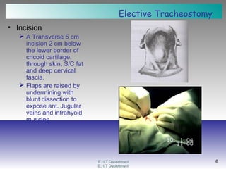
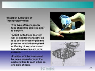
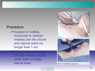
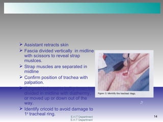
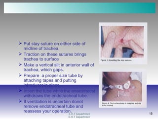
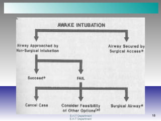
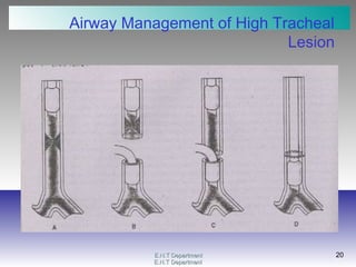
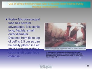
2. E.N.T DepartmentE.N.T Department
2
E.N.T DepartmentE.N.T Department
2
Laryngotomy
Emergency Procedure
“Laryngotomy is opening the airway through
the cricothyroid membrane”
• It is used for acute complete airway
obstruction when endotracheal intubation/
ventilation is not possible.
• The procedure can be accomplished in 15
to 30 seconds.
3. E.N.T DepartmentE.N.T Department
3
E.N.T DepartmentE.N.T Department
3
• Position: Supine with neck
extended.
• Skin Incision:
A small vertical incision is made in
midline over the thyroid and cricoid
cartilages.
Wound spread apart with finger
dissection to identify cricothyroid
membrane.
• Cricothyroid membrane
incision:
Membrane is incised horizontally
as close to cricoid as possible.
• Widening of opening and
placing a tube.
4. E.N.T DepartmentE.N.T Department
4
E.N.T DepartmentE.N.T Department
4
TRACHEOSTOMY
•Environment:
•Best performed as an elective
procedure under endotracheal
anaesthesia, in an adequately
equipped operation theatre and aseptic
measures.
•Position:
•Supine position with a sandbag under
patient’s shoulders to give extension
of head and prominence to the trachea
and larynx.
•Under local anaesthesia a
compromised position of extension
will have to be found.
5. E.N.T DepartmentE.N.T Department
5
E.N.T DepartmentE.N.T Department
5
• Anaesthesia:
Endotracheal anaesthesia
or
Local anaesthesia (in
obstructive pathologies)
obtained by injection of
skin and subcutaneous
tissues with Xylocaine 2%
1:200000 adrenaline
solution.
Drugs which depress resp.
system better avoided.
6. E.N.T DepartmentE.N.T Department
6
E.N.T DepartmentE.N.T Department
6
Elective Tracheostomy
• Incision
A Transverse 5 cm
incision 2 cm below
the lower border of
cricoid cartilage,
through skin, S/C fat
and deep cervical
fascia.
Flaps are raised by
undermining with
blunt dissection to
expose ant. Jugular
veins and infrahyoid
muscles.
7. E.N.T DepartmentE.N.T Department
7
E.N.T DepartmentE.N.T Department
7
•Separation of Infrahyoid
Muscles
•The fibrous median
raphe b/w the
sternohyoid muscles is
defined and separated
with blunt dissection
•The sternothyroid
muscles on a deeper
plane are identified and
retracted laterally.
8. E.N.T DepartmentE.N.T Department
8
E.N.T DepartmentE.N.T Department
8
•Identification of Thyroid Isthmus:
•Anatomical variations in size
and position of thyroid isthmus
should be expected
•The thyroid isthmus may be
small and not interfere with the
approach but in most patients it
is of sufficient size to need
dividing.
•A small horizontal incision is
made in the pretracheal fascia
•Pull thyroid isthmus up or
down or
•Divide the thyroid isthmus b/w
large haemostats and ligate or
are over sewn
9. E.N.T DepartmentE.N.T Department
9
E.N.T DepartmentE.N.T Department
9
•Opening of the Trachea:
•Trachea is retracted in
an anterio-superior
direction by a tracheal
hook below the cricoid
•A transverse incision
into intercartilaginous
membrane below the 2nd
or 3rd
ring and converted
into a circular opening.
10. E.N.T DepartmentE.N.T Department
10
E.N.T DepartmentE.N.T Department
10
•Insertion & fixation of
Tracheostomy tube:
•The type of tracheosomy
tube should be selected prior
to surgery.
•A Soft cuffed tube (ported)
will be needed if anaesthesia
is to be continued or positive
pressure ventilation required
or if entry of secretions and
blood into trachea are to be
avoided.
•Position of tube is retained
by tapes passed around the
neck and tied to each other on
one side of neck.
11. E.N.T DepartmentE.N.T Department
11
E.N.T DepartmentE.N.T Department
11
• Wound Closure and
Dressing:
Wound loosely
approximated with skin
sutures and sterile
sponge trachesotomy
dressing is done around
the tube.
There should be
sufficient space
remaining around the
tube to minimize the
danger of subcutaneous
emphysema.
12. E.N.T DepartmentE.N.T Department
12
E.N.T DepartmentE.N.T Department
12
PaediatricTracheostomy
• Tracheostomy in children and babies
causes anxiety to all concerned.
• Needs to be carried out with precision
and in controlled conditions.
• It needs to be done under general
anaesthesia with:
Endotracheal intubation
Face mask or laryngeal mask with PPV
Bronchoscope
• Local with vasoconstrictor not required.
• Slight extension of neck to avoid
thoracic trachea coming up into neck.
13. E.N.T DepartmentE.N.T Department
13
E.N.T DepartmentE.N.T Department
13
Procedure
Incision in midline,
horizontal or vertical
midway b/w the cricoid
and sternal notch no
longer than 1 cm.
Pickup subcutaneous
fat and remove a small
circle down to deep
fascial layer.
14. E.N.T DepartmentE.N.T Department
14
E.N.T DepartmentE.N.T Department
14
Assistant retracts skin
Fascia divided vertically in midline
with scissors to reveal strap
muslces.
Strap muscles are separated in
midline
Confirm position of trachea with
palpation.
If thyroid isthmus is bulky it can be
divided in midline with diathermy,
or moved up or down out of the
way.
Identify cricoid to avoid damage to
1st
tracheal ring.
15. E.N.T DepartmentE.N.T Department
15
E.N.T DepartmentE.N.T Department
15
Put stay suture on either side of
midline of trachea.
Traction on these sutures brings
trachea to surface
Make a vertical slit in anterior wall of
trachea, which gaps.
Prepare a proper size tube by
attaching tapes and putting
introducer in place.
Insert the tube while the anaesthetist
withdraws the endotracheal tube.
If ventilation is uncertain donot
remove endotracheal tube and
reassess your operation.
17. E.N.T DepartmentE.N.T Department
17
E.N.T DepartmentE.N.T Department
17
When to do What?
(Non Surgical Versus Surgical Airway)
Dr.Raza RathoreDr.Raza Rathore
Head of Dept of AnaesthesiaHead of Dept of Anaesthesia
Capital HospitalCapital Hospital
22. E.N.T DepartmentE.N.T Department
22
E.N.T DepartmentE.N.T Department
22
Anaesthetic airway management in lower
tracheal stenosis.
• Airway management is of paramount importance in
tracheal stenosis.
• It is necessary to keep the patient spontaneously
breathing until airway is secured.
• Appropriate ventilation technique needs to be employed
when trachea is opened for resection:
Jet ventilation
Multiple risks. Accurate measurement of end tidal CO2 and tidal volume
is impossible.
Distal tracheal intubation
Spontaneous ventilation, veno – venous extracorporeal
membrane oxygenator and Special Equipment & expertise.
Cardiopulmonary bypass.
23. E.N.T DepartmentE.N.T Department
23
E.N.T DepartmentE.N.T Department
23
Use of portex microlaryngeal tube to intubate lower trachea during
tracheal reconstruction.
Portex Microlaryungeal
tube has several
advantages. It is sterile,
long, flexible, small
outer diameter.
Distance from tip to top
of cuff is 3.5 cm so can
be easily placed in Left
main bronchus without
causing left upper lobe
collapse.
( Case Report: Anaesthetic Management of Lower
tracheal Reconstruction by Muhammad Hamid, fazal
Hameed Khan & Zafar Mohuddin Omar, JCPSP 2003,
Vol.13 (12): 715-6)