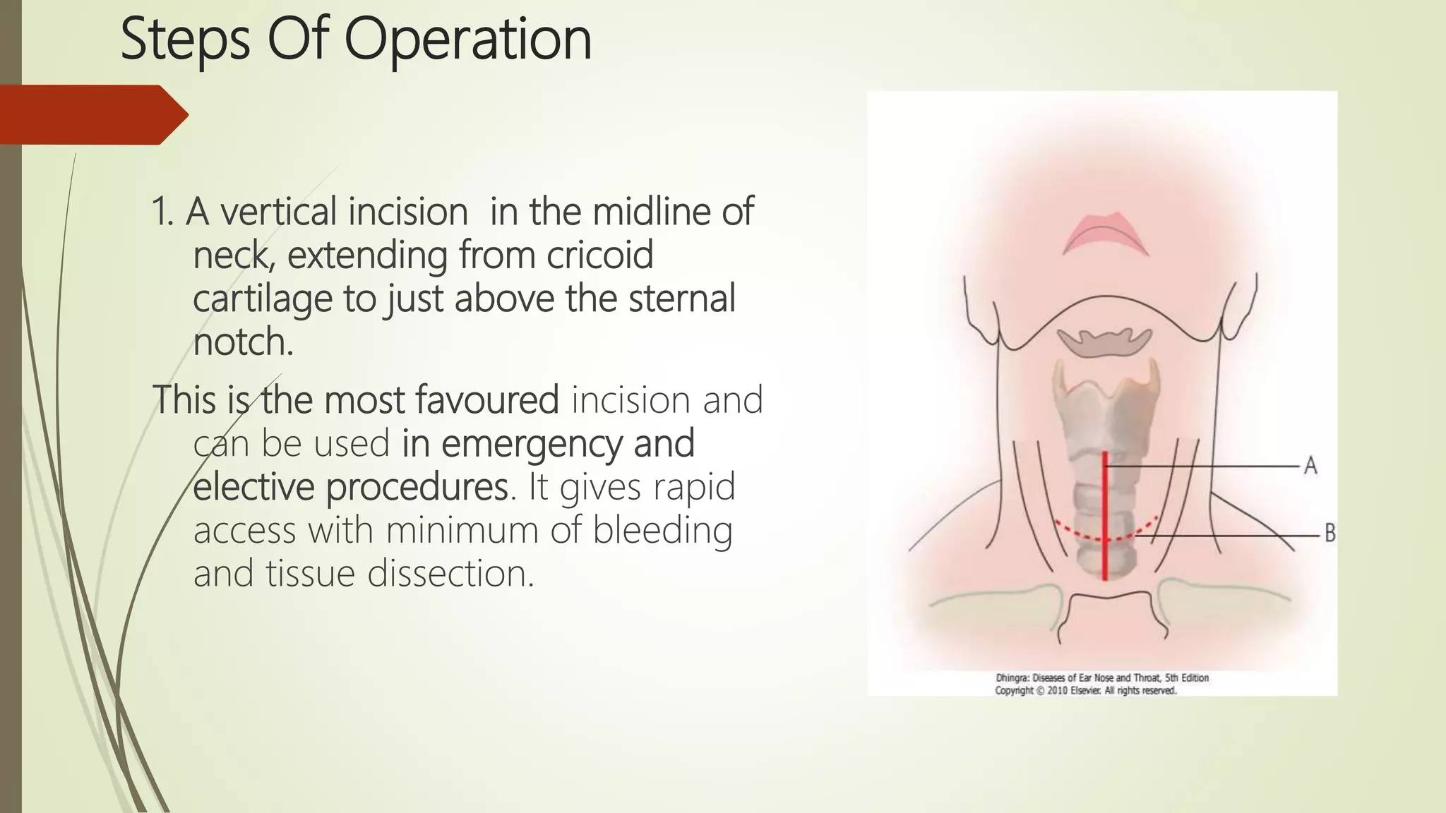
TRACHEOSTOMY
S.SUKRUTH
HISTORY OF TRACHEOSTOMY
 TRACHEOSTOMY WAS PORTRAYED ON EGYPTIAN
TABLETS DATED BACK TO 3600 BC
 Asclepiades of Persia is credited as the first person to
perform a tracheostomy in 100 BC
 The first successful tracheostomy was performed by
Brasovala in the 15th century.
 Antonio Musa Brasavola , an Italian physician,
performed the first documented case of a successful
tracheostomy.
 He published his account in 1546.
 The patient, who suffered from a laryngeal abscess and
recovered from the procedure.
 The tracheostomy has gone by several different names, including pharyngotomy,
laryngotomy, bronchotomy, tracheostomy and tracheotomy.
 The word tracheostomy first appeared in print in 1649,
 Tracheostomies were originally used for emergency treatment of upper airway
obstruction, but with little success. Upper airway obstruction in children was first
discussed as a clinical entity in 1765.
 Tracheostomies were used in the early 1800's for airway inflammation in children
due to Diphtheria.
 The first documented successful tracheostomy performed on a child was
reported in 1808.
 In 1909, a lower tracheostomy technique was introduced in which the tracheal
incision extends to the 4th or 5th tracheal ring. This operative technique was
refined by Chevalier Jackson when faced with the challenge of the polio epidemic
of the 1940's. This technique is basically the same today.
Definition
A ‘tracheostomy’ is a artificial
(usually) surgically created airway
fashioned by making a hole in the
anterior wall of the trachea and the
insertion of a tracheostomy tube,
which may or may not be
permanent
The term ‘Tracheotomy’ means
opening the trachea which is a step
in tracheostomy operation
Functions of Tracheostomy
1. Alternative pathway for breathing
2. Improves alveolar ventilation In cases of respiratory insufficiency :
(a) Decreasing the dead space by 30-50% (normal dead space is 150
ml).
(b) Reducing the resistance to airflow.
3. Protects the airways By using cuffed tube, tracheobronchial tree is
protected against aspiration of:
(a) Pharyngeal secretions, as in case of bulbar paralysis or coma.
(b) Blood, as in haemorrhage from pharynx, larynx or maxillofacial
injuries. With tracheostomy, pharynx and larynx can also be packed to
control bleeding.
4. Permits removal of tracheobronchial secretions
When patient is unable to cough as in coma, head injuries,
respiratory paralysis; or
when cough is painful, as in chest injuries or upper
operations, the tracheobronchial airway can be kept clean of
secretions by repeated suction through the tracheostomy,
avoiding need for repeated bronchoscopy or intubation
is not only traumatic but requires expertise.
5. Intermittent positive pressure respiration (IPPR)
If IPPR is required beyond 72 hours, tracheostomy is
superior to intubation.
6. To administer anaesthesia
laryngopharyngeal growths or trismus.
Indications of Tracheostomy
A. Respiratory obstruction.
B. Retained secretions.
C. Respiratory insufficiency.
Types of Tracheostomy
 Emergency tracheostomy
 Elective or tranquil tracheostomy
 Permanent tracheostomy
 Percutaneous dilatational tracheostomy
 Mini tracheostomy (cricothyroidotomy)
1. Emergency tracheostomy
 It is employed when airway obstruction is complete or
almost complete and
 There is an urgent need to establish the airway.
 Intubation or laryngotomy are either not possible or feasible
in such cases.
2. Elective tracheostomy
(syn. tranquil, orderly or routine tracheostomy)
 This is a planned, unhurried procedure
 It is of two types:
(a) Therapeutic: to relieve respiratory obstruction, remove
tracheobronchial secretions or give assisted ventilation.
(b) Prophylactic: to guard against anticipated respiratory obstruction or
aspiration of blood or pharyngeal secretions such as in extensive surgery of tongue,
floor of mouth, mandibular resection or laryngofissure
3. Permanent tracheostomy
 Required for case of bilateral abductor paralysis or laryngeal
stenosis.
Technique
 Whenever possible, endotracheal
intubation should be done before
tracheostomy. This is specially important in
infants and children.
 Position
Supine with a pillow under the shoulders so
that neck is extended.
Anesthesia
2 % lignocaine & 1 in 2 lakh adrenaline
injected into incision line
Steps Of Operation
1. A vertical incision in the midline of
neck, extending from cricoid
cartilage to just above the sternal
notch.
This is the most favoured incision and
can be used in emergency and
elective procedures. It gives rapid
access with minimum of bleeding
and tissue dissection.
 2. After incision, tissues are dissected in the midline. Dilated veins are
either displaced or ligated.
 3. Strap muscles are separated in the midline and retracted laterally.
 4. Thyroid isthmus is displaced upwards or divided between the clamps, and suture-ligated.
5. Trachea is fixed with a hook and opened with a vertical incision in the region
of 3rd and 4th or 3rd and 2nd rings.
This is then converted into a circular opening. The first tracheal ring is never
divided as perichondritis of cricoid cartilage with stenosis can result
6. Tracheostomy tube of appropriate size is inserted and secured by tapes
Lubricated tracheostomy tube inserted into trachea
Confirm presence of tube in trachea with help of ambu bag & auscultation
 7. Skin incision should
not be sutured or
packed tightly as it may
lead to development of
subcutaneous
emphysema.
8. Gauze dressing is
placed between the skin
and flange of the tube
around the stoma
Pic - Betadine soaked gauze or Sofratulle put
around the tracheostomy opening.
9.Tapes of tracheostomy tube tied around the neck keeping a space
for 1 finger. Neck kept flexed.
Skin incision closed loosely to avoid surgical emphysema.
TRACHEOSTOMY IN CHILDREN &
INFANTS
 Soft and compressible trachea ,so difficult to identify and may get
displaced & injure recurrent laryngeal nerve
 So always useful to have an endotracheal tube or a bronchoscope
inserted into trachea before operation
 Preferably in general anaesthesia
 Don’t extend neck too much as pleura , innominate vessels , thymus
may get injured
 Before incising trachea , silk cultures are places in the trachea on
either side of midline
 tracheal lumen is small, do not insert knife too deep bcoz injury to
post. Tracheal wall or esophagus
 Selection of tube is important .it should be of proper diameter , length &
curvature
 A long tube impinges inti the carina or right bronchus
 With high curvature , lower end of tube impinges on anterior tracheal wall
while upper part compresses the tracheal rings or cricoid
 Trachea is simply incised without excising a circular piece of tracheal wall
 Avoid infolding of ant. Tracheal wall when inserting the tracheostomy tube
 Post operative x-ray of the neck to know position of the tube
 Use of soft silastic and portex tube
Post Operative Care
 1.Constant Supervision
 2.Suction
 3.Tracheostomy tube care
 4. Others
 5.Prevention of crusting and tracheitis
Decannulation
 Adult: plug or seal tube opening & if tolerated for 24 hrs, remove tube.
 Child: Sequentially reduce size of tube. After tube removal  close wound.
Healing occurs within 1 week. Secondary closure after freshening the wound
margin is required rarely.
 Infant or a young child
-Decannulate in operation theatre
-Equipment for re-intubation should ne available like good headlight,
laryngoscope, proper sized endotracheal tubes and a tracheostomy tray
-After decannulation observe for respiratory distress,t achycardia, colour.
-Oximetry is useful
Complications of tracheostomy
IMMEDIATE
(at time of operation)
INTERMEDIATE
(first few days or days)
LATE
(prolonged use of tube)
Haemorrhage Bleeding Haemorrhage
Apnoea Displacement of tube Laryngeal stenosis
Pneumothorax Blocking of tube Tracheal Stenosis
Injury to recurrent
laryngeal nerves
Subcutaneous
Emphysema
Tracheooesophageal
Fistula
Aspiration of blood Tracheitis &
Tracheobronchitis
Problems of
decannulation
Injury to oesophagus Atelectasis & lung abcess Problems of
tracheostomy scar
Local wound infections &
granulations
Persistent
tracheocutaneous fistula
THANK YOU

TRACHEOSTOMY

  • 1.
  • 2.
    HISTORY OF TRACHEOSTOMY TRACHEOSTOMY WAS PORTRAYED ON EGYPTIAN TABLETS DATED BACK TO 3600 BC  Asclepiades of Persia is credited as the first person to perform a tracheostomy in 100 BC  The first successful tracheostomy was performed by Brasovala in the 15th century.  Antonio Musa Brasavola , an Italian physician, performed the first documented case of a successful tracheostomy.  He published his account in 1546.  The patient, who suffered from a laryngeal abscess and recovered from the procedure.
  • 3.
     The tracheostomyhas gone by several different names, including pharyngotomy, laryngotomy, bronchotomy, tracheostomy and tracheotomy.  The word tracheostomy first appeared in print in 1649,  Tracheostomies were originally used for emergency treatment of upper airway obstruction, but with little success. Upper airway obstruction in children was first discussed as a clinical entity in 1765.  Tracheostomies were used in the early 1800's for airway inflammation in children due to Diphtheria.  The first documented successful tracheostomy performed on a child was reported in 1808.  In 1909, a lower tracheostomy technique was introduced in which the tracheal incision extends to the 4th or 5th tracheal ring. This operative technique was refined by Chevalier Jackson when faced with the challenge of the polio epidemic of the 1940's. This technique is basically the same today.
  • 4.
    Definition A ‘tracheostomy’ isa artificial (usually) surgically created airway fashioned by making a hole in the anterior wall of the trachea and the insertion of a tracheostomy tube, which may or may not be permanent The term ‘Tracheotomy’ means opening the trachea which is a step in tracheostomy operation
  • 5.
    Functions of Tracheostomy 1.Alternative pathway for breathing 2. Improves alveolar ventilation In cases of respiratory insufficiency : (a) Decreasing the dead space by 30-50% (normal dead space is 150 ml). (b) Reducing the resistance to airflow. 3. Protects the airways By using cuffed tube, tracheobronchial tree is protected against aspiration of: (a) Pharyngeal secretions, as in case of bulbar paralysis or coma. (b) Blood, as in haemorrhage from pharynx, larynx or maxillofacial injuries. With tracheostomy, pharynx and larynx can also be packed to control bleeding.
  • 6.
    4. Permits removalof tracheobronchial secretions When patient is unable to cough as in coma, head injuries, respiratory paralysis; or when cough is painful, as in chest injuries or upper operations, the tracheobronchial airway can be kept clean of secretions by repeated suction through the tracheostomy, avoiding need for repeated bronchoscopy or intubation is not only traumatic but requires expertise. 5. Intermittent positive pressure respiration (IPPR) If IPPR is required beyond 72 hours, tracheostomy is superior to intubation. 6. To administer anaesthesia laryngopharyngeal growths or trismus.
  • 7.
    Indications of Tracheostomy A.Respiratory obstruction. B. Retained secretions. C. Respiratory insufficiency.
  • 8.
    Types of Tracheostomy Emergency tracheostomy  Elective or tranquil tracheostomy  Permanent tracheostomy  Percutaneous dilatational tracheostomy  Mini tracheostomy (cricothyroidotomy)
  • 9.
    1. Emergency tracheostomy It is employed when airway obstruction is complete or almost complete and  There is an urgent need to establish the airway.  Intubation or laryngotomy are either not possible or feasible in such cases.
  • 10.
    2. Elective tracheostomy (syn.tranquil, orderly or routine tracheostomy)  This is a planned, unhurried procedure  It is of two types: (a) Therapeutic: to relieve respiratory obstruction, remove tracheobronchial secretions or give assisted ventilation. (b) Prophylactic: to guard against anticipated respiratory obstruction or aspiration of blood or pharyngeal secretions such as in extensive surgery of tongue, floor of mouth, mandibular resection or laryngofissure
  • 11.
    3. Permanent tracheostomy Required for case of bilateral abductor paralysis or laryngeal stenosis.
  • 12.
    Technique  Whenever possible,endotracheal intubation should be done before tracheostomy. This is specially important in infants and children.  Position Supine with a pillow under the shoulders so that neck is extended. Anesthesia 2 % lignocaine & 1 in 2 lakh adrenaline injected into incision line
  • 13.
    Steps Of Operation 1.A vertical incision in the midline of neck, extending from cricoid cartilage to just above the sternal notch. This is the most favoured incision and can be used in emergency and elective procedures. It gives rapid access with minimum of bleeding and tissue dissection.
  • 14.
     2. Afterincision, tissues are dissected in the midline. Dilated veins are either displaced or ligated.
  • 15.
     3. Strapmuscles are separated in the midline and retracted laterally.  4. Thyroid isthmus is displaced upwards or divided between the clamps, and suture-ligated.
  • 16.
    5. Trachea isfixed with a hook and opened with a vertical incision in the region of 3rd and 4th or 3rd and 2nd rings. This is then converted into a circular opening. The first tracheal ring is never divided as perichondritis of cricoid cartilage with stenosis can result 6. Tracheostomy tube of appropriate size is inserted and secured by tapes Lubricated tracheostomy tube inserted into trachea Confirm presence of tube in trachea with help of ambu bag & auscultation
  • 17.
     7. Skinincision should not be sutured or packed tightly as it may lead to development of subcutaneous emphysema. 8. Gauze dressing is placed between the skin and flange of the tube around the stoma Pic - Betadine soaked gauze or Sofratulle put around the tracheostomy opening.
  • 18.
    9.Tapes of tracheostomytube tied around the neck keeping a space for 1 finger. Neck kept flexed. Skin incision closed loosely to avoid surgical emphysema.
  • 19.
    TRACHEOSTOMY IN CHILDREN& INFANTS  Soft and compressible trachea ,so difficult to identify and may get displaced & injure recurrent laryngeal nerve  So always useful to have an endotracheal tube or a bronchoscope inserted into trachea before operation  Preferably in general anaesthesia  Don’t extend neck too much as pleura , innominate vessels , thymus may get injured  Before incising trachea , silk cultures are places in the trachea on either side of midline  tracheal lumen is small, do not insert knife too deep bcoz injury to post. Tracheal wall or esophagus
  • 20.
     Selection oftube is important .it should be of proper diameter , length & curvature  A long tube impinges inti the carina or right bronchus  With high curvature , lower end of tube impinges on anterior tracheal wall while upper part compresses the tracheal rings or cricoid  Trachea is simply incised without excising a circular piece of tracheal wall  Avoid infolding of ant. Tracheal wall when inserting the tracheostomy tube  Post operative x-ray of the neck to know position of the tube  Use of soft silastic and portex tube
  • 21.
    Post Operative Care 1.Constant Supervision  2.Suction  3.Tracheostomy tube care  4. Others  5.Prevention of crusting and tracheitis
  • 23.
    Decannulation  Adult: plugor seal tube opening & if tolerated for 24 hrs, remove tube.  Child: Sequentially reduce size of tube. After tube removal  close wound. Healing occurs within 1 week. Secondary closure after freshening the wound margin is required rarely.  Infant or a young child -Decannulate in operation theatre -Equipment for re-intubation should ne available like good headlight, laryngoscope, proper sized endotracheal tubes and a tracheostomy tray -After decannulation observe for respiratory distress,t achycardia, colour. -Oximetry is useful
  • 25.
    Complications of tracheostomy IMMEDIATE (attime of operation) INTERMEDIATE (first few days or days) LATE (prolonged use of tube) Haemorrhage Bleeding Haemorrhage Apnoea Displacement of tube Laryngeal stenosis Pneumothorax Blocking of tube Tracheal Stenosis Injury to recurrent laryngeal nerves Subcutaneous Emphysema Tracheooesophageal Fistula Aspiration of blood Tracheitis & Tracheobronchitis Problems of decannulation Injury to oesophagus Atelectasis & lung abcess Problems of tracheostomy scar Local wound infections & granulations Persistent tracheocutaneous fistula
  • 26.