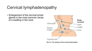
4. BRANCHIAL CYSTS
• Branchial is derived from Greek word branchia that refers to gills.
• Branchial cleft cysts are congenital epithelial cysts, which arise
on the lateral part of the neck from a failure of obliteration of the
second branchial cleft in embryonic development.
• Branchial cleft cysts are the most common cause of congenital
neck masses.
• There is a tendency for cases to cluster in families.
• They are congenital, but may not present until later in life
usually early adulthood.
5.
6. FEATURES
• The cyst usually presents in the upper neck in early or middle
adulthood and is found at the junction of the upper third and middle
third of the sternomastoid muscle at its anterior border.
• Many branchial cleft cysts are asymptomatic.
• It is a fluctuant swelling that may transilluminate and is often soft in
its early stages so that it may be difficult to palpate.
• Depending on the size and the anatomical extension of the
mass, local symptoms, such as dysphagia, dysphonia,
dyspnea, and stridor, may occur.
7. FEATURES 2
• They may become tender, enlarged, or inflamed, or they may
develop abscesses, especially during periods of upper
respiratory tract infection, due to the lymphoid tissue located
beneath the epithelium.
• Spontaneous rupture of an abscessed branchial cleft cyst may
result in a purulent draining sinus to the skin or the pharynx.
8. DDX
• Lymphadenopathy (reactive, neoplastic, lymphoma, metastasis)
• Vascular neoplasms and malformations
• Capillary hemangioma
• Carotid body tumour
• Lymphatic malformation (cystic hygroma)
• Ectopic thyroid tissue
• Ectopic salivary tissue
• Hydatid cyst of the neck
9. EXAMINATION OF THE MASS
• Size
• Site
• Shape
• Surface
• Consistency
• Fixation: deep/superficial
• Pulsatility
• Compressibility
• Transillumination
• Bruit
11. MANAGEMENT
• Antibiotics are used to treat infections or abscesses related to
branchial cysts.
• Surgical management is the definitive management.
• Branchial cleft cyst surgery is best delayed until the patient is at
least age 3 months. Definitive branchial cleft cyst surgery
should not be attempted during an episode of acute infection or
if an abscess is present. Surgical incision and drainage of
abscesses is indicated if present, usually along with concurrent
antimicrobial therapy.
• In patients over 40 years of age, a high index of suspicion for a
necrotic metastatic lymph should exists and malignancy should be
excluded before excision.
12. Complications
• Untreated branchial cleft cyst lesions are prone to recurrent
infection and abscess formation with resultant scar formation
and possible compromise to local structures.
• Complications of surgical excision of branchial cleft cysts result
from damage to nearby vascular or neural structures, which
include carotid vessels and the facial, hypoglossal, vagus, and
lingual nerves.
• There are rare case reports of malignancies having been
identified in branchial cleft lesions, including branchiogenic
carcinoma and papillary thyroid carcinoma.
13. prognosis
• Following surgical excision of branchial cleft cysts, recurrence is
uncommon, with a risk estimated at 3%, unless previous
surgery or recurrent infection has occurred, in which case, it
may be as high as 20%.
14. BRANCHIAL FISTULA
• A branchial fistula is thought to represent a persistent second
branchial cleft.
• It may be unilateral or bilateral.
• The external orifice is nearly always situated in the lower third of the
neck near the anterior border of the sternocleidomastoid, while the
internal orifice is located on the anterior aspect of the posterior
faucial pillar just behind the tonsil.
15. Branchial fistula
• The internal aspect of the tract may, however, end blindly at or close
to the lateral pharyngeal wall, constituting a sinus rather than a
fistula.
• The tract is lined by ciliated columnar epithelium and, as such, there
may be a small amount of recurrent mucopurulent discharge onto the
neck.
• The tract follows the same path as a branchial cyst and requires
complete excision to avoid recurrence.
16.
17. Cystic hygroma
• Lymphangiomas are benign malformations of the lymphatic
system and can occur anywhere in the body.
• Cystic hygromas are macrocystic lymphangiomas that occur in
the neck and less frequently in the cheek, axilla, groin and
mediastinum.
• Other types of lymphangiomas include capillary & cavernous.
18. AETIOLOGY
• They are usually congenital, but can be acquired as a result of trauma,
inflammation or lymphatic drainage obstruction.
• Karyotropic abnormalities present in 25-70% of children with
lympangiomas e.g. Turner, Klinefelter, Trisomy 21, trisomy 18,
trisomy 13.
• There is also an association with intrauterine alcohol exposure.
•
19. Pathophysiology
• Lymphangiomas are thought to arise from the following:
1. Sequestration of lymphatic tissue from lymphatic sacs during
development
2. Failure of these tissues to communicate with the lymphatic or
venous system
3. Tissue dilation resulting in the cystic morphology
• These lymphatic rests can penetrate adjacent structures or
dissect along fascial planes and eventually become canalized.
Because of the lack of an outflow tract, these spaces retain their
secretions and develop cystic components.
• The nature of the surrounding tissue determines whether the
20. presentation
• Cystic hygromas usually present in the neonate or in early infancy,
and occasionally may present at birth and be so large as to obstruct
labour.
• The cysts are filled with clear lymph and lined by a single layer of
epithelium with a mosaic appearance.
• Swelling usually occurs in the neck and may involve the parotid,
submandibular, tongue and floor of mouth areas. The swelling may be
bilateral and is soft and partially compressible, visibly increasing in
size when the child coughs or cries. The characteristic that
distinguishes it from all other neck swellings is that it is brilliantly
translucent.
22. diagnosis
Intrauterine
• Transabdominal USS by ten weeks or via transvaginal USS
• Fast spin MRI
• Elevated alpha-fetoprotein levels in amniocentesis fluid have
been reported in pregnancies.
Flexible laryngoscopy performed by an otolaryngologist can be
a useful adjunct to evaluate vocal fold mobility as well as airway
patency.
MRI, CT & USS of the neck
23.
24. MANAGEMENT
• Watchful waiting in asymptomatic patients.
• Use of sclerosing agents like such as OK-432 (an inactive strain of
group A Streptococcus pyogenes), bleomycin, sodium
tetradecyl sulfate.
• IV antibiotics in case of infection
• Mainstay is surgical excision.
• The exceptions to excision at the time of diagnosis include
premature infants who are small and those with involvement of
crucial neurovascular structures that are small and difficult to
identify (e.g. facial nerve). If no airway obstruction is present,
surgery can be delayed until the child is aged 2 years or older,
28. CBT a.k.a Chemodectoma
• Carotid body tumors (CBTs) are rare.
• These tumors develop within the adventitia of the medial aspect
of the carotid bifurcation.
• The carotid body originates in the neural crest. (function…
29. classification
Three classes of CBT
• Familial, more common in younger people
• Sporadic, most common
• Hyperpastic, common in patients with chronic hypoxia such as
those living at high altitudes, copd, cyanotic heart disease.
30. pathophysiology
• Genetically, genes encoding the subunits of the enzyme
succinate dehydrogenase complex, which is part of the Kreb's
cycle. Defective succinate dehydrogenase has been postulated
to cause an increase in the intracellular concentration of
molecular hypoxia mediators and the vascular endothelial
growth factor (VEGF) thus resulting in hyperplasia,
angiogenesis, and neoplasia.
• Chronic hypoxic conditions lead to hypertrophy, hyperplasia,
and neoplasia of the chief cells.
• CBTs can be occasionally coupled with non paraganglonic
tumors in syndromes, including MEN type II, von Hippel-Lindau
syndrome, and neurofibromatosis type 1.
31. presentation
• CBTs present most commonly as an asymptomatic palpable
neck mass in the anterior triangle of the neck.
• They are slow-growing tumors that can remain asymptomatic
for many years. The doubling time (TD) as estimated by Jansen
et al using sequential imaging, was 7.13 years with a median
growth rate of 0.83 mm/year.
• Approximately 10% of the cases present with cranial nerve
palsy with paralysis of the hypoglossal, glossopharyngeal,
recurrent laryngeal, or spinal accessory nerve, or involvement
of the sympathetic chain.
• CBTs may, therefore, be associated with pain, hoarseness,
dysphagia, Horner syndrome, or shoulder drop
32. Presentation 2
• As the tumor enlarges and compresses the carotid artery and
the surrounding nerves, other symptoms may also be present,
such as pain, tongue paresis, hoarseness, Horner syndrome,
and dysphagia.
• In cases of functional carotid body tumors, symptoms similar to
those of pheochromocytoma, such as paroxysmal hypertension,
palpitations, and diaphoresis, are seen.
33. On examination
• the mass is typically vertically fixed because of its attachment to
the bifurcation of the common carotid (Fontaine sign).
• The mass is firm, rubbery, pulsatile, mobile from side to side but not
up and down, and can sometimes be emptied by firm pressure, after
which it slowly refills in a pulsatile manner.
• A bruit can be felt; however, the absence of a bruit does not rule
out a carotid body tumor.
• Vagal body tumors are more cranially located and sometimes
project into the lateral pharynx as a pulsatile mass.
34. Diagnosis
• USS with colour doppler to assess vascularity of the neck mass.
• CT of head & neck usually revealing hypervascularity of tumor
between external and internal carotid arteries.
• MRI with classic salt & pepper appearance in T1 weighted
• MR angiography
• Fine needle aspiration and biopsy should be avoided
35. staging
Shamblin describes 3 different types or stages of carotid body
tumors.
• Type I consists of a small tumor that is easily dissected from the
adjacent vessels in a periadventitial plane.
• Type II tumors are larger and more adherent and partially
surround the vessel.
• Type III tumors are large and completely surround the carotid
bifurcation.
As described, types II and III tumors are more likely to require
carotid resection.
36.
37. MANAGEMENT
• Choice is made between radiotherapy and surgery and should
consider factors such as,
presence of other paragangliomas
presence of bilateral carotid body tumors
age
health of the patient
patient's preference.
39. causes
• Inflammatory e.g. Reactive hyperplasia
• Infective
Viral For example, infectious mononucleosis, HIV
Bacterial Streptococcus, Staphylococcus Actinomycosis Tuberculosis
Brucellosis
Protozoan e.g. Toxoplasmosis
• Neoplastic
Malignant Primary (e.g. lymphoma)
Secondary (e.g. squamous cell carcinoma
40. inflammatory
• Non-specific inflammatory lymphadenopathy commonly follows
recurrent bouts of tonsillitis, especially if the attacks have been
treated inadequately. This is common in children less than 1o
years.
• Non-specific reactive lymphoid hyperplasia can follow any
inflammatory process or be associated with skin conditions,
particularly of the scalp, when it is termed dermatopathic
lymphadenopathy.
42. REFERENCES
• Andrew W. McCaskie et. Al., Bailey & love’s Short Practice of Surgery
27th edition 2017 CRC Press Chapter 47
• Medscape
https://emedicine.medscape.com/article/1575155-treatment#d1
• Norman L. Browse et. Al., Browse’s Introduction to The Symptoms
and Signs of Surgical Disease.
• F. Charles et. Al., Schwartz’s Principles of Surgery 11th edition
Editor's Notes
At the fourth week of embryonic life, the development of 4 branchial (or pharyngeal) clefts results in 5 ridges known as the branchial (or pharyngeal) arches, which contribute to the formation of various structures of the head, the neck, and the thorax. The second arch grows caudally and, ultimately, covers the third and fourth arches. The buried clefts become ectoderm-lined cavities, which normally involute around week 7 of development. If a portion of the cleft fails to involute completely, the entrapped remnant forms an epithelium-lined cyst with or without a sinus tract to the overlying skin.
commonly presents as a solitary, painless mass in the neck of a child or a young adult. A history of intermittent swelling and tenderness of the lesion during upper respiratory tract infection may exist.
Contrast enhanced CT at the level of the hyoid bone reveals ill defined non enhancing water attenuation mass (g
S sternoclaidomastoid
Although the anterior aspect of the cyst is easy to dissect, it may pass backwards and upwards through the bifurcation of the common carotid artery as far as the pharyngeal constrictors. It passes superficial to the hypoglossal and glossopharyngeal nerves, but deep to the posterior belly of the digastric. These structures and the spinal accessory nerve must be positively identified to avoid damage.
A branchial fistula is an abnormal canal that opens internally into the tonsillar sinus (fossa) and externally on the side of the neck . Saliva may drip from the fistula, which may become infected. This uncommon cervical canal results from persistence of remnants
of the 2nd pharyngeal pouch and 2nd pharyngeal groove. The fistula ascends from its cervical opening, usually along the anterior border of the SCM
in the inferior third of the neck. It first passes through the subcutaneous tissue, platysma, and fascia of the neck to enter the carotid sheath. It then passes between the internal and the external carotid arteries on its way to its opening in the tonsillar sinus. Its course can be demonstrated by radiography.
Rarely, children with LMs display symptoms of newly onset obstructive sleep apnea syndrome (OSAS). This situation may involve children with LM or other space-occupying lesions of the supraglottic or paraglottic region. Suprahyoid lymphangiomas tend to cause more breathing difficulties than infrahyoid lesions.
Potentially life-threatening airway compromise that manifests as noisy breathing (stridor) and cyanosis is a possible symptom of lymphangiomas.
Feeding difficulties, as well as failure to thrive, may alert the clinician to a potential lymphangioma. This is especially true when the lesion affects structures of the upper aerodigestive tract
OE.. It is doughy
CT and MRI reveal a ringlike margin enhancement with sharp demarcation of cystic areas. The cystic areas tend to appear circumscribed and discrete.
Contrast helps to enhance cyst-wall visualization and the relation to surrounding blood vessels.
Bleomycin, a cytotoxic antibiotic, has been considered a poor choice because of its toxicity (pulmonary fibrosis
the mainstay of treatment of LMs is surgical excision. [32] Although surgery is the standard treatment, LM is a benign lesion. If acute infection occurs prior to resection, surgery should be delayed at least 3 months.
The surgical team should attempt to remove the LM completely or, failing that, to remove as much as possible, sparing all vital neurovascular structures.
The carotid body, which originates in the neural crest, is important in the body's acute adaptation to fluctuating concentrations of oxygen, carbon dioxide, and pH. The carotid body protects the organs from hypoxic damage by releasing neurotransmitters that increase the ventilatory rate when stimulated.
Relevant Anatomy
The carotid body is a small, reddish-brown, oval structure, located in the posteromedial aspect of the carotid artery bifurcation. The healthy gland measures 3-5 mm in diameter and weighs less than 15 mg on average. [23] The vast majority of the literature states that the gland is located in the adventitia near the carotid artery bifurcation. However, according to Maxwell et al, most surgeons experienced with carotid body dissection maintain that it is more peripherally located, within periadventitial tissue. This distinction is critical, as dissections in the deeper planes of the carotid artery are associated with higher risk for complications from vessel injury. [24]
The gland is highly vascular and receives its blood supply from feeder vessels running through the Mayer ligaments, primarily from the external carotid artery, typically the ascending pharyngeal artery. It is innervated by the Hering nerve, originating from the glossopharyngeal nerve about 1.5 cm distal to the jugular foramen.
Von hi … A rare, inherited disorder that causes tumors and cysts to grow in certain parts of the body, including the brain, spinal cord, eyes, inner ear, adrenal glands, pancreas, kidney, and reproductive tract. The tumors are usually benign (not cancer), but some may be malignant (cancer
The reticuloses are common in children and
young adults.
Sex Males are affected more often than females.
Symptoms The most common presenting symptom
is a painless lump in the neck, which is noticed by
chance and grows slowly.
Malaise, weight loss and pallor are common
symptoms.
Itching of the skin (pruritus) is an unexplained
but distinctive complaint.
There may be fever with rigors, occurring in a
periodic fashion (Pel–Ebstein fever).
Lymphomatous infiltration of the skeleton may
cause pains in the bones, and there may be abdominal
pain after drinking alcohol.
If there are large masses of lymph glands in the
mediastinum, they may occlude the superior vena
cava, causing venous congestion in the neck and
the development of collateral veins across the chest
wall.
Large masses in the abdomen can obstruct the
inferior vena cava and cause oedema of both legs.