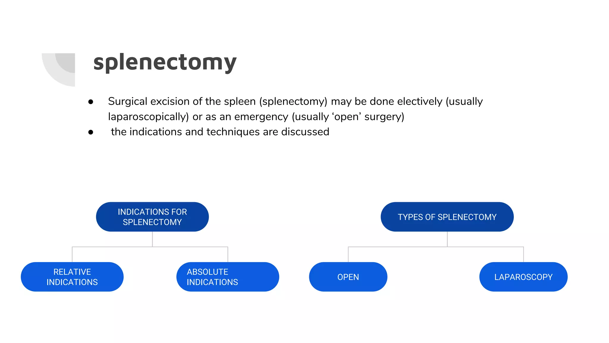
Indication for
splenectomy
Surgical department At Wolfson medical Center
Dr. Nasim Badarna - internship
The spleen - description
❖ Immunologic organ without distinct lobes or segments that weighs about 100– 175 g.
The spleen is responsible for the removal of old red blood cells and bacteria from the
blood circulation.
❖ Macroscopically, the cut surface of the organ reveals a ‘red pulp’, within which are
scattered numerous white nodules (‘white pulp’).
Anatomy
❖ The spleen is the largest single mass of lymphoid tissue in the body
❖ invested in peritoneum, in the left upper quadrant.
❖ The healthy spleen is the size of a closed fist and is not palpable on clinical
examination. Hypertrophy of the organ can occur in disease states and enlargement is
seen in an inferior and medial direction, towards the umbilicus and right iliac fossa.
ANATOMIC BOUNDARIES
Left upper quadrant (LUQ) of the abdomen, between the 8th and 11th ribs
Superior: Left diaphragm leaf.
Inferior: Colon, splenic flexure,
and phrenocolic ligament.
Medial: Pancreas (tail) and stomach.
Lateral: Rib cage.
Anterior: Rib cage, stomach.
Posterior: Rib cage.
Ligaments of the spleen
● Gastrosplenic
● Splenonephric
● Splenocolic
● Splenophernic
Blood supply of the spleen
receives most of its arterial supply from the splenic
artery.
- arises from the coeliac trunk, running laterally
along the superior aspect of the pancreas,
within the splenorenal ligament.
- These arterial branches do not anastomose
with each other – giving rise to vascular
segments of the spleen. This enables a surgeon
to remove one of these segments without
affecting the others: subtotal splenectomy.
Blood supply of the spleen
❖ The splenic vein receives blood from the spleen and
stomach via short gastric and left gastroepiploic veins.
❖ the splenic vein, having received the inferior mesenteric
vein, joins the superior mesenteric vein to form the portal
vein
Functions
❖ Blood filtration and immune responses
❖ Blood sequestration
➢ damaged cells are recognized in the splenic cords and are
phagocytosed, removing them from the circulation.
■ Howell–Jolly bodies (remnants of nuclear DNA)
■ Heinz bodies (denatured haemoglobin)
❖ Haemopoiesis: the production of blood cells by the spleen is restricted
to the first five months of intrauterine life.The spleen retains a
haemopoietic role in some disease states.
splenectomy
● Surgical excision of the spleen (splenectomy) may be done electively (usually
laparoscopically) or as an emergency (usually ‘open’ surgery)
● the indications and techniques are discussed
INDICATIONS FOR
SPLENECTOMY
ABSOLUTE
INDICATIONS
RELATIVE
INDICATIONS
TYPES OF SPLENECTOMY
LAPAROSCOPYOPEN
Absolute indications
indications
INDICATIONS FOR SPLENECTOMY
Most common indications for splenectomy:
1. Trauma
2. Idiopathic thrombocytopenic purpura (ITP) refractory to steroids
Haematological indications:
In a number of conditions, red cell morphology is altered such that the cells are
phagocytosed causing anaemia and splenomegaly.
● Rarely cures the disease
● Alleviates symptoms
● Corrects hematologic abnormalities
● Staging & Diagnosis
Haematological indications:
● hereditary spherocytosis
○ Most common type of hemolytic anemia Splenomegaly & Gall
stones. Dx by (+) spherocytes in the blood
○ Splenectomy in the 4th year of life +/- cholecystectomy if (+)
cholelithiasis
● Autoimmune Hemolytic Anemia (AIHA)
○ Splenectomy Splenectomy (80% favorable clinical response) if:
medical tx fails, Intolerance to steroids or its side-effects
● Sickle Cell Disease (SCD)
● Thalassemia
○ Splenectomy indicated if: Symptomatic splenomegaly
Haematological indications: ITP
Immune thrombocytopenia purpura s the commonest haematological
indication for elective splenectomy.
➔ Excessive destruction of platelets due to synthesis of antiplatelet
antibody within the spleen can lead to low concentrations of platelets
such that spontaneous bleeding
Initial treatment: corticosteroid therapy; 20% complete response -->no
further treatment. Patients with refractory thrombocytopenia may benefit
from splenectomy.
Neoplasia
splenectomy was previously undertaken to aid staging of
haematological malignancies (e.g. lymphomas, leukaemias), but
modern cross-sectional imaging has made such surgery redundant
(although resection of the spleen is sometimes done for
symptomatic reasons (pain) or to ameliorate the hypersplenism
associated with an enlarged organ).
Overall, resection of the spleen for neoplasia has declined markedly
in recent years. Massively enlarged spleens (i.e. chronic lymphocytic
leukaemia) should be treated by open splenectomy.
Splenic rupture
● Trauma
○ The spleen is the most commonly injured organ in blunt abdominal
trauma, and trauma is the most common reason for splenectomy.
○ Rib fractures on the left - especially the 9th and 10th, which make up
20% of cases.
● Spontaneous rupture
○ Children are at greater risk of splenic rupture, as well as patients with
pathological splenomegaly (e.g. mononucleosis)
Splenic trauma
SIGNS AND SYMPTOMS
History: Check for preexisting diseases that cause
splenomegaly (these patients are more vulnerable to splenic
injury), details of injury mechanism.
Exam: Look for peritoneal irritation, Kehr’s sign,
left-sided lower rib fractures, external signs of injury.
● Thirty percent of patients with splenic injury will
present with hypotensive shock due to hemorrhage.
Managment
In the emergency situation, trauma patients should be treated in
accordance with the ABC principles of the Advanced Trauma and
Life Support™
❖ Initial Airway, breathing, circulation (ABCs).
❖ It can be very difficult to decide whether a patient needs an
emergency splenectomy after trauma, particularly when the
patient is haemodynamically stable and has minimal signs
of abdominal injury.
Managment
Special investigations do not provide absolute answers and the risk of
delayed and unnecessary laparotomies will remain.
● Ultrasound (US): May be used for initial assessment to detect
hemoperitoneum as a part of focused abdominal sonography for trauma
(FAST) exam.
● Computed tomographic (CT) scan: Able to define injury precisely.
● Diagnostic peritoneal lavage (DPL): Not specific for splenic injury but
will show hemoperitoneum.
● Angiogram: May be able to use therapeutically in the stable patient
(embolization of CT-identified injury).
Hemodynamically unstable
Focused assessment with sonography for
trauma (FAST) is an excellent investigation for
haemoperitoneum in blunt trauma with a
sensitivity of 88%.
An emergency laparotomy is indicated for a
positive FAST in the shocked patient.
FAST exam
● 4 views
● Each view 30 -60 sec
● Total exam time usually
takes 3-5 min
● Fresh blood
Anechoic (Black)
● Coagulated blood
First hypoechoic
Later hyperechoic
Radiographic signs on US
● Perisplenic fluid
● enlarged spleen
● irregular borders
● abnormal position
● increase in size over
time.
Hemodynamically stable
CT scan is the single most useful investigation
in the hemodynamically stable patient as it can
assess:
1. intraperitoneal fluid
2. solid organ injury
3. retroperitoneal haematoma.
Repeated scanning (ultrasound or CT) have
been found particularly helpful in assessing
splenic bleeding or haematoma especially if
patient is slowly dropping her haemoglobin.
AAST spleen injury scale
The American Association for the Surgery of Trauma (AAST)
splenic injury scale, most recently revised in 2018, is currently
the most widely used grading system for splenic trauma
In haemodynamically stable patients, it is more
likely that a subcapsular splenic haematoma is
formed rather than a free rupture into the
peritoneal cavity
Grade 1
Management
Definitive treatment
operative Nonoperative
Operative vs Non-operative
Operative management indications:
● Unstable
● Signs and symptoms of ongoing
hemorrhage.
● Injury ≥ grade III
● Failure of nonoperative management
○ usually within 48–72 hours.
○ vascular blush on CT scan are
likely to fail nonoperative
management.
Nonoperative management criteria
● Stable
● Injury grade I or II
● No evidence of injury to other
intra-abdominal organs.
*Patients who are stable or who
stabilize with fluid resuscitation may
be considered for conservative
management.
non-operative management
Many of these patients (65–95% of adults and 87–98% of children) can be treated
conservatively.
Consists of:
1. bed rest,
2. nasogastric tube (NGT) decompression
3. Fluid resusitation
4. Close observation - monitored setting, serial exam, Hematocrits.
a. Close observation; watch for secondary haemorrhage or continual slow ooze
affects some patients, prompting a laparotomy.
b. observes patients for 7 days post-injury
c. most delayed haemorrhage declare themselves within 72 hours.
Operative management
● the spleen is the organ most at risk of damage after blunt trauma to
the abdomen, yet fewer than 10% of patients require a laparotomy.
● Unstable patients with abdominal distension, peritonitis and
hypotension despite fluid resuscitation require transfer to the
Operating Suite for an emergency laparotomy.
Splenectomy: surgical techniques
Open splenectomy
Immediate resuscitation and rapid transfer to the operating theatre is
necessary to control haemorrhage if the patient is haemodynamically
unstable.
Once the spleen is removed, care is taken to avoid haemorrhage from the
splenic bed and also the tail of the pancreas, which is intimately related to
the splenic hilum. A drain is often placed up to the left upper quadrant after
surgery.
Patients are placed in a supine, crucifix position on the operating table to
allow optimal venous and arterial access for monitoring and fluid
replacement. Placement of a nasogastric tube and urinary catheter
An upper midline incision is done and the abdominal viscera and liver
examined.
Laparoscopic splenectomy
The patient is placed in a right-lateral decubitus
position with the left arm elevated; the position is
maintained with lateral supports or pneumatic
devices.
The lateral position allows the spleen to ‘hang
down’ from its diaphragmatic attachments to
allow access to the hilar vessels.
Splenectomy Complications
● LLL atelectasis, pneumonia, effusion
● Hemorrhage (mostly with laparoscopic, and mostly first 24 hours PO)
● Intraabdominal abscess (LUQ)
● Gastric dilation
○ rare due to the manipulation of the stomach during splenectomy. Placement of a
nasogastric tube during surgery can prevent this complication in most cases
● Pancreatitis or fistula formation
○ amylase content of drain fluid should be measured if this is suspected.
● Thrombocytosis
○ many of the platelets that were sequestered in the spleen are now out in the
circulation.
Complications of splenectomy
● OPSI :overwhelming post-splenectomy infection is a rare but rapidly fatal infection.
typically characterized by either meningitis or sepsis
○ Incidence ≈1% rare (up to 5%); 50% Mortality
○ Risk factors
■ Children < 15 yrs old
■ Immunosupression
■ Hematologic dz (thalassemia, SCD, etc)
■ Highest within the first 2 yrs post splenectomy
○ Pnemococcus, H.influenza, meningococcus
■ vaccination 7-14 days prior to splenectomy
■ Pneumovax booster Q5yrs and annual H.influenza immunizations
■ Abx prophylaxis for children x2 yrs post splenectomy
overwhelming post-splenectomy infection (OPSI)
Thank you for your attention
Reference
● upToDate
● First aid clerkship for surgery
● Radiopedia - https://radiopaedia.org/articles/aast-spleen-injury-scale?lang=us
Davies JM, Barnes R, Milligan D. British Committee for Standards in Haematology. Working
Party of the Haematology/Oncology Task Force. Update of guidelines for the prevention and
treatment of infection in patients with an absent or dysfunctional spleen. Clin Med 2002; 2:
440–3.

indication for splenectomy

  • 1.
    Indication for splenectomy Surgical departmentAt Wolfson medical Center Dr. Nasim Badarna - internship
  • 2.
    The spleen -description ❖ Immunologic organ without distinct lobes or segments that weighs about 100– 175 g. The spleen is responsible for the removal of old red blood cells and bacteria from the blood circulation. ❖ Macroscopically, the cut surface of the organ reveals a ‘red pulp’, within which are scattered numerous white nodules (‘white pulp’).
  • 3.
    Anatomy ❖ The spleenis the largest single mass of lymphoid tissue in the body ❖ invested in peritoneum, in the left upper quadrant. ❖ The healthy spleen is the size of a closed fist and is not palpable on clinical examination. Hypertrophy of the organ can occur in disease states and enlargement is seen in an inferior and medial direction, towards the umbilicus and right iliac fossa.
  • 4.
    ANATOMIC BOUNDARIES Left upperquadrant (LUQ) of the abdomen, between the 8th and 11th ribs Superior: Left diaphragm leaf. Inferior: Colon, splenic flexure, and phrenocolic ligament. Medial: Pancreas (tail) and stomach. Lateral: Rib cage. Anterior: Rib cage, stomach. Posterior: Rib cage.
  • 5.
    Ligaments of thespleen ● Gastrosplenic ● Splenonephric ● Splenocolic ● Splenophernic
  • 6.
    Blood supply ofthe spleen receives most of its arterial supply from the splenic artery. - arises from the coeliac trunk, running laterally along the superior aspect of the pancreas, within the splenorenal ligament. - These arterial branches do not anastomose with each other – giving rise to vascular segments of the spleen. This enables a surgeon to remove one of these segments without affecting the others: subtotal splenectomy.
  • 7.
    Blood supply ofthe spleen ❖ The splenic vein receives blood from the spleen and stomach via short gastric and left gastroepiploic veins. ❖ the splenic vein, having received the inferior mesenteric vein, joins the superior mesenteric vein to form the portal vein
  • 8.
    Functions ❖ Blood filtrationand immune responses ❖ Blood sequestration ➢ damaged cells are recognized in the splenic cords and are phagocytosed, removing them from the circulation. ■ Howell–Jolly bodies (remnants of nuclear DNA) ■ Heinz bodies (denatured haemoglobin) ❖ Haemopoiesis: the production of blood cells by the spleen is restricted to the first five months of intrauterine life.The spleen retains a haemopoietic role in some disease states.
  • 9.
    splenectomy ● Surgical excisionof the spleen (splenectomy) may be done electively (usually laparoscopically) or as an emergency (usually ‘open’ surgery) ● the indications and techniques are discussed INDICATIONS FOR SPLENECTOMY ABSOLUTE INDICATIONS RELATIVE INDICATIONS TYPES OF SPLENECTOMY LAPAROSCOPYOPEN
  • 10.
  • 11.
  • 12.
    INDICATIONS FOR SPLENECTOMY Mostcommon indications for splenectomy: 1. Trauma 2. Idiopathic thrombocytopenic purpura (ITP) refractory to steroids
  • 13.
    Haematological indications: In anumber of conditions, red cell morphology is altered such that the cells are phagocytosed causing anaemia and splenomegaly. ● Rarely cures the disease ● Alleviates symptoms ● Corrects hematologic abnormalities ● Staging & Diagnosis
  • 14.
    Haematological indications: ● hereditaryspherocytosis ○ Most common type of hemolytic anemia Splenomegaly & Gall stones. Dx by (+) spherocytes in the blood ○ Splenectomy in the 4th year of life +/- cholecystectomy if (+) cholelithiasis ● Autoimmune Hemolytic Anemia (AIHA) ○ Splenectomy Splenectomy (80% favorable clinical response) if: medical tx fails, Intolerance to steroids or its side-effects ● Sickle Cell Disease (SCD) ● Thalassemia ○ Splenectomy indicated if: Symptomatic splenomegaly
  • 15.
    Haematological indications: ITP Immunethrombocytopenia purpura s the commonest haematological indication for elective splenectomy. ➔ Excessive destruction of platelets due to synthesis of antiplatelet antibody within the spleen can lead to low concentrations of platelets such that spontaneous bleeding Initial treatment: corticosteroid therapy; 20% complete response -->no further treatment. Patients with refractory thrombocytopenia may benefit from splenectomy.
  • 16.
    Neoplasia splenectomy was previouslyundertaken to aid staging of haematological malignancies (e.g. lymphomas, leukaemias), but modern cross-sectional imaging has made such surgery redundant (although resection of the spleen is sometimes done for symptomatic reasons (pain) or to ameliorate the hypersplenism associated with an enlarged organ). Overall, resection of the spleen for neoplasia has declined markedly in recent years. Massively enlarged spleens (i.e. chronic lymphocytic leukaemia) should be treated by open splenectomy.
  • 17.
    Splenic rupture ● Trauma ○The spleen is the most commonly injured organ in blunt abdominal trauma, and trauma is the most common reason for splenectomy. ○ Rib fractures on the left - especially the 9th and 10th, which make up 20% of cases. ● Spontaneous rupture ○ Children are at greater risk of splenic rupture, as well as patients with pathological splenomegaly (e.g. mononucleosis)
  • 18.
    Splenic trauma SIGNS ANDSYMPTOMS History: Check for preexisting diseases that cause splenomegaly (these patients are more vulnerable to splenic injury), details of injury mechanism. Exam: Look for peritoneal irritation, Kehr’s sign, left-sided lower rib fractures, external signs of injury. ● Thirty percent of patients with splenic injury will present with hypotensive shock due to hemorrhage.
  • 19.
    Managment In the emergencysituation, trauma patients should be treated in accordance with the ABC principles of the Advanced Trauma and Life Support™ ❖ Initial Airway, breathing, circulation (ABCs). ❖ It can be very difficult to decide whether a patient needs an emergency splenectomy after trauma, particularly when the patient is haemodynamically stable and has minimal signs of abdominal injury.
  • 20.
    Managment Special investigations donot provide absolute answers and the risk of delayed and unnecessary laparotomies will remain. ● Ultrasound (US): May be used for initial assessment to detect hemoperitoneum as a part of focused abdominal sonography for trauma (FAST) exam. ● Computed tomographic (CT) scan: Able to define injury precisely. ● Diagnostic peritoneal lavage (DPL): Not specific for splenic injury but will show hemoperitoneum. ● Angiogram: May be able to use therapeutically in the stable patient (embolization of CT-identified injury).
  • 22.
    Hemodynamically unstable Focused assessmentwith sonography for trauma (FAST) is an excellent investigation for haemoperitoneum in blunt trauma with a sensitivity of 88%. An emergency laparotomy is indicated for a positive FAST in the shocked patient.
  • 23.
    FAST exam ● 4views ● Each view 30 -60 sec ● Total exam time usually takes 3-5 min ● Fresh blood Anechoic (Black) ● Coagulated blood First hypoechoic Later hyperechoic
  • 25.
    Radiographic signs onUS ● Perisplenic fluid ● enlarged spleen ● irregular borders ● abnormal position ● increase in size over time.
  • 26.
    Hemodynamically stable CT scanis the single most useful investigation in the hemodynamically stable patient as it can assess: 1. intraperitoneal fluid 2. solid organ injury 3. retroperitoneal haematoma. Repeated scanning (ultrasound or CT) have been found particularly helpful in assessing splenic bleeding or haematoma especially if patient is slowly dropping her haemoglobin.
  • 27.
    AAST spleen injuryscale The American Association for the Surgery of Trauma (AAST) splenic injury scale, most recently revised in 2018, is currently the most widely used grading system for splenic trauma
  • 30.
    In haemodynamically stablepatients, it is more likely that a subcapsular splenic haematoma is formed rather than a free rupture into the peritoneal cavity
  • 31.
  • 32.
  • 33.
    Operative vs Non-operative Operativemanagement indications: ● Unstable ● Signs and symptoms of ongoing hemorrhage. ● Injury ≥ grade III ● Failure of nonoperative management ○ usually within 48–72 hours. ○ vascular blush on CT scan are likely to fail nonoperative management. Nonoperative management criteria ● Stable ● Injury grade I or II ● No evidence of injury to other intra-abdominal organs. *Patients who are stable or who stabilize with fluid resuscitation may be considered for conservative management.
  • 34.
    non-operative management Many ofthese patients (65–95% of adults and 87–98% of children) can be treated conservatively. Consists of: 1. bed rest, 2. nasogastric tube (NGT) decompression 3. Fluid resusitation 4. Close observation - monitored setting, serial exam, Hematocrits. a. Close observation; watch for secondary haemorrhage or continual slow ooze affects some patients, prompting a laparotomy. b. observes patients for 7 days post-injury c. most delayed haemorrhage declare themselves within 72 hours.
  • 35.
    Operative management ● thespleen is the organ most at risk of damage after blunt trauma to the abdomen, yet fewer than 10% of patients require a laparotomy. ● Unstable patients with abdominal distension, peritonitis and hypotension despite fluid resuscitation require transfer to the Operating Suite for an emergency laparotomy.
  • 36.
    Splenectomy: surgical techniques Opensplenectomy Immediate resuscitation and rapid transfer to the operating theatre is necessary to control haemorrhage if the patient is haemodynamically unstable. Once the spleen is removed, care is taken to avoid haemorrhage from the splenic bed and also the tail of the pancreas, which is intimately related to the splenic hilum. A drain is often placed up to the left upper quadrant after surgery.
  • 37.
    Patients are placedin a supine, crucifix position on the operating table to allow optimal venous and arterial access for monitoring and fluid replacement. Placement of a nasogastric tube and urinary catheter An upper midline incision is done and the abdominal viscera and liver examined.
  • 38.
    Laparoscopic splenectomy The patientis placed in a right-lateral decubitus position with the left arm elevated; the position is maintained with lateral supports or pneumatic devices. The lateral position allows the spleen to ‘hang down’ from its diaphragmatic attachments to allow access to the hilar vessels.
  • 40.
    Splenectomy Complications ● LLLatelectasis, pneumonia, effusion ● Hemorrhage (mostly with laparoscopic, and mostly first 24 hours PO) ● Intraabdominal abscess (LUQ) ● Gastric dilation ○ rare due to the manipulation of the stomach during splenectomy. Placement of a nasogastric tube during surgery can prevent this complication in most cases ● Pancreatitis or fistula formation ○ amylase content of drain fluid should be measured if this is suspected. ● Thrombocytosis ○ many of the platelets that were sequestered in the spleen are now out in the circulation.
  • 41.
    Complications of splenectomy ●OPSI :overwhelming post-splenectomy infection is a rare but rapidly fatal infection. typically characterized by either meningitis or sepsis ○ Incidence ≈1% rare (up to 5%); 50% Mortality ○ Risk factors ■ Children < 15 yrs old ■ Immunosupression ■ Hematologic dz (thalassemia, SCD, etc) ■ Highest within the first 2 yrs post splenectomy ○ Pnemococcus, H.influenza, meningococcus ■ vaccination 7-14 days prior to splenectomy ■ Pneumovax booster Q5yrs and annual H.influenza immunizations ■ Abx prophylaxis for children x2 yrs post splenectomy
  • 42.
  • 43.
    Thank you foryour attention
  • 44.
    Reference ● upToDate ● Firstaid clerkship for surgery ● Radiopedia - https://radiopaedia.org/articles/aast-spleen-injury-scale?lang=us Davies JM, Barnes R, Milligan D. British Committee for Standards in Haematology. Working Party of the Haematology/Oncology Task Force. Update of guidelines for the prevention and treatment of infection in patients with an absent or dysfunctional spleen. Clin Med 2002; 2: 440–3.

Editor's Notes

  • #13  Note: In the past it was staging for Hodgkin’s disease, but now splenectomy is not required for this.
  • #22 ULTRASOUND TRAUMA - elsevier Ultrasound in Med. & Biol., Vol. 41, No. 10, pp. 2543–2561, 2015 Copyright 2015 World Federation for Ultrasound in Medicine & Biology Printed in the USA. All rights reserved
  • #24 Laboratory for percutaneous surgery(the perk lab) Queens university 2015
  • #25 Ultrasound in Med. & Biol., Vol. 41, No. 10, pp. 2543–2561, 2015 Copyright 2015 World Federation for Ultrasound in Medicine & Biology Printed in the USA. All rights reserved
  • #28 https://radiopaedia.org/articles/aast-spleen-injury-scale?lang=us The 2018 update incorporates "vascular injury" (i.e. pseudoaneurysm, arteriovenous fistula) into the imaging criteria for visceral injury 4.
  • #29 Case courtesy of Dr Sachintha Hapugoda, Radiopaedia.org, rID: 51434
  • #32 https://radiopaedia.org/articles/aast-spleen-injury-scale