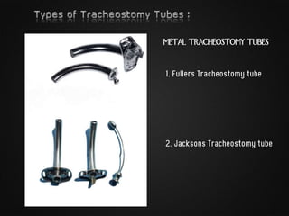
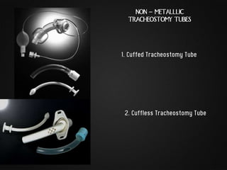
TRACHEOTOMY – Operative procedure that creates an
artificial opening in the trachea.
TRACHEOSTOMY – Converting this opening to a
permanent or semi permanent stoma on the skin
surface.
The first correct description of the tracheotomy operation -
- Ibn Zuhr in the 12th century.
The currently used surgical Tracheostomy technique
-Dr. Chevalier Jackson from Pittsburgh, Pennsylvania.
1. Emergency – Urgent establishment of Airway
2. Elective
i. Therapeutic
 Respiratory obstruction
 Tracheobronchial Secretions
 Assisted Ventilation
ii. Prophylactic
 Anticipated obstruction or aspiration
3. Permanent
 Bilateral Abductor paralysis
 Laryngeal Stenosis
Other types :
1) High Tracheostomy –
 Above thyroid isthmus
 Perichondritis & Subglottic Stenosis
2) Mid Tracheostomy
 Most preferred
3) Low Tracheostomy
 Below level of isthmus
 Large blood vessels
 Deep seated trachea
 Tube impinges on Suprasternal notch
 Whenever possible – Endotracheal intubation.
 Position – Supine with neck extended
to bring trachea forward.
Anesthesia – 1-2 % Lignocaine with Epinephrine
Not needed in Emergency & Unconscious
patients.
1. Vertical skin incision along
relaxed skin tension lines 1 cm below the cricoid two
finger breadths above the sternal notch.
Or
Transverse incision 5cm long, 2 finger
breadth above sternal notch.
2. Retractors are placed, the
skin & tissues are dissected
in the midline.
Dilated veins are ligated.
3. The strap muscles are visualized in the midline & the
muscles are divided along the raphe, then retracted
laterally
4. The thyroid isthmus lies in the field of the dissection.
Typically, the isthmus is 5 to 10 mm in its vertical dimension,
mobilize it away from the trachea and retract it or divide the
thyroid isthmus between the ligatures.
5. Few drops of 4% Lignocaine injected.
6. Then place the tracheal incision in the second or third
tracheal interspace. A vertical incision is converted to circular
opening.
An inferior based flap, or
Björk flap, (through 2nd & 3rd
Tracheal rings) is commonly
used.
7. The windpipe and surrounding
area is completely suctioned of
all secretions and blood.
8. Insert Tracheostomy tube (with
concomitant withdrawal of endotracheal
tube), secure with tape around neck.
9. Gauze dressing
Guide wire and catheter are advanced
together into the trachea as far as the
skin positioning marks on the guide
catheter to the skin.
Guide wire introduction, with
removal of sheath
Guide wire, guide catheter,
and dilator unit are advanced
together into the trachea to
the skin positioning mark
The tracheotomy tube is loaded onto a dilator
and advanced into the trachea over the guide
wire and catheter. The guide wire and
catheter are removed, leaving only the
tracheostomy tube in the trachea
1.Ciaglia - the sequential
insertion and removal of
a series (usually 4-5) of
increasing larger dilators
over the wire into the
trachea.
2.Griggs - insertion of
a specially designed
pair of guide-wire
forceps along the wire
into the trachea and
then are opened to
complete the dilation
in one step.
3.Rhino - insertion of a
single large tapered
dilator over a plastic
guidewire
reinforcement.
4.Frova Percutwist -
insertion of a specially
designed screw of
increasing diameter
which rotates to create
the dilatation.
METAL TRACHEOSTOMY TUBES
1. Fullers Tracheostomy tube
2. Jacksons Tracheostomy tube
1. Cuffed Tracheostomy Tube
NON - METALLLIC
TRACHEOSTOMY TUBES
2. Cuffless Tracheostomy Tube
Neonatal Tracheostomy Tubes ( Portex & Silastic )
Humidifier Speaking valve
Tracheostomy

Tracheostomy

  • 2.
    TRACHEOTOMY – Operativeprocedure that creates an artificial opening in the trachea. TRACHEOSTOMY – Converting this opening to a permanent or semi permanent stoma on the skin surface. The first correct description of the tracheotomy operation - - Ibn Zuhr in the 12th century. The currently used surgical Tracheostomy technique -Dr. Chevalier Jackson from Pittsburgh, Pennsylvania.
  • 3.
    1. Emergency –Urgent establishment of Airway 2. Elective i. Therapeutic  Respiratory obstruction  Tracheobronchial Secretions  Assisted Ventilation ii. Prophylactic  Anticipated obstruction or aspiration 3. Permanent  Bilateral Abductor paralysis  Laryngeal Stenosis
  • 4.
    Other types : 1)High Tracheostomy –  Above thyroid isthmus  Perichondritis & Subglottic Stenosis 2) Mid Tracheostomy  Most preferred 3) Low Tracheostomy  Below level of isthmus  Large blood vessels  Deep seated trachea  Tube impinges on Suprasternal notch
  • 5.
     Whenever possible– Endotracheal intubation.  Position – Supine with neck extended to bring trachea forward. Anesthesia – 1-2 % Lignocaine with Epinephrine Not needed in Emergency & Unconscious patients.
  • 6.
    1. Vertical skinincision along relaxed skin tension lines 1 cm below the cricoid two finger breadths above the sternal notch. Or Transverse incision 5cm long, 2 finger breadth above sternal notch.
  • 7.
    2. Retractors areplaced, the skin & tissues are dissected in the midline. Dilated veins are ligated.
  • 8.
    3. The strapmuscles are visualized in the midline & the muscles are divided along the raphe, then retracted laterally
  • 9.
    4. The thyroidisthmus lies in the field of the dissection. Typically, the isthmus is 5 to 10 mm in its vertical dimension, mobilize it away from the trachea and retract it or divide the thyroid isthmus between the ligatures. 5. Few drops of 4% Lignocaine injected.
  • 10.
    6. Then placethe tracheal incision in the second or third tracheal interspace. A vertical incision is converted to circular opening. An inferior based flap, or Björk flap, (through 2nd & 3rd Tracheal rings) is commonly used.
  • 11.
    7. The windpipeand surrounding area is completely suctioned of all secretions and blood. 8. Insert Tracheostomy tube (with concomitant withdrawal of endotracheal tube), secure with tape around neck.
  • 12.
  • 13.
    Guide wire andcatheter are advanced together into the trachea as far as the skin positioning marks on the guide catheter to the skin. Guide wire introduction, with removal of sheath
  • 14.
    Guide wire, guidecatheter, and dilator unit are advanced together into the trachea to the skin positioning mark The tracheotomy tube is loaded onto a dilator and advanced into the trachea over the guide wire and catheter. The guide wire and catheter are removed, leaving only the tracheostomy tube in the trachea
  • 15.
    1.Ciaglia - thesequential insertion and removal of a series (usually 4-5) of increasing larger dilators over the wire into the trachea. 2.Griggs - insertion of a specially designed pair of guide-wire forceps along the wire into the trachea and then are opened to complete the dilation in one step.
  • 16.
    3.Rhino - insertionof a single large tapered dilator over a plastic guidewire reinforcement. 4.Frova Percutwist - insertion of a specially designed screw of increasing diameter which rotates to create the dilatation.
  • 17.
    METAL TRACHEOSTOMY TUBES 1.Fullers Tracheostomy tube 2. Jacksons Tracheostomy tube
  • 18.
    1. Cuffed TracheostomyTube NON - METALLLIC TRACHEOSTOMY TUBES 2. Cuffless Tracheostomy Tube
  • 19.
    Neonatal Tracheostomy Tubes( Portex & Silastic ) Humidifier Speaking valve