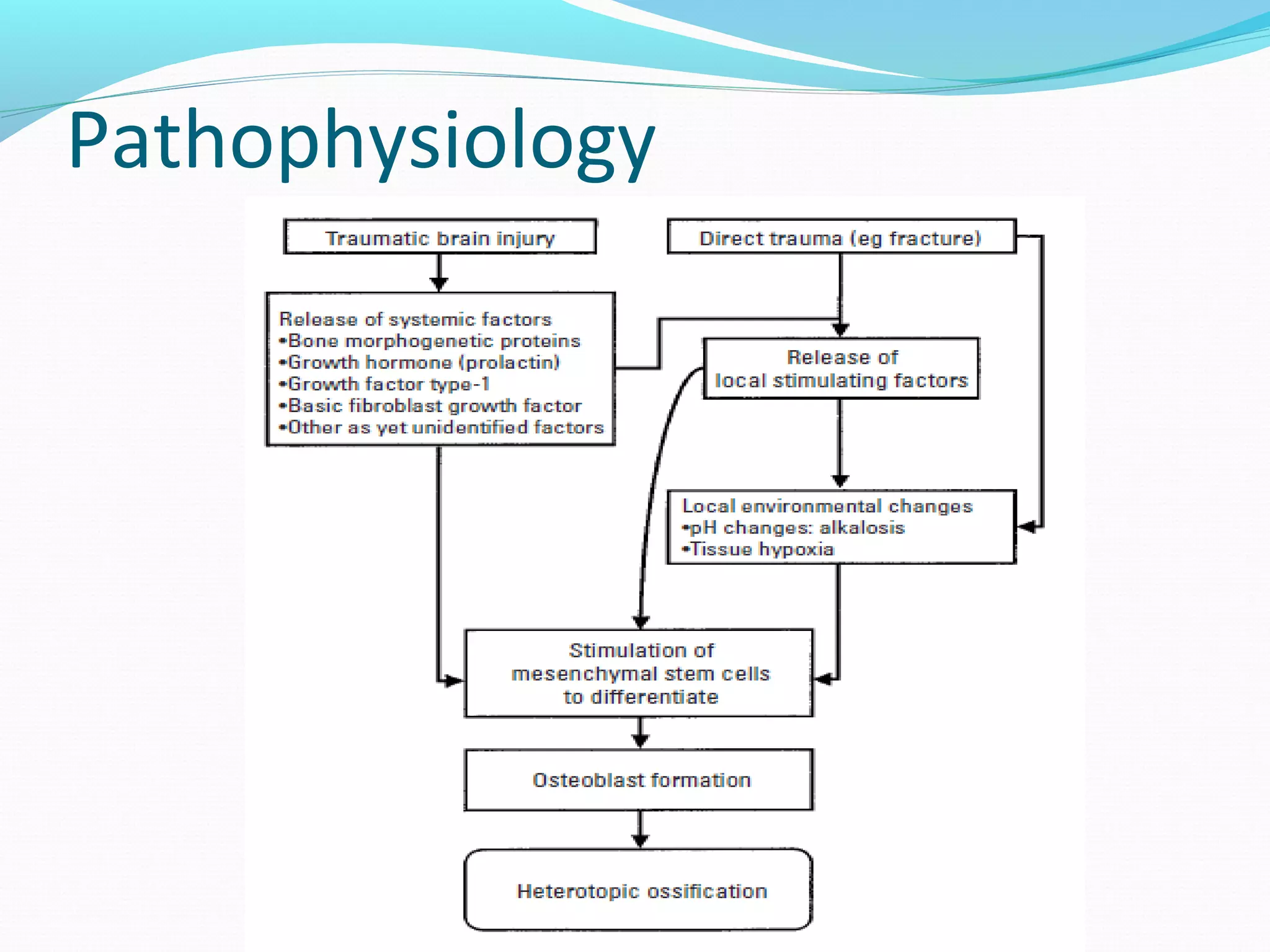
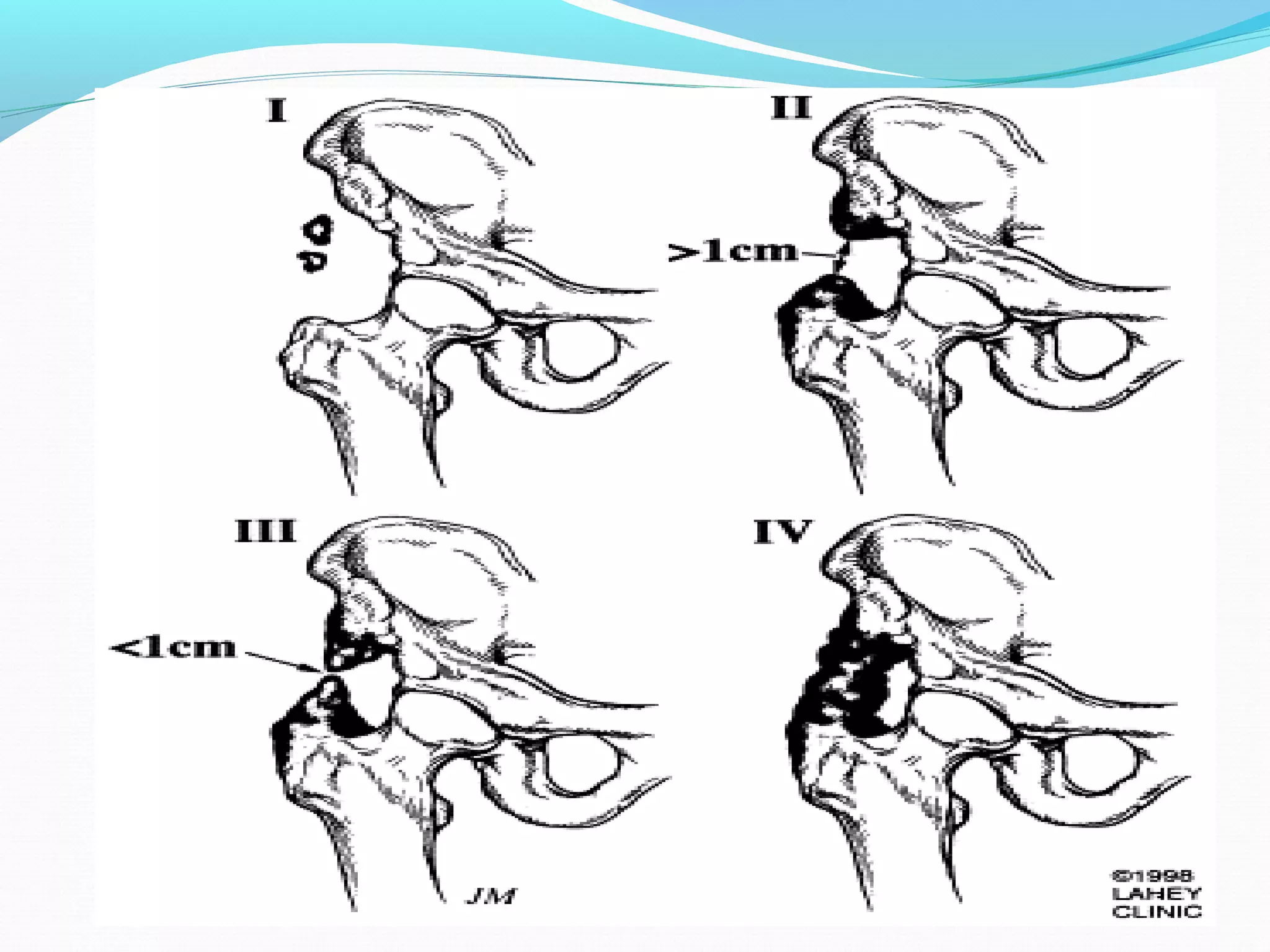
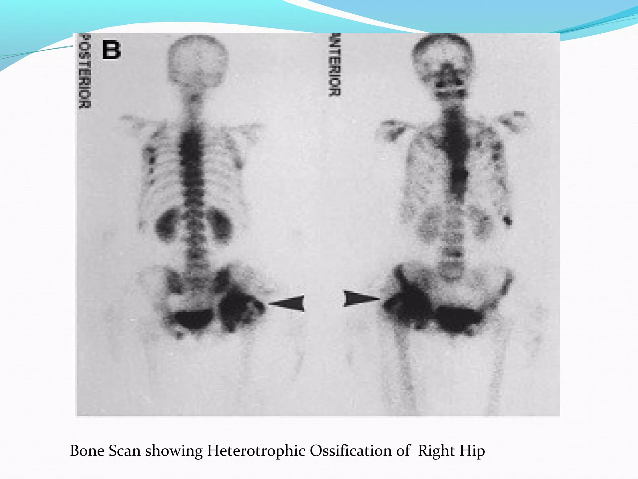
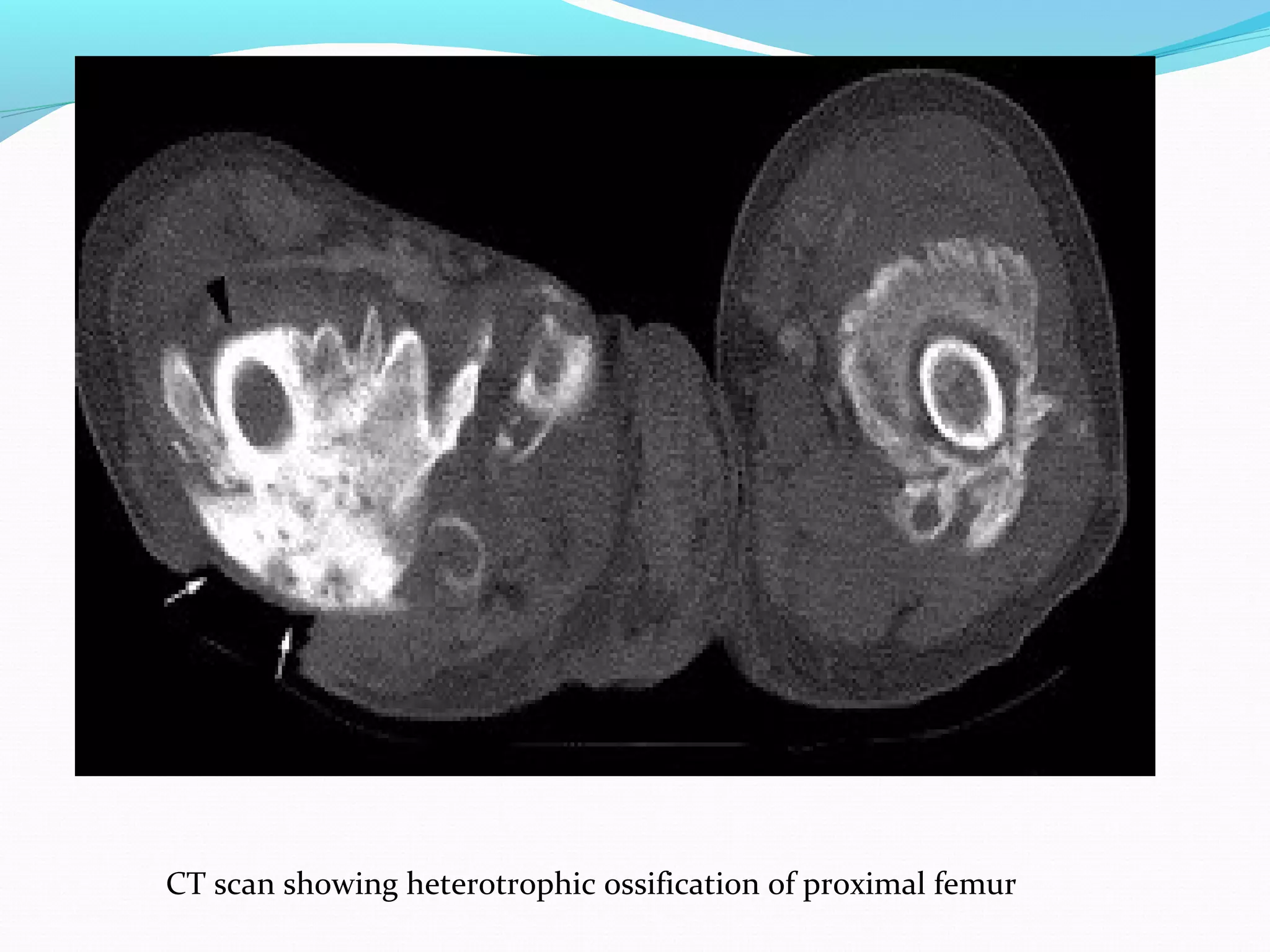
By Dr Ann Noble Zachariah
MD Resident
Department of Physical Medicine and Rehabilitation
Definition
HO is the formation of lamellar bone within the soft
tissue surrounding the joint
Can occur in skin, subcutaneous tissue , skeletal
muscles and fibrous tissue surrounding the bone.
First described by Patin in 1692 – while working with
children with myositis ossifican progressiva
In 1918, Dejerine & Ceillier detailed the anatomical,
clinical, and histological features of HO
Site of HO
Only the joints below the Neurological level of Injury
will develop HO
The most common sites are
- Hip
- Knee
- Shoulders
 Other sites also include spine, elbow, wrist.
Incidence of HO
Following lower extremity amputee about 7%→
Following traumatic brain injuries 11% (ranges→
reported from 10-20%)
Following spinal cord injuries 20% (ranges reported→
from 20-40%)
Following THR 53% (ranges reported from 0.6-90%)→
Following TKR 15% (ranges reported from 1-42%)→
Risk Factors
- Older age (children and adolescents have a lower
incidence)
-Neurological complete lesions
- Male gender
- Spasticity
- DVT
- Pressure sores
- Fractures
Etiology
 Exact etiopathophysiology is unknown
Bone morphogenic protein ( released in TBI ) plays a
key role
Damage to the sympathetic tracts in the spinal cord -
promote HO by increasing the local vascularity and
blood perfusion around the joint.
 When passive ROM is delayed more than 1 week
after injury.
 Forced ROM of contracted limb may cause micro
trauma and bleeding, leading to HO
Pathophysiology
Classification of HO
HO in Hip HO in Knee
Clinical features
Clinical signs and symptoms usually appear with 3 –
12 weeks
 Symptoms include
- Pain
- Malaise
- Low grade fever
- Warmth of the joint
- Increase in spasticity
 the initial inflammatory phase of HO may mimic
other pathologies such as cellulitis, thrombophlebitis,
osteomyelitis, or a tumorous process
Associated Comorbidities
 Traumatic Brain Injury
Traumatic Spinal Cord injury
Ankylosing spondylitis
Rhuematoid arthritis
Hypertrophic osteoarthitis
Diffuse idiopathic skeletal hyperostosis
Paget's disease
Differential Diagnosis
 Tumours – Osteosarcoma, Osteochrondroma
 Infections- Osteomyelitis
 Inflammation – Cellulitis
 Circulatory complications – Deep Vein Thrombosis ,
Thromphoblebitis
Investigations
 Alkaline Phosphatase is frequently used in early
detection of HO. It has high sensitivity, low
specificity.Serum ALP levels can be dependent on renal and
hepatic function, so they are not always useful.
An elevation of serum creatinine phosphokinase(CPK)
may be a more reliable predictor of HO
 Elevated levels of nonspecific markers of inflammation
such as C-reactive protein (CRP) and the erythrocyte
sedimentation rate (ESR) can be useful
CRP is a more reliable predictor of disease activity, with
normalization of the CRP correlating with resolution of the
inflammatory phase of HO
Prostoglandin E2:
- monitior PGE2 excretion in 24-hour urinalysis
- PGE2 felt to be reliable bone marker for early
detection and determining treatment efficacy
- A sudden increase is an indication for bone
scintigraphy
Urinary excretion of hydroxyproline and collagen
metabolites correlates with alkaline phosphatase
levels and can also serve as indirect markers for the
presence of HO.
Radiological Imaging
Three Phase bone scintigraphy
both diagnostic and therapeutic follow-up purposes
most sensitive imaging modality for early detection
Phases 1 and 2 are indicative of hyperaemia and blood
pooling (precursors to process of HO)
Usually positive after 2-4 weeks
Serial bone scans used to monitor metabolic activity of
HO to determine optimal timing for surgical resection
and to predict postoperative occurrence
Bone Scan showing Heterotrophic Ossification of Right Hip
Radiography, MRI, CT Scan:
all have low specificity in early stage of disease process
better for detection in later stages when ossification is
more pronounced
MRI and CT are valuable pre-operative tools to determine
relationship to blood vessels and peripheral nerve
structures
HO demonstrable on radiographs 4-6 weeks post-injury
MRI better than radiograph at detecting HO in early
phases
Plain film radiographs are most commonly used due to
their simplicity and low cost.
Typical radiologic presentation of HO is circumfrential
ossification with a lucent center
CT scan showing heterotrophic ossification of proximal femur
Ultrasonography:
earlier detection than conventional radiograph
local signs of inflammation in SCI patients are
suggestive of HO
high sensitivity and specificity for early detection of
HO 1-week post-THR.
Medical Management
The two types of medications shown to have both prophylactic and
treatment benefits are as follows:
Non-steroidal anti-inflammatory drugs: NSAIDS
Indomethacin
-inhibition of the differentiation of mesenchymal cells into osteogenic
cells.
- inhibition of post-traumatic bone remodelling by suppression of
prostaglandin-mediated response
Dose – 25 mg TID
Bisphosphonates:
- inhibition of calcium phosphate precipitation
- slowing of hydroxyapatite crystal aggregation
-inhibition of the transformation of calcium phosphate to
hydroxyapatite
Treatment with bisphosphonates
- decrease the rate of new bone formation in patients with
HO; however, it has no effect on bone which has already
been deposited.
-Disodium etidronate inhibits osteoclastic activity and
conversion of calcium phosphate to hydroxyapatite.
- Although IV administration of etidronate reportedly led
to quicker resolution of edema with less rebound formation
after the medication was discontinued, it is no longer
available.
- Current recommendation is for oral administration of
etidronate 20 mg/kg/d for 6 months if the CPK level is
elevated at the time of diagnosis or 20 mg/kg/d for 3
months, followed by 10 mg/kg/d for an additional 3 months
if the initial CPK level is normal
Radiotherapy
The use of radiotherapy as a prophylactic treatment
comes mainly from the literature concerning total hip
arthroplasty.
Radiating pluripotential mesenchymal cells may
effectively prevent the development of HO.
A dose of 700-800 cGy of local radiation in the first
four post-operative days has shown to prevent HO
formation in patients who are at high risk.
Surgical management
The two main goals of sugical intervention are to alter
the position of the affected joint or improve its range
of motion (ROM).
Clinicians must determine if the lesion has reached
maturation before surgical excision to decrease the
risk of intraoperative complications such as
hemorrhage, and the reoccurance of the ectopic
lesion.
The use of bone scans to determine metabolic activity
of the lesion and serum ALP levels are common aids
in this decision making process.
Criteria for surgical removal of HO
The criteria are as follows:
significantly limited ROM of involved joint (should have <
50o
ROM); for most patients, progression to joint ankylosis is
the most serious complication of heterotopic ossification.
Absence of local fever, swelling, erythema, or other
clinical findings of acute heterotopic ossification.
Normal serum alkaline phosphate levels.
Return of bone scan findings to normal or near normal; if
serial quantitative bone scans are obtained, there should be
a sharply decreasing trend followed by a steady state for 2-3
months.
Rehabilitation Measures
Physical therapy along with pharmacotherapy has
been shown to benefit patients suffering from
heterotopic ossification.
Pre-operative PT can be used to help preserve the
structures around the lesion.
ROM exercises (PROM, AAROM, AROM) and
strengthening will help prevent muscle atrophy and
preserve joint motion.
Therapy which is too aggressive can aggrevate the
condition and lead to inflammation, erythema,
hemorrhage, and increased pain.
 Post-operative rehabilitation has also shown benefit
patients with recent surgical resection of
heterotopic ossification.
The post-op management of HO is similar to pre-op
treatment but much more emphasis is placed on
edema control, scar management, and infection
prevention.
Phases of Rehab
Phase I (Week 1)
Goals
Prevent infection
Protect and decrease stress on surgical site
Decrease pain
Control and decrease edema
ROM to 80% of affected joint
Maintain ROM of joint proximal and distal to surgical
site
 Phase II ( 2 – 8 weeks )
 Goals
 Reduce pain
Manage edema
Encourage limited ADL performances
Promote scar mobility and proper remodeling
Promote full ROM of affected joint
Encourage quality muscle contractions
Phase III (9-24 weeks)
Goals
Self-manage pain
Prevent flare-up with functional activities
Improve strength
Improve ROM (if still limited)
Return to previous levels of activity
Heterotrophic ossification

Heterotrophic ossification

  • 1.
    By Dr AnnNoble Zachariah MD Resident Department of Physical Medicine and Rehabilitation
  • 2.
    Definition HO is theformation of lamellar bone within the soft tissue surrounding the joint Can occur in skin, subcutaneous tissue , skeletal muscles and fibrous tissue surrounding the bone. First described by Patin in 1692 – while working with children with myositis ossifican progressiva In 1918, Dejerine & Ceillier detailed the anatomical, clinical, and histological features of HO
  • 3.
    Site of HO Onlythe joints below the Neurological level of Injury will develop HO The most common sites are - Hip - Knee - Shoulders  Other sites also include spine, elbow, wrist.
  • 4.
    Incidence of HO Followinglower extremity amputee about 7%→ Following traumatic brain injuries 11% (ranges→ reported from 10-20%) Following spinal cord injuries 20% (ranges reported→ from 20-40%) Following THR 53% (ranges reported from 0.6-90%)→ Following TKR 15% (ranges reported from 1-42%)→
  • 5.
    Risk Factors - Olderage (children and adolescents have a lower incidence) -Neurological complete lesions - Male gender - Spasticity - DVT - Pressure sores - Fractures
  • 6.
    Etiology  Exact etiopathophysiologyis unknown Bone morphogenic protein ( released in TBI ) plays a key role Damage to the sympathetic tracts in the spinal cord - promote HO by increasing the local vascularity and blood perfusion around the joint.  When passive ROM is delayed more than 1 week after injury.  Forced ROM of contracted limb may cause micro trauma and bleeding, leading to HO
  • 7.
  • 8.
  • 11.
    HO in HipHO in Knee
  • 12.
    Clinical features Clinical signsand symptoms usually appear with 3 – 12 weeks  Symptoms include - Pain - Malaise - Low grade fever - Warmth of the joint - Increase in spasticity  the initial inflammatory phase of HO may mimic other pathologies such as cellulitis, thrombophlebitis, osteomyelitis, or a tumorous process
  • 13.
    Associated Comorbidities  TraumaticBrain Injury Traumatic Spinal Cord injury Ankylosing spondylitis Rhuematoid arthritis Hypertrophic osteoarthitis Diffuse idiopathic skeletal hyperostosis Paget's disease
  • 14.
    Differential Diagnosis  Tumours– Osteosarcoma, Osteochrondroma  Infections- Osteomyelitis  Inflammation – Cellulitis  Circulatory complications – Deep Vein Thrombosis , Thromphoblebitis
  • 15.
    Investigations  Alkaline Phosphataseis frequently used in early detection of HO. It has high sensitivity, low specificity.Serum ALP levels can be dependent on renal and hepatic function, so they are not always useful. An elevation of serum creatinine phosphokinase(CPK) may be a more reliable predictor of HO  Elevated levels of nonspecific markers of inflammation such as C-reactive protein (CRP) and the erythrocyte sedimentation rate (ESR) can be useful CRP is a more reliable predictor of disease activity, with normalization of the CRP correlating with resolution of the inflammatory phase of HO
  • 16.
    Prostoglandin E2: - monitiorPGE2 excretion in 24-hour urinalysis - PGE2 felt to be reliable bone marker for early detection and determining treatment efficacy - A sudden increase is an indication for bone scintigraphy Urinary excretion of hydroxyproline and collagen metabolites correlates with alkaline phosphatase levels and can also serve as indirect markers for the presence of HO.
  • 17.
    Radiological Imaging Three Phasebone scintigraphy both diagnostic and therapeutic follow-up purposes most sensitive imaging modality for early detection Phases 1 and 2 are indicative of hyperaemia and blood pooling (precursors to process of HO) Usually positive after 2-4 weeks Serial bone scans used to monitor metabolic activity of HO to determine optimal timing for surgical resection and to predict postoperative occurrence
  • 18.
    Bone Scan showingHeterotrophic Ossification of Right Hip
  • 19.
    Radiography, MRI, CTScan: all have low specificity in early stage of disease process better for detection in later stages when ossification is more pronounced MRI and CT are valuable pre-operative tools to determine relationship to blood vessels and peripheral nerve structures HO demonstrable on radiographs 4-6 weeks post-injury MRI better than radiograph at detecting HO in early phases Plain film radiographs are most commonly used due to their simplicity and low cost. Typical radiologic presentation of HO is circumfrential ossification with a lucent center
  • 20.
    CT scan showingheterotrophic ossification of proximal femur
  • 21.
    Ultrasonography: earlier detection thanconventional radiograph local signs of inflammation in SCI patients are suggestive of HO high sensitivity and specificity for early detection of HO 1-week post-THR.
  • 22.
    Medical Management The twotypes of medications shown to have both prophylactic and treatment benefits are as follows: Non-steroidal anti-inflammatory drugs: NSAIDS Indomethacin -inhibition of the differentiation of mesenchymal cells into osteogenic cells. - inhibition of post-traumatic bone remodelling by suppression of prostaglandin-mediated response Dose – 25 mg TID Bisphosphonates: - inhibition of calcium phosphate precipitation - slowing of hydroxyapatite crystal aggregation -inhibition of the transformation of calcium phosphate to hydroxyapatite
  • 23.
    Treatment with bisphosphonates -decrease the rate of new bone formation in patients with HO; however, it has no effect on bone which has already been deposited. -Disodium etidronate inhibits osteoclastic activity and conversion of calcium phosphate to hydroxyapatite. - Although IV administration of etidronate reportedly led to quicker resolution of edema with less rebound formation after the medication was discontinued, it is no longer available. - Current recommendation is for oral administration of etidronate 20 mg/kg/d for 6 months if the CPK level is elevated at the time of diagnosis or 20 mg/kg/d for 3 months, followed by 10 mg/kg/d for an additional 3 months if the initial CPK level is normal
  • 24.
    Radiotherapy The use ofradiotherapy as a prophylactic treatment comes mainly from the literature concerning total hip arthroplasty. Radiating pluripotential mesenchymal cells may effectively prevent the development of HO. A dose of 700-800 cGy of local radiation in the first four post-operative days has shown to prevent HO formation in patients who are at high risk.
  • 25.
    Surgical management The twomain goals of sugical intervention are to alter the position of the affected joint or improve its range of motion (ROM). Clinicians must determine if the lesion has reached maturation before surgical excision to decrease the risk of intraoperative complications such as hemorrhage, and the reoccurance of the ectopic lesion. The use of bone scans to determine metabolic activity of the lesion and serum ALP levels are common aids in this decision making process.
  • 26.
    Criteria for surgicalremoval of HO The criteria are as follows: significantly limited ROM of involved joint (should have < 50o ROM); for most patients, progression to joint ankylosis is the most serious complication of heterotopic ossification. Absence of local fever, swelling, erythema, or other clinical findings of acute heterotopic ossification. Normal serum alkaline phosphate levels. Return of bone scan findings to normal or near normal; if serial quantitative bone scans are obtained, there should be a sharply decreasing trend followed by a steady state for 2-3 months.
  • 27.
    Rehabilitation Measures Physical therapyalong with pharmacotherapy has been shown to benefit patients suffering from heterotopic ossification. Pre-operative PT can be used to help preserve the structures around the lesion. ROM exercises (PROM, AAROM, AROM) and strengthening will help prevent muscle atrophy and preserve joint motion. Therapy which is too aggressive can aggrevate the condition and lead to inflammation, erythema, hemorrhage, and increased pain.
  • 28.
     Post-operative rehabilitationhas also shown benefit patients with recent surgical resection of heterotopic ossification. The post-op management of HO is similar to pre-op treatment but much more emphasis is placed on edema control, scar management, and infection prevention.
  • 29.
    Phases of Rehab PhaseI (Week 1) Goals Prevent infection Protect and decrease stress on surgical site Decrease pain Control and decrease edema ROM to 80% of affected joint Maintain ROM of joint proximal and distal to surgical site
  • 30.
     Phase II( 2 – 8 weeks )  Goals  Reduce pain Manage edema Encourage limited ADL performances Promote scar mobility and proper remodeling Promote full ROM of affected joint Encourage quality muscle contractions
  • 31.
    Phase III (9-24weeks) Goals Self-manage pain Prevent flare-up with functional activities Improve strength Improve ROM (if still limited) Return to previous levels of activity