
Recommended
More Related Content
Similar to 1periprosthetic fracture around hip.pptx
Similar to 1periprosthetic fracture around hip.pptx (20)
Recently uploaded
Recently uploaded (20)
1periprosthetic fracture around hip.pptx
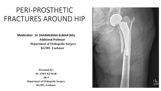
- 1. PERI-PROSTHETIC FRACTURES AROUND HIP Moderator: Dr. DHARMENDRA KUMAR (MS) Additional Professor Department of Orthopedic Surgery KGMU, Lucknow Presented by: Dr. AMIT KUMAR JR-3 Department of Orthopedic Surgery KGMU, Lucknow
- 2. OUTLINE 1. INTRODUCTION • DEFINATION • EPIDEMOLOGY • RISK FACTORS 2. GENRAL PRINCIPLES OF MANAGEMENT • DIAGNOSIS • CLASSIFICATION • TREATMENT • COMPLICATIONS
- 3. Introduction • Peri-implant Fracture: fracture around implant (plate, rod, prosthesis) • Peri-prosthetic Fracture: is a type of peri-implant fracture which occurse around joint replacement prosthesis. • They occur due to : 1. Trauma 2. Osteolysis 3. Osteoprosis It occur mostly in old age with osteoporosis making standard fixation technique difficult
- 4. Epidemology • The largest series of peri-prosthetic fracture (Total hip Arthroplasty) I. 1% after primary and 4% after revision THA II. 75% are due to low energy Trauma More commonly seen in • Females • Old age patient
- 5. • Low energy falls account for mechanism of injury in most patient with Peri- prosthetic fracture in both lower limb • Lower limb fractures occur most commonly Post-operatively where as upper limb fracture most commonly occur in Intra Operatively • 75% of all prei-prosthetic femur fracture occur post operatively with low energy trauma • Peri-prosthetic fracture most commonly post revision surgery then primary surgery because of reduced bone stock and due to incorporation of bone cement with medullary canal. • Risk of peri-prosthetic increases when there is mismatch between shape of long prosthesis stem & the shape of bone
- 6. Risk Factors Poor bone quality • Major Risk factors:- female, Revision surgery, elder age group.higher BMI • Osteopaenia, Osteolysis, Osteoporosis • Medication related such as Chronic steroid • Diabetes • OA • Inflammatory arthritis RA • Infection • Pagets disease • Stiffness • Neurological condition:- Epilepsy, Parkinson’s, Ataxia, Myasthenia. • Infection Surgery related • Inadequate exposure • Under-reaming • Overzealous Reaming • Heavy impaction • Mal-positioning of prosthesis • Cemented Arthroplasty correlate with low prevalence • Over resection • Cement mantle fracture • Lucency at cement-bone/cement mantle
- 7. Intra-operative risks: • Risk factors to intra-operative # • Under ream >2mm • Impaired bone quality • Cementless component • Dysplastic bone • Signs of intra-operative # • Sound of crack • Sudden change in resistance • Abnormal movement
- 8. Sign and Symptoms • Start up of abrupt pain • Increase difficulty with ambulation • Progressive limb shortening • Increasing deformity of the extremity
- 9. Investigations • Radiologic • X-ray • CT scan • MRI • DEXA • Laboratory • Routine blood investigations • ESR • CRP • Calcium profile
- 10. DIAGNOSIS • If trauma is absent /trivial – suggestive of Osteopaenia / Osteolysis • Skiagram of joint involved in AP and Lat view and full length of bone above and below the joint Evaluate prosthesis relative to fracture and prosthesis relative to native bone
- 11. Tells about 1. Prosthesis loosening 2. Presence of bone loss 3. Osteolysis Prosthetic and limb alignment
- 12. Classification • There are many classification for peri-prosthetic fractures : 1. American Academy of Orthopaedic surgeons (AAOS) 2. Cooke and newman (modified Bethea) 3. Johansson Classification
- 13. 4. Vancouver Classification • Most widely used • Based on location of fracture relative to prosthesis • Stability of prosthesis • Quality of surrounding bone
- 14. Vancouver Classification A: fracture involve the trochanteric area • A(G): greater trochanter • A(L): lesser trochanter B: #around the stem or just below it • B1: stem stable • B2: stem loose • B3: stem loose, bone stock inadequate C: fracture well below the stem
- 15. GOALS of treatment • Timely and uncomplicated fracture union • Restoration of alignment • Return of pre injury level of pain and function • Stability of prosthesis • Restoration of adequate bone stock to maximize potential success
- 16. Intra-operative Principles • Stable Intra-operative • Observation • Bracing • Use of cast • Protected weight bearing • Unstable intra-operative • Revision with screws/ exchange of cup / exchange of implant • Open reduction and internal fixation • Bone graft application • Protected weight bearing
- 17. Intra-operative Principles Cont’d • Revision Principle • Use the fracture for access to remove implant • Bypass the fracture with long stem • Stabilize fracture • Get stable implant fixation
- 18. Vancouver Classification - Intraoperative Periprosthetic Fracture
- 20. Management : Intraoperative fractures
- 21. Management : postoperative fractures
- 24. TREATMENT Vancouver Type A • Peri-prosthetic femur fracture around trochanteric areas • Usually non displaced or minimally displaced Stabilized by opposite pull and continuity of soft tissue sleeve connecting abductors and vastus lateralis Can be managed conservatively with symptomatic management and partial weight bearing with regular follow up
- 25. • Widly Displaced of Unstable type of A (gt) type Associated with minimal pain ; weakness ; limp ORIF with Claw Plating
- 26. • A (L) : large fracture involving segment of proximal medial femoral cortex associated with tapered press fit stem design • Treated with CERCALAGE wires/ Cables with or without Revision of prosthesis
- 27. • Vancouver type B • Identified mostly intra-operatively and treated mostly with intervention Indication of conservative management • Stable femoral stem and non displaced diaphyseal fractures • Proximal fragment related to osteolysis with adequate distal stem fixation • Minimally displaced trochanteric fractured
- 28. Indication of surgical treatment 1. Loose implant 2. Proximal Metaphyseal fracture with proximal fit stem 3. Displaced diaphyseal fracture or distal fractures 4. Widely displaced GT fractures with alter abductor function
- 29. • TYPE B1 – it has well fixed prosthesis So can be treated with 1. Wires or cables 2. Plate and screws or cables 3. Cortical allograft 4. Combination of above methods
- 30. • Type B2 – its prosthesis is unstable • So the treatment options available are 1. Revision Arthroplasty + ORIF 2. Replacement with Long Stem Prosthesis 3. Cemented prosthesis
- 31. • Type B3 – it has unstable prosthesis with Poor bone stock • Available options are 1. Proximal femoral Reconstruction 2. Composite allograft 3. Proximal femoral replacement
- 32. Treatment for VANCOUVER TYPE C • Fracture line well distal to Stem so its treatment is irrespective of Stem by 1. ORIF WITH plating + screws 2. Cables
- 33. COMPLICATIONS 1. Extensive soft tissue stripping during reduction 2. Extensive soft tissue destruction during cable application 3. Mismatch between plate contour and bone causing mal-reduction 4. Inadequate proximal fragment fixation 5. Inadequate stability
- 35. Complications • Dislocation • Infection • Instability • Re-fracture • Non-union • Aseptic loosening • Implant failure • Scar complications
- 37. Acetabular # Rare: 0.07 % (Peterson et al 1996) Disastrous complication of THA Usually intraoperative Seen with Cementless THR Rare in Cemented THR
- 38. Acetabular Fracture • The incidence is 0.2%. cemented THA • The incidence has increased Cementless • Under-reaming of the acetabulum • Press-fit stability with a cementless. • Under-reaming by as much as 4 mm is acceptable • Now agree that 2 mm or less is safer
- 39. Classification (Acetabular #) Many classifications have been proposed: Peterson and Lewallen AAOS Unified classification system (UCS) Della Valle Comprehensive Reproducible
- 40. Treatment Goals: Rigid fixation for bony union Stable integration of component Re-establishing: Offset Limb length
- 41. Treatment… Surgical Options: Impaction bone grafting Plating & column screws Cup screw augmentation Highly-porous metal cups Antiprotrusio cages Jumbo cups Cup/cage constructs
- 42. Treatment (Acetabular #) Intraoperative undisplaced Stable #s, stable implants Manage nonoperatively If there is concern about stability; additional screws fixation of the component & postop protected weight bearing is advised!
- 43. Treatment (Acetabular #) Intraoperative Displaced # Unstable Prosthesis Plating of the posterior column +/- Bone grafting or metal augments Cup with multiple screws +/- Protrusion cage / Jumbo cups
- 44. Treatment (Acetabular #) Traumatic Nondisplaced # Stable prosthesis Protected weight bearing for 6 – 8wks Healed fractures in 80 % Closed radiologic monitoring for 2yrs High rate of loosening even in # union CT Scan is mandatory for decision!
- 46. Treatment (Acetabular #) Traumatic displaced Loose prosthesis ORIF (Posterior or Bicolumnar) Revision of the acetabular component ± Antiprotrusio cage
- 48. Treatment (Acetabular #) Pelvic discontinuity due to osteolysis Small defect / Good bone quality Contained ant. & post. Acetabular rim ORIF with posterior column plate + Bone grafting + Revision cup
- 49. Treatment (Acetabular #) Pelvic discontinuity due to osteolysis Large defect / Good bone quality Bicolumnar plating + Bone grafting + Highly porous tantalum shell Alternatively: Protrusion ring + Bone grafting
- 51. Treatment (Acetabular #) Pelvic discontinuity due to osteolysis Large defect / Poor defect A cup-cage construct augment Reconstruction ring + Highly porous cup + Cemented Polyethylene cup Can be single or 2-Staged
- 54. Conclusion • Principle of management of PF is still an evolving technique • Fracture pattern, Patient factors, and healing potential must be considered • Emphasis on simultaneously creating strong, durable mechanical construct • Optimization of the biologic environment for fracture healing
- 55. Thank You