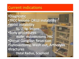
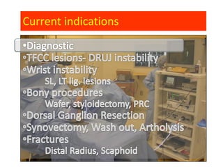
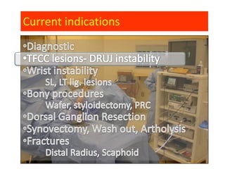
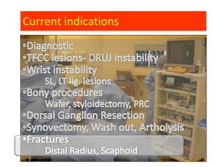
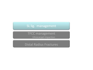
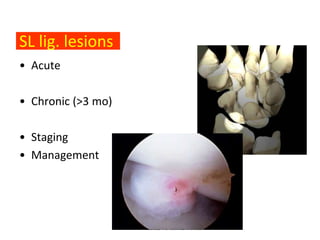
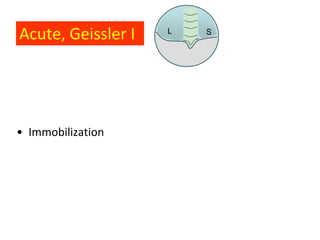
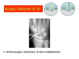
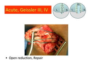
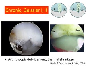
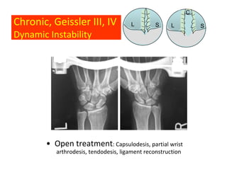
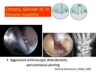
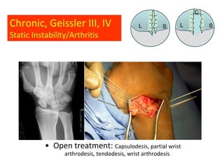
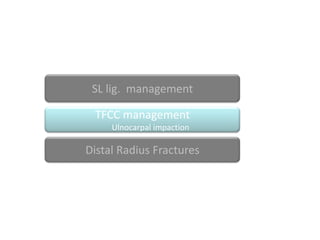
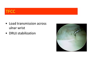
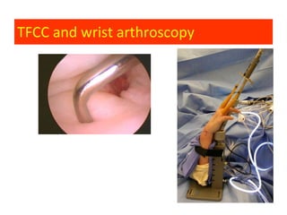
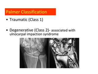
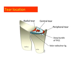
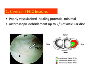
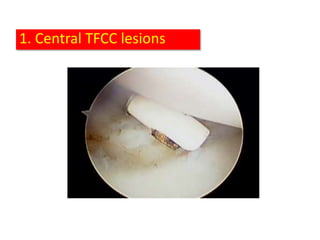
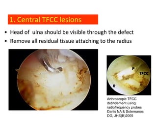
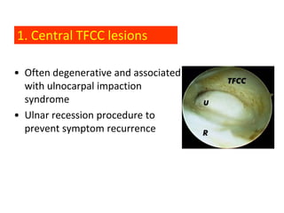
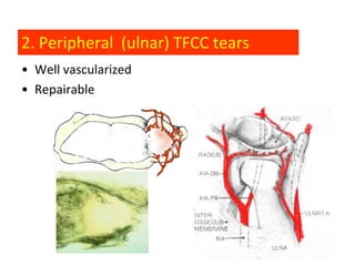
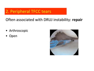
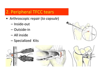
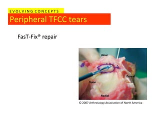
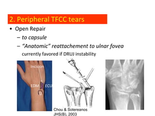
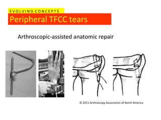
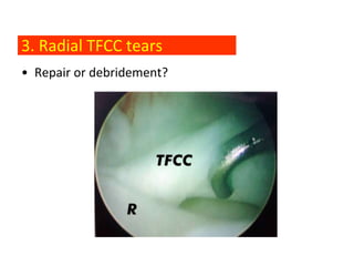
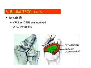
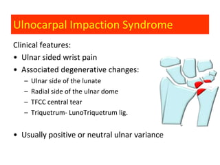
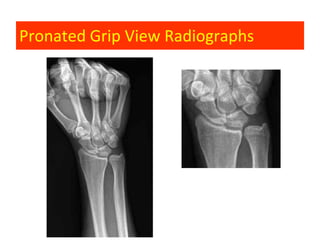
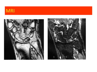
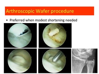
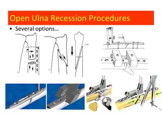
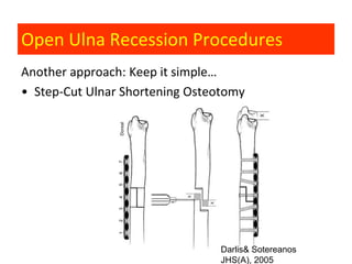
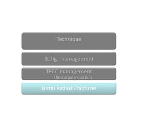
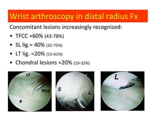
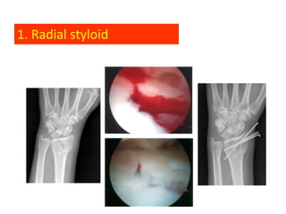
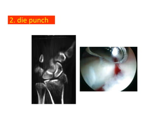
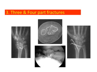
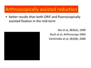
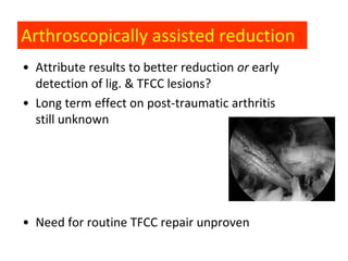
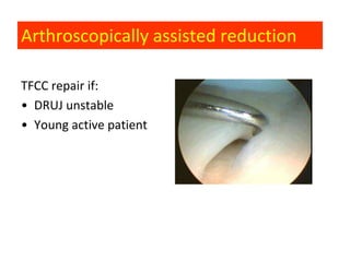
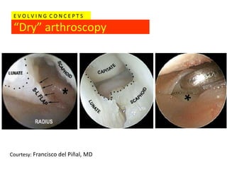
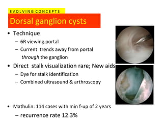
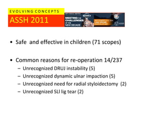
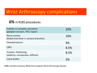
Wrist Arthroscopy
Clinical applications
        Nickolaos A. Darlis, MD, PhD
Current indications
Current indications
Current indications
Current indications
Current indications
Current indications
Current indications
Current indications
SL lig. management

 TFCC management
     Ulnocarpal impaction

Distal Radius Fractures
SL lig. lesions
• Acute

                    •Δυναμική Αζηάθεια
• Chronic (>3 mo)
                    •Σηαηική Αζηάθεια
                    •Αρθρίηιδα (SLAC)
• Staging
• Management
Geissler classification

Type I

  L         S
Geissler classification

Type II

  L         S
Geissler classification

Type III

  L         S
Geissler classification

Type IV
          C


L             S
Geissler classification
Type IV
Acute, Geissler I   L   S


• Immobilization
Acute, Geissler II, III           L     S       L   S


• Arthroscopic reduction, K-wire stabilization
C

Acute, Geissler III, IV    L   S   L       S


• Open reduction, Repair
Chronic, Geissler I, II         L        S        L         S


• Arthroscopic debridement, thermal shrinkage
                             Darlis & Sotereanos, JHS(A), 2005
C
Chronic, Geissler III, IV             L         S     L          S
Dynamic Instability


       • Open treatment: Capsulodesis, partial wrist
          arthrodesis, tendodesis, ligament reconstruction
C
Chronic, Geissler III, IV     L       S      L          S
Dynamic Instability


• Aggressive arthroscopic debridement,
            percutaneous pinning
                             Darlis & Sotereanos, JHS(A), 2006
C

Chronic, Geissler III, IV                     L         S     L       S
Static Instability/Arthritis


           • Open treatment: Capsulodesis, partial wrist
                 arthrodesis, tendodesis, wrist arthrodesis
SL lig. management

 TFCC management
     Ulnocarpal impaction

Distal Radius Fractures
TFCC

• Load transmission across
  ulnar wrist
• DRUJ stabilization
TFCC and wrist arthroscopy
Palmer Classification
• Traumatic (Class 1)

• Degenerative (Class 2)- associated with
  ulnocarpal impaction syndrome
Tear location

           Radial tear          Central tear

                                               Peripheral tear)


                                               Deep bundle
                                               of TFCC

            radius
                                         Volar radioulnar lig.
                         ulna
TFCC tears
 Pre-styloid recess
EVOLVING CONCEPTS

Volar & Dorsal RadioUlnar lig.
1. VRUL, DRUL major contributors to DRUJ stability
EVOLVING CONCEPTS

Deep Bundle
2. Deep bundle of TFCC attaching to ulnar fovea crucial
   for DRUJ stability
1. Central TFCC lesions
• Poorly vascularized- healing potential minimal
• Arthroscopic debridement up to 2/3 of articular disc
1. Central TFCC lesions
• Head of ulna should be visible through the defect
• Remove all residual tissue attaching to the radius


                                                Arthroscopic TFCC
                                                debridement using
                                                radiofrequency probes
                                                Darlis NA & Sotereanos
            Darlis & Sotereanos, JHS(A), 2005   DG, JHS(B)2005
1. Central TFCC lesions

• Often degenerative and associated
  with ulnocarpal impaction
  syndrome
• Ulnar recession procedure to
  prevent symptom recurrence
2. Peripheral (ulnar) TFCC tears
• Well vascularized
• Repairable
2. Peripheral TFCC tears
Often associated with DRUJ instability: repair

• Arthroscopic
• Open
2. Peripheral TFCC tears
• Arthroscopic repair (to capsule)
   – Inside-out
   – Outside-in
   – All inside
   – Specialized Kits
EVOLVING CONCEPTS

Peripheral TFCC tears
  FasT-Fix® repair


                     © 2007 Arthroscopy Association of North America
2. Peripheral TFCC tears
• Open Repair
  – to capsule
  – “Anatomic” reattachement to ulnar fovea
    currently favored if DRUJ instability

        Incision


        EDM        ECU
              U


                         Chou & Sotereanos
                         JHS(B), Chou, Sarris, Sotereanos, JHS(B), 2003
                                 2003
EVOLVING CONCEPTS

Peripheral TFCC tears
  Arthroscopic-assisted anatomic repair


                         © 2011 Arthroscopy Association of North America
3. Radial TFCC tears
• Repair or debridement?
3. Radial TFCC tears
• Repair if:
   – VRUL or DRUL are involved
   – DRUJ instability
Ulnocarpal Impaction Syndrome
Clinical features:
• Ulnar sided wrist pain
• Associated degenerative changes:
   –   Ulnar side of the lunate
   –   Radial side of the ulnar dome
   –   TFCC central tear
   –   Triquetrum- LunoTriquetrum lig.


• Usually positive or neutral ulnar variance
Pronated Grip View Radiographs
MRI
Arthroscopic Wafer procedure
• Preferred when modest shortening needed
Open Ulna Recession Procedures
• Several options…
Open Ulna Recession Procedures
Another approach: Keep it simple…
• Step-Cut Ulnar Shortening Osteotomy


                                 Darlis& Sotereanos
                                 JHS(A), 2005
Technique


 SL lig. management

 TFCC management
     Ulnocarpal impaction

Distal Radius Fractures
Wrist arthroscopy in distal radius Fx
Concomitant lesions increasingly recognized:
• ΤFCC ≈60% (43-78%)
• SL lig.≈ 40% (32-75%)
• LT lig. ≈20% (15-61%)
• Chondral lesions ≈20% (19-32%)
Arthroscopically assisted reduction
• Currently indicated in selected injuries
   –   Radial styloid Fx
   –   Die Punch Fx
   –   Three & Four part Fx
   –   DRUJ instability


Especially in young, high demand patients
1. Radial styloid
2. die punch
3. Three & Four part fractures
Arthroscopically assisted reduction
• better results than both ORIF and fluoroscopically
  assisted fixation in the mid-term

                                     Doi et al, JBJS(A), 1999
                               Ruch et al, Arthroscopy 2004
                              Varitimidis et al, JBJS(B), 2008
Arthroscopically assisted reduction
• Attribute results to better reduction or early
  detection of lig. & TFCC lesions?
• Long term effect on post-traumatic arthritis
  still unknown


• Need for routine TFCC repair unproven
Arthroscopically assisted reduction

TFCC repair if:
• DRUJ unstable
• Young active patient
EVOLVING CONCEPTS

 “Dry” arthroscopy


Courtesy: Francisco del Piñal, MD
EVOLVING CONCEPTS

 Dorsal ganglion cysts
• Technique
   – 6R viewing portal
   – Current trends away from portal
     through the ganglion
• Direct stalk visualization rare; New aids
   – Dye for stalk identification
   – Combined ultrasound & arthroscopy


• Mathulin: 114 cases with min f-up of 2 years
   – recurrence rate 12.3%
EVOLVING CONCEPTS

ASSH 2011

• Safe and effective in children (71 scopes)

• Common reasons for re-operation 14/237
   –   Unrecognized DRUJ instability (5)
   –   Unrecognized dynamic ulnar impaction (5)
   –   Unrecognized need for radial styloidectomy (2)
   –   Unrecognized SLl lig tear (2)
Wrist Arthroscopy complications
     6% in 9185 procedures
  Inability to complete procedure                                  20%
  (ganglion excision, TFCC repair)
  Nerve Lesions                                                    10%
  (Radial and Ulnar n. sensory branches)
  Chondral lesions                                                 9%
  CRPS                                                             8.5%
  Traction- Potitioning                                            8.5%
  (oedema, neurapraxia, stiffness)
  Loose bodies                                                     5%
2008, Caroline Leclercq, MD & the European Wrist Arthroscopy Society
European Wrist
Arthroscopy Society
www.geap.org
Thank you

Wrist arthroscopy metsovo 2011