SlideShare a Scribd company logo
Bilateral sagittal split osteotomy
(BSSO)
Presented by
KANIMOZHIY SENGUTTUVAN
2nd year post graduate
Thaimoogambigai Dental College and Hospital
• Bilateral sagittal split osteotomy (BSSO)
of the mandible is one of the most
frequently performed surgical
procedures.
• This is a very popular, most versatile
procedure performed on the mandibular
ramus and body.
• First described by Obwegeser and
Trauner and later modified by Dal Pont,
Hunsuck and Epker.
INTRODUCTION
• The osteotomy splits the ramus and the posterior body of the mandible sagittally, which
allows either setback or advancement.
• This is a highly cosmetic procedure, as it is done intraorally.
• For mandibular advancement, there is no need for bone grafts.
• Thus donor site morbidity and second operative site for the bone graft is totally avoided.
• BSSO gives excellent results. Only drawback is the technique demands high level of
operative skill and experience to minimize the surgical complications.
• In 1957, Obwegeser and Trauner first
described the sagittal split osteotomy of the
ramus region, only by placing buccal and
lingual cortical horizontal cuts.
• The procedure has undergone numerous
modifications over the years.
HUGO OBWEGESER & TRAUNER 1957
HISTORY
• In 1961, Dal Pont changed the lower
horizontal cut to a vertical cut on the
buccal cortex between the first and the
second molars, thereby obtaining
broader contact surfaces and requiring
minimal muscular displacement with
improved access.
DALPONT (1961)
• In 1968, Hunsuck further modified the
technique, advocating a shorter, horizontal
medial cut, just posterior to the lingula, to
minimize soft tissue dissection.
• His anterior vertical cut was similar to
Dal Pont’s.
HUNSUCK (1968)
• In 1977, Epker suggested several
modifications.
• These include minimal stripping of
the masseter muscle and limited
medial dissection.
• These modifications helped to reduce
postoperative swelling, oedema,
haemorrhage
• Epker et al described completing the
osteotomy through the inferior
border of the mandible.
INDICATIONS
1.Mandibular deficiency
-with normal or short face,
-with long face- increase maxillary vertical dimension
-excessive chin height
-for correction of sleep apnea
Limitation- Additional surgery for most dentofacial deformity
2.Mandibular prognathism
Limitation -Large setbacks of more than 7 -8 mm, IVRO/ inverted L osteotomy should be
considered
3.Mandibular asymmetry
- Hemi mandibular hypertrophy
-Hemi mandibular elongation
4. Open bite
5. Cross bite
CONTRAINDICATIONS
• Severe decreased posterior mandibular body height
• Extremely thin medial –lateral width of ramus
• Severe ramus hypoplasia and
• Severe asymmetries
Surgical procedure
Incision & Dissection
• The incision begins at the anterior aspect of the ramus at a
midpoint between upper and lower molars.
• The incision is about 2 cm long.
• It runs down the lateral crest of the external oblique ridge and
ends in the facial vestibule at approximately the first molar region.
• After sharp dissection of the mucosa, the buccinator muscle over
the ramus is dissected off at its medial aspect so as to keep as
much soft tissue laterally as possible.
• Then, the cut is completed through periosteum to bone.
Osteotomy cut
• The procedure starts with three corticotomies.
• The first cut is made through the lingual cortex just
above the mandibular foramen parallel to the
occlusion.
• The corticotomy is extended from the anterior
border of the ramus to just behind the entrance of
the inferior alveolar canal (lingula).
• The second corticotomy is made
through the buccal cortex in a vertical
direction at the level of the first or
second molar.
• We place the vertical cut at the horizontal
ramus between the first and second molars
and extend it downward to the inferior
border, curving around it.
• Cortical bone below the second molar is
thick; this thickness protects the
neurovascular bundle.
• Nevertheless, the neurovascular bundle lies
just below the cortical bone; thus, the
osteotomy is required to go only through
cortical bone.
• The third corticotomy connects the
first two osteotomy lines along the
anterior border of the ascending
ramus.
The sagittal split
• The final split is completed with a thin
osteotome, splitting the entire
ascending ramus from the anterior to
the posterior border of the ramus.
Mobilization
• A special bone spreader can be used
to mobilize the segments
• After the bilateral split is completed
the large tooth bearing segment can
be moved three dimensionally.
• Once the osteotomy has been completed on
both sides, a prefabricated interocclusal
acrylic wafer splint is placed.
• We achieve maxillo-mandibular fixation
(MMF) with three wire ligatures.
• Some centers do not use an occlusal wafer
splint.
• In this case, the teeth must be placed in good
three-point contact for a stable MMF to avoid
any shift when screws or plate and screws are
set.
Positioning of the tooth bearing segment
• Care must be taken to maintain the normal
fossa-condyle relation (see upper insert) and
to avoid condylar displacement (see lower
insert).
• Usually this is achieved by manual
positioning of the condyle bearing segment
superiorly into the glenoid fossa
Positioning of condyle bearing segment
A. Custom splint that lies along the lateral surface of the maxillary teeth is placed before
osteotomy. The splint has a guide wire attached, extending along the lateral surface of the
ramus. A saw is used to score a groove in the bone along this guide wire.
B. After the osteotomy, the distal segment is wired into the final splint (MMF). The condylar
positioning splint is reapplied and alignment of the scored mark in the ramus with the
guide wire is examined. In this illustration, the ramus has rotated counterclockwise.
C. The ramus can be rotated around the condyle until the scored groove aligns with the guide
wire. Fixation between the proximal and distal segments can then be performed.
Condylar positioning technique described in 1976 by Leonard.’
Condylar positioning device connecting the maxillary dentition to
the ramus with a stiff wire.
A.Placement of device before osteotomy. The device is removed, the
osteotomy performed, and the distal segment wired into the final splint
(MMF).
B. The positioning device is replaced and fixation between the proximal and
distal segments can be performed. Such devices prevent rotation of the
proximal segment but do not provide three-dimensional control.
Condylar positioning technique that produce three-dimensional control of the
proximal segment.
A.A bone plate is adapted and secured between the proximal segment and
zygomaticomaxillary buttress. The screws in the buttress are removed, the
osteotomy performed, and the distal segment wired into the final splint
(MMF).
B. The screws are then replaced in the buttress, repositioning the ramus into
its preosteotomy position.
Fixation techniques
• With wire at upper and lower border
• Lag screws/Bicortical screws
• Mini plates
• Bioresorbable plates and screws
• In the past, osteosynthesis was achieved with upper border wire, lower border
wire, or circum-ramus body wire.
• Today, this inveterate method is no longer justifiable for use on a routine basis
because MMF is mandatory for weeks following surgery with this type of fixation.
UPPER AND LOWER BORDER OR CIRCUM-RAMUS WIRING
• Lag screws are indicated only when passive bone
contact with no tension or torque to the condyle
is sufficient.
• Two or three 2.0-mm position screws are used for
stabilization.
• These are placed transorally at the superior
border of the osteotomy site.
Lag Screws/ position screws
• It is important to place the first screw where bone contact is sufficient and passive
with no tension, to avoid displacement of the condyle.
• The proximal segment is positioned slightly into the fossa and is held with a ramus
pusher.
• For placement of a 2.0-mm screw, a 1.5-mm drill hole is positioned through
the proximal and distal segment.
• The two segments must be kept aligned and are held together as the first
hole is drilled with a 1.5-mm wire-passing bur.
• Then, a 2-mm self-tapping screw engages both segments of cortical bone.
Osteosythesis With Miniplates
• Fixation can be accomplished by bridging the osteotomy
with a bone plate.
• Usually, a four-hole bone plate with two screws on each
side of the cut is sufficient for stable fixation.
• Self retaining monocortical screws provide adequate
stability.
• The plates we use are 2.3-mm rigid standard titanium
plates (Martin).
• More flexible plates such as low-profile plates with 1.8-
mm diameter do not provide enough strength for
function.
• Therefore, if we use non-rigid plates for fixation, we add
a second plate or position screws to give additional
stability.
• Usually, two screws on each side of the cut is sufficient.
• In unusual splits (bad splits), it is sometimes necessary
to use longer plates to stabilize the osteotomy.
• After the segments have been stabilized
• Before the osteotomy site is closed, it is wise to proof the occlusion.
• Therefore, the MMF is released, the splint is removed, and functions such as extent of
mouth opening and left and right lateral and protrusive functions are checked and
compared with original data.
• The need for screws or plates and screws is temporary until the sagittal split has
healed.
• Some centers remove metallic hardware on a routine base; others do so only if
symptoms are present.
• The wounds are then irrigated meticulously with saline.
• For wound closure, we use a 4.0 non-resorbable suture in a horizontal mattress
suture technique; this suture is removed 10 to 12 days after completion of surgery.
Osteosynthesis With Resorbable
Fixation Implants
• The use of biologically inert resorbable implants
eliminates the need for a second operation to
remove fixation material.
• Polyglycolic acid, polylactic acid, and
polyester polyparadioxanon are organic
macromolecular compounds that are
degradable and absorbable by the body.
• These compounds possess physical properties such as rigidity and tensile
and flex strength that are necessary for their use as internal fixation
devices in orthognathic surgery.
• As experiments have shown, the degradation process in itself does not
imply immediate absorption of an implant.
• About 70% of the material in the implant (depending on
the material) remains in situ for about 3 months before
the degradation process starts.
• The rate of degradation is dependant on several factors:
molecular weight, crystalline, and thermal history and
geometry of the implant.
COMPLICATIONS
Injury to the Inferior Alveolar Nerve (IAN)
• Postoperative neurosensory deficit generally is considered to be
caused by mechanical damage of the IAN (open injuries).
• Even in cases in which the IAN is not visibly traumatized during surgery,
postoperative neurosensory deficit can occur (closed injury).
• This may result from edema, hematoma, or direct damage to
cancellous bone fragments after stabilization or incorrect placement of
screws.
• Nerve injuries are described clinically as “open” or “closed.”
• Open injuries are those observed by the surgeon at the time they occur.
• Immediate primary repair by suturing of the nerve guarantees the best result.
• Closed injuries are not visualized at the time of their occurrence (e.g., laceration of nerves by
osteosynthesis screws), and they may or may not be suspected.
• Closed injuries are followed by repeat examination every 4 weeks.
• If unacceptable loss of sensation or painful or unpleasant sensation occurs, microsurgical
repair is the only way to regain sensation of the nerve
• The IAN is occasionally lacerated during osteotomy at the site of its
entrance into the mandible.
• This can be prevented by protecting this site during osteotomy with a
freer.
• Lesions of the IAN in the anterior vertical part of the osteotomy are
most often due to the close relationship of the mandibular canal to
the lateral mandibular cortex in the second molar.
• The risk for neurosensory disturbance is significantly greater if the
distance between the mandibular canal and the buccal cortex of the
mandible is 2 mm or less.
• Tear of the IAN can be the consequence of forced
manipulation during the splitting process.
• To avoid this, only rotating instruments and saws may be
used to open the mandibular cortex.
• Therefore, the osteotome may penetrate only very
superficially into the mandible during splitting.
• The IAN is variable with respect to its anatomic location in
the mandible.
• However, some mandibular deformities seem to be
associated with certain abnormal locations of the IAN.
• The presence of an unerupted third molar during surgery
has been proposed as a factor that increases the risk of
neural deficit.
• In addition, roots of third molars are frequently located close
to the mandibular canal and tear the nerve upon
mobilization.
• Paresthesia is an expression of increased manipulation that is
performed if the neurovascular bundle has to be dissected from
the proximal segment or released from the bone canal in the
proximal segment.
Injury to the Lingual Nerve
• Occasionally, a temporary disturbance of the lingual nerve occurs.
• Two well-known reasons have been put forth for lingual nerve disturbance:
bicortical screw placement and pressure of a hematoma at the lingual side of the
mandible.
• Bicortical screws that are placed near the superior border of the mandible in the
region of the third molar must not extend beyond the inner cortex of the mandible
because they may damage the lingual nerve in this region.
• Hematoma following severe bleeding at the inner side of
the mandible exerts pressure on the lingual nerve.
• This can lead to temporary paresthesia of the tongue that is
characterized by sensible defects of the affected side of the
tongue
Displacement of the Condyle
• Failure to seat the proximal segments properly into the fossa
during surgery may result in condylar displacement.
• This is the main reason for TMJ symptoms after BSSO
• Rotation of the proximal segment
• Condylar sag
• Condylar torque
Condylar Sag
• Condylar sag can be defined as an immediate or late caudal
movement of the condyle in the glenoid fossa after surgical
establishment of a pre-planned occlusion and rigid fixation of
the bone fragments, leading to a change in the occlusion.
CENTRAL CONDYLAR SAG
• In central condylar sag, the condyle is
positioned inferiorly in the glenoid fossa and
makes no contact with any part of the fossa
• After removal of the IMF and in the absence of
intracapsular edema or hemarthrosis (causing
hydraulic pressure), the condyle will move
superiorly after removal of maxillo-mandibular
fixation, leading to a malocclusion
PERIPHERAL CONDYLAR SAG - I
• Two types of peripheral condylar sag may occur.
• In Type 1, the condyle is positioned inferiorly, with
some fossa contact (lateral, medial, posterior, or
anterior) with the maxillo-mandibular fixation in
position (teeth in occlusion) and rigid fixation placed.
• This type of condylar malpositioning provides physical
support to the occlusion.
• Postoperative resorption or change in condylar shape
will lead to late relapse.
PERIPHERAL CONDYLAR SAG - II
• In Type II, the condyle is positioned correctly in the fossa with the maxillo-
mandibular fixation in position (teeth in occlusion); however, with the
placement of rigid fixation, a torqueing force is applied to the condyle and
ramus of the mandible.
• The tension on the ramus is released when the maxillo-mandibular
fixation is removed, and the condyle will move either laterally or medially
and slide inferiorly in the fossa.
• The incidence of condylar resorption has
been reported at between 1% and 31%.
Resorption of Condyle
• Predisposing factors for condyle resorption include the following:
• High mandibular plane angle, condyle with posterior inclination, and a low
posterior-to-anterior face/height ratio are predisposing skeletal factors for condylar
resorption.
• Contributing are surgical factors such as counter-clockwise rotation of the
proximal segment, preoperative TMJ dysfunction, and vascular necrosis of the
condyle that occurs as a consequence of traumatic stress during surgery.
• Proper positioning of the proximal segment into the fossa after the mandible is
split is of utmost importance for avoiding postoperative TMJ problems such as
condylar resorption.
UNFAVORABLE BONY SPLIT
• Poor split of the distal segment most often affects the lingual
cortical bone posterior to the third molar.
• This may result in sequestration of the fragment, delayed union,
malunion of the osteotomy site, or even infection.
• Stabilization of the unfavourable fracture prevents postsurgical
instability.
• Unanticipated splits of the proximal segment may occur before
or after splitting of the mandible
• Often, the reason for a poor split is improper wedging with osteotome, especially in cases
where bone is used as a fulcrum during splitting.
• Therefore, careful inspection of the inferior border and the outer cortex is mandatory.
• Predisposing factors include
• difficult anatomy,
• poor osteotomy design, and
• the presence of impacted third molars.
• Anatomic features such as width and thickness of the ascending ramus and the
relationship between positions in the canal have to be studied previously.
• Lack of cancellous bone between the two cortical bone layers makes the split more difficult
• Correct cuts through cortical bone with the use of a reciprocating saw or a bur are
essential for avoiding a bad split.
• This is especially true for the cut at the inner side of the ascending ramus and the
inferior border prior to application of the chisel to the osteotomy.
• An increased risk of unanticipated fracture during BSSO has been associated
with the presence of impacted third molars, which have been postulated to
compromise the anatomic structure of the mandible, causing adverse
proximal and distal segment splits.
• Stabilization is more challenging than in non-fractured splits, and more
osteosynthesis material is required.
• Plates, bicortical screws, or a combination of these can be used to stabilize
fractured bony segments.
EARLY DIAGNOSIS
LATE DIAGNOSIS
(a)The buccal cortex fractures separately from the lower border, while the fracture
runs superiorly toward the coronoid notch. The splitting maneuver is stopped and
the lower border captured to form part of the proximal segment.
(b) The buccal cortex fracture continues superiorly and includes the coronoid
process. The buccal osteotomy is redefined and the lower border captured. Rigid
fixation is achieved using a bone plate and by securing the fractured cortex by
means of bicortical screws,
NERVE CANAL IS STILL ATTACHED TO THE PROXIMAL BONE SEGMENT
RETROMOLAR BONE FRACTURE
• Reason for bleeding in this phase is inadvertent laceration of the masseter muscle during
incision of the vestibular mucosa.
• This can be avoided by meticulous cutting of the mucosa all the way through to the
periosteum.
HEMORRHAGE
• Uncontrolled hemorrhage is a rare condition in mandibular surgery.
• It occurs in most cases as the consequence of clumsy use of rotating instruments or chisels,
which reflects obvious inexperience of the surgeon in performing the surgical procedure.
• Bleeding from the pterygoideus medialis muscle may occur during osteotomy at the lingual
surface of the ascending mandibular ramus, if the periosteum is not completely released
from the bone.
• Bleedings from the facial artery may occur during removal of soft tissue from the
mandibular border; this must be treated with a suture.
• Bleeding from the inferior alveolar artery can occur when bone fragments after
osteotomy are separated via forced use of a bibeveled osteotome.
• This complication can be avoided by a cautious split performed under good vision.
• This can be achieved by placement of the suction tip into the bottom of the split. In
addition, careful preparation and release of vessels (and nerves) from the proximal
fragment is necessary
INFECTION
• Wound dehiscence and/or infection is now a rare complication and is
most often related to poor oral hygiene.
• Tobacco smoking seems to be an additional risk factor.
• Contributing factors include long duration of the surgical procedure,
the patient’s age, the presence of foreign bodies, sequestering after
bone fracture, and hematoma.
• The mandatory use of prophylactic antibiotics during and after surgery
has substantially reduced the risk of infection.
• Relapse is a multifactorial problem that cannot be attributed to a single
cause.
• Proper diagnoses, adequate treatment planning, correct and skillful
surgical technique are essential contributions of the surgeon.
• Soft tissue and muscle tension may influence postoperative stability.
• Factors that have an effect on relapse include type of fixation, magnitude
and type of mandibular movement, position of proximal segment, and
disk displacement.
Relapse
Stability depends on :
• Adequate presurgical orthodontics.
• Long-term maxillomandibular fixation (MMF).
• Non-rigid fixation that allow muscular adaptation.
• Minimal muscle alteration, Good bony contact, and control of the proximal
segment
Factors :
• Magnitude of mandibular advancement or setback,
• Stretch of surrounding soft tissue,
• Positioning of mandibular condyles
• Method of fixation
• Growth of mandible
• skeletal behaviour among hyper/ hypo divergent skeletal patterns
• Obligate relapse after mandibular advancements >7mm
• Mandibular setback >12 mm - skeletal relapse
• Closure of anterior open bite with only mandibular osteotomies
How to reduce/avoid :
• Counter-clockwise rotation of the mandible be avoided
• Mandibular advancement limited to < 7mm
• Bimaxillary surgery
REFERENCES
• FONSECA -Oral and Maxillofacial Surgery, Second edition Volume 3,
Orthognathic surgery Esthetic surgery.
• Johan P. Reyneke, Essential of orthognathic surgery
• Textbook of Oral and Maxillofacial Surgery, Third Edition- Neelima Anil Malik.
• Intraoperative diagnosis of condylar sag after bilateral sagittal split ramus
osteotomy, Johan. P. Reyneke, C. Ferretti , British Journal of Oral and
Maxillofacial Surgery (2002) 40, 285–292
• Condylar Positioning Devices For Orthognathic Surgery, EDWARD ELLIS III, DDS,
MS, J Oral Maxillofacial Surgery 52 536.552(1994).
THANK YOU !

More Related Content

What's hot

mandibular molar Impactions
mandibular molar Impactionsmandibular molar Impactions
mandibular molar ImpactionsNishant Tewari
 
Maxillary Osteotomy Procedures
Maxillary Osteotomy ProceduresMaxillary Osteotomy Procedures
Maxillary Osteotomy Procedures
dr.nikil נαιη
 
Management of condylar fractures
Management of condylar fracturesManagement of condylar fractures
Management of condylar fracturesdralimohammedhasan
 
Mandibular third moalr impaction
Mandibular third moalr impactionMandibular third moalr impaction
Mandibular third moalr impaction
Ashish Soni
 
Vestibuloplasty- ridge extension procedures
Vestibuloplasty- ridge extension proceduresVestibuloplasty- ridge extension procedures
Vestibuloplasty- ridge extension procedures
Zeeshan Arif
 
Vestibuloplasty
VestibuloplastyVestibuloplasty
Vestibuloplasty
vrushupatel
 
Zygomatic Complex Fracture- ZMC
Zygomatic Complex Fracture- ZMCZygomatic Complex Fracture- ZMC
Zygomatic Complex Fracture- ZMC
Himanshu Soni
 
Mpds
MpdsMpds
Orthognathic surgery
Orthognathic surgeryOrthognathic surgery
Orthognathic surgery
Mohammed Rhael
 
Mandibular fractures
Mandibular fracturesMandibular fractures
Mandibular fractures
Arjun Shenoy
 
Odontogenic Keratocyst (OKC)
Odontogenic Keratocyst (OKC)Odontogenic Keratocyst (OKC)
Odontogenic Keratocyst (OKC)
Avinandan Jana
 
Impaction
ImpactionImpaction
Impaction
Dr. swati sahu
 
Le fort 1
Le fort 1Le fort 1
Le fort 1
Sarahdentist
 
Zygomatic arch fracture
Zygomatic arch fractureZygomatic arch fracture
Zygomatic arch fracture
mostafa heeba
 
Cephalometrics
CephalometricsCephalometrics
Cephalometrics
Piyush Verma
 
Mandibular Angle Fractures
Mandibular Angle FracturesMandibular Angle Fractures
Mandibular Angle Fractures
Ahmed Adawy
 
Resective osseous surgery
Resective osseous surgeryResective osseous surgery
Resective osseous surgery
Shilpa Shiv
 
Mandibular osteotomies
Mandibular osteotomiesMandibular osteotomies
Mandibular osteotomies
Ram Yadav
 

What's hot (20)

mandibular molar Impactions
mandibular molar Impactionsmandibular molar Impactions
mandibular molar Impactions
 
Maxillary Osteotomy Procedures
Maxillary Osteotomy ProceduresMaxillary Osteotomy Procedures
Maxillary Osteotomy Procedures
 
Management of condylar fractures
Management of condylar fracturesManagement of condylar fractures
Management of condylar fractures
 
Mandibular third moalr impaction
Mandibular third moalr impactionMandibular third moalr impaction
Mandibular third moalr impaction
 
Vestibuloplasty- ridge extension procedures
Vestibuloplasty- ridge extension proceduresVestibuloplasty- ridge extension procedures
Vestibuloplasty- ridge extension procedures
 
Vestibuloplasty
VestibuloplastyVestibuloplasty
Vestibuloplasty
 
Zygomatic Complex Fracture- ZMC
Zygomatic Complex Fracture- ZMCZygomatic Complex Fracture- ZMC
Zygomatic Complex Fracture- ZMC
 
Mpds
MpdsMpds
Mpds
 
Orthognathic surgery
Orthognathic surgeryOrthognathic surgery
Orthognathic surgery
 
Impaction
Impaction Impaction
Impaction
 
Mandibular fractures
Mandibular fracturesMandibular fractures
Mandibular fractures
 
Odontogenic Keratocyst (OKC)
Odontogenic Keratocyst (OKC)Odontogenic Keratocyst (OKC)
Odontogenic Keratocyst (OKC)
 
Impaction
ImpactionImpaction
Impaction
 
Le fort 1
Le fort 1Le fort 1
Le fort 1
 
Zygomatic arch fracture
Zygomatic arch fractureZygomatic arch fracture
Zygomatic arch fracture
 
Cephalometrics
CephalometricsCephalometrics
Cephalometrics
 
Mandibular Angle Fractures
Mandibular Angle FracturesMandibular Angle Fractures
Mandibular Angle Fractures
 
Gingivectomy
Gingivectomy Gingivectomy
Gingivectomy
 
Resective osseous surgery
Resective osseous surgeryResective osseous surgery
Resective osseous surgery
 
Mandibular osteotomies
Mandibular osteotomiesMandibular osteotomies
Mandibular osteotomies
 

Similar to Bsso

SPLINT FABRICATION AND POST SURGICAL ORTHODONTICS
SPLINT FABRICATION AND POST SURGICAL ORTHODONTICSSPLINT FABRICATION AND POST SURGICAL ORTHODONTICS
SPLINT FABRICATION AND POST SURGICAL ORTHODONTICS
Shehnaz Jahangir
 
Fixed func 2
Fixed  func 2Fixed  func 2
Fixed func 2
Indian dental academy
 
SUM MANDIBULAR.pptx
SUM MANDIBULAR.pptxSUM MANDIBULAR.pptx
SUM MANDIBULAR.pptx
Drsumayyakhan
 
Fracture Fixation Techniques.pptx
Fracture Fixation Techniques.pptxFracture Fixation Techniques.pptx
Fracture Fixation Techniques.pptx
Rekha Pathak
 
MANDIBULAR OSTEOTOMIES.pptx
MANDIBULAR OSTEOTOMIES.pptxMANDIBULAR OSTEOTOMIES.pptx
MANDIBULAR OSTEOTOMIES.pptx
DR DAVIS NADAKKAVUKARAN
 
Mandibular osteotomy and genioplasty
Mandibular osteotomy and genioplastyMandibular osteotomy and genioplasty
Mandibular osteotomy and genioplasty
Dr Rohie Jawarker
 
Osteotomy around elbow
Osteotomy around elbowOsteotomy around elbow
Osteotomy around elbow
Sushil Sharma
 
Total elbow arthroplasty
Total elbow arthroplastyTotal elbow arthroplasty
Total elbow arthroplasty
Sudheer Kumar
 
Total elbow arthroplasty
Total elbow arthroplastyTotal elbow arthroplasty
Total elbow arthroplasty
Anil Kumar Prakash
 
Dental Implant
Dental ImplantDental Implant
Dental Implant
Saleh Bakry
 
Preparation of surgical splints in bi jaw surgery
Preparation of surgical splints in bi jaw surgeryPreparation of surgical splints in bi jaw surgery
Preparation of surgical splints in bi jaw surgery
Dr.Sai kiran Kovoor
 
Cortical bone repositioning technique for horizontal alveolar bone augmentati...
Cortical bone repositioning technique for horizontal alveolar bone augmentati...Cortical bone repositioning technique for horizontal alveolar bone augmentati...
Cortical bone repositioning technique for horizontal alveolar bone augmentati...
MD Abdul Haleem
 
Revision tha
Revision thaRevision tha
Revision tha
Asish Rajak
 
Fixed functional appliances
Fixed  functional appliancesFixed  functional appliances
Fixed functional appliances
Indian dental academy
 
Principle of internal and external fixation slideshare
Principle of internal and external fixation slidesharePrinciple of internal and external fixation slideshare
Principle of internal and external fixation slideshare
KisanNepali
 
immediate implant
immediate implantimmediate implant
immediate implant
Sheeba Chowdary
 
DR KAPIL.pptx
DR KAPIL.pptxDR KAPIL.pptx
DR KAPIL.pptx
Oralmaxillofacialsur2
 
Dental Implant Designs
Dental Implant DesignsDental Implant Designs
Dental Implant Designs
Dr.Richa Sahai
 
Pearls and pitfalls with im nailing of proximal tibia fractures
Pearls and pitfalls with im nailing of proximal tibia fracturesPearls and pitfalls with im nailing of proximal tibia fractures
Pearls and pitfalls with im nailing of proximal tibia fractures
BipulBorthakur
 

Similar to Bsso (20)

SPLINT FABRICATION AND POST SURGICAL ORTHODONTICS
SPLINT FABRICATION AND POST SURGICAL ORTHODONTICSSPLINT FABRICATION AND POST SURGICAL ORTHODONTICS
SPLINT FABRICATION AND POST SURGICAL ORTHODONTICS
 
Fixed func 2
Fixed  func 2Fixed  func 2
Fixed func 2
 
SUM MANDIBULAR.pptx
SUM MANDIBULAR.pptxSUM MANDIBULAR.pptx
SUM MANDIBULAR.pptx
 
Fracture Fixation Techniques.pptx
Fracture Fixation Techniques.pptxFracture Fixation Techniques.pptx
Fracture Fixation Techniques.pptx
 
MANDIBULAR OSTEOTOMIES.pptx
MANDIBULAR OSTEOTOMIES.pptxMANDIBULAR OSTEOTOMIES.pptx
MANDIBULAR OSTEOTOMIES.pptx
 
Mandibular osteotomy and genioplasty
Mandibular osteotomy and genioplastyMandibular osteotomy and genioplasty
Mandibular osteotomy and genioplasty
 
Osteotomy around elbow
Osteotomy around elbowOsteotomy around elbow
Osteotomy around elbow
 
Total elbow arthroplasty
Total elbow arthroplastyTotal elbow arthroplasty
Total elbow arthroplasty
 
Total elbow arthroplasty
Total elbow arthroplastyTotal elbow arthroplasty
Total elbow arthroplasty
 
Dental Implant
Dental ImplantDental Implant
Dental Implant
 
Preparation of surgical splints in bi jaw surgery
Preparation of surgical splints in bi jaw surgeryPreparation of surgical splints in bi jaw surgery
Preparation of surgical splints in bi jaw surgery
 
Cortical bone repositioning technique for horizontal alveolar bone augmentati...
Cortical bone repositioning technique for horizontal alveolar bone augmentati...Cortical bone repositioning technique for horizontal alveolar bone augmentati...
Cortical bone repositioning technique for horizontal alveolar bone augmentati...
 
Revision tha
Revision thaRevision tha
Revision tha
 
Fixed functional appliances
Fixed  functional appliancesFixed  functional appliances
Fixed functional appliances
 
Principle of internal and external fixation slideshare
Principle of internal and external fixation slidesharePrinciple of internal and external fixation slideshare
Principle of internal and external fixation slideshare
 
immediate implant
immediate implantimmediate implant
immediate implant
 
DR KAPIL.pptx
DR KAPIL.pptxDR KAPIL.pptx
DR KAPIL.pptx
 
Dental Implant Designs
Dental Implant DesignsDental Implant Designs
Dental Implant Designs
 
Pearls and pitfalls with im nailing of proximal tibia fractures
Pearls and pitfalls with im nailing of proximal tibia fracturesPearls and pitfalls with im nailing of proximal tibia fractures
Pearls and pitfalls with im nailing of proximal tibia fractures
 
Alignment and leveling
Alignment and levelingAlignment and leveling
Alignment and leveling
 

Recently uploaded

Superficial & Deep Fascia of the NECK.pptx
Superficial & Deep Fascia of the NECK.pptxSuperficial & Deep Fascia of the NECK.pptx
Superficial & Deep Fascia of the NECK.pptx
Dr. Rabia Inam Gandapore
 
Hemodialysis: Chapter 3, Dialysis Water Unit - Dr.Gawad
Hemodialysis: Chapter 3, Dialysis Water Unit - Dr.GawadHemodialysis: Chapter 3, Dialysis Water Unit - Dr.Gawad
Hemodialysis: Chapter 3, Dialysis Water Unit - Dr.Gawad
NephroTube - Dr.Gawad
 
263778731218 Abortion Clinic /Pills In Harare ,
263778731218 Abortion Clinic /Pills In Harare ,263778731218 Abortion Clinic /Pills In Harare ,
263778731218 Abortion Clinic /Pills In Harare ,
sisternakatoto
 
Phone Us ❤85270-49040❤ #ℂall #gIRLS In Surat By Surat @ℂall @Girls Hotel With...
Phone Us ❤85270-49040❤ #ℂall #gIRLS In Surat By Surat @ℂall @Girls Hotel With...Phone Us ❤85270-49040❤ #ℂall #gIRLS In Surat By Surat @ℂall @Girls Hotel With...
Phone Us ❤85270-49040❤ #ℂall #gIRLS In Surat By Surat @ℂall @Girls Hotel With...
Savita Shen $i11
 
micro teaching on communication m.sc nursing.pdf
micro teaching on communication m.sc nursing.pdfmicro teaching on communication m.sc nursing.pdf
micro teaching on communication m.sc nursing.pdf
Anurag Sharma
 
Alcohol_Dr. Jeenal Mistry MD Pharmacology.pdf
Alcohol_Dr. Jeenal Mistry MD Pharmacology.pdfAlcohol_Dr. Jeenal Mistry MD Pharmacology.pdf
Alcohol_Dr. Jeenal Mistry MD Pharmacology.pdf
Dr Jeenal Mistry
 
ARTIFICIAL INTELLIGENCE IN HEALTHCARE.pdf
ARTIFICIAL INTELLIGENCE IN  HEALTHCARE.pdfARTIFICIAL INTELLIGENCE IN  HEALTHCARE.pdf
ARTIFICIAL INTELLIGENCE IN HEALTHCARE.pdf
Anujkumaranit
 
Pharynx and Clinical Correlations BY Dr.Rabia Inam Gandapore.pptx
Pharynx and Clinical Correlations BY Dr.Rabia Inam Gandapore.pptxPharynx and Clinical Correlations BY Dr.Rabia Inam Gandapore.pptx
Pharynx and Clinical Correlations BY Dr.Rabia Inam Gandapore.pptx
Dr. Rabia Inam Gandapore
 
Physiology of Special Chemical Sensation of Taste
Physiology of Special Chemical Sensation of TastePhysiology of Special Chemical Sensation of Taste
Physiology of Special Chemical Sensation of Taste
MedicoseAcademics
 
Charaka Samhita Sutra sthana Chapter 15 Upakalpaniyaadhyaya
Charaka Samhita Sutra sthana Chapter 15 UpakalpaniyaadhyayaCharaka Samhita Sutra sthana Chapter 15 Upakalpaniyaadhyaya
Charaka Samhita Sutra sthana Chapter 15 Upakalpaniyaadhyaya
Dr KHALID B.M
 
The Normal Electrocardiogram - Part I of II
The Normal Electrocardiogram - Part I of IIThe Normal Electrocardiogram - Part I of II
The Normal Electrocardiogram - Part I of II
MedicoseAcademics
 
Are There Any Natural Remedies To Treat Syphilis.pdf
Are There Any Natural Remedies To Treat Syphilis.pdfAre There Any Natural Remedies To Treat Syphilis.pdf
Are There Any Natural Remedies To Treat Syphilis.pdf
Little Cross Family Clinic
 
Surat @ℂall @Girls ꧁❤8527049040❤꧂@ℂall @Girls Service Vip Top Model Safe
Surat @ℂall @Girls ꧁❤8527049040❤꧂@ℂall @Girls Service Vip Top Model SafeSurat @ℂall @Girls ꧁❤8527049040❤꧂@ℂall @Girls Service Vip Top Model Safe
Surat @ℂall @Girls ꧁❤8527049040❤꧂@ℂall @Girls Service Vip Top Model Safe
Savita Shen $i11
 
How STIs Influence the Development of Pelvic Inflammatory Disease.pptx
How STIs Influence the Development of Pelvic Inflammatory Disease.pptxHow STIs Influence the Development of Pelvic Inflammatory Disease.pptx
How STIs Influence the Development of Pelvic Inflammatory Disease.pptx
FFragrant
 
Couples presenting to the infertility clinic- Do they really have infertility...
Couples presenting to the infertility clinic- Do they really have infertility...Couples presenting to the infertility clinic- Do they really have infertility...
Couples presenting to the infertility clinic- Do they really have infertility...
Sujoy Dasgupta
 
Report Back from SGO 2024: What’s the Latest in Cervical Cancer?
Report Back from SGO 2024: What’s the Latest in Cervical Cancer?Report Back from SGO 2024: What’s the Latest in Cervical Cancer?
Report Back from SGO 2024: What’s the Latest in Cervical Cancer?
bkling
 
NVBDCP.pptx Nation vector borne disease control program
NVBDCP.pptx Nation vector borne disease control programNVBDCP.pptx Nation vector borne disease control program
NVBDCP.pptx Nation vector borne disease control program
Sapna Thakur
 
Knee anatomy and clinical tests 2024.pdf
Knee anatomy and clinical tests 2024.pdfKnee anatomy and clinical tests 2024.pdf
Knee anatomy and clinical tests 2024.pdf
vimalpl1234
 
Ocular injury ppt Upendra pal optometrist upums saifai etawah
Ocular injury  ppt  Upendra pal  optometrist upums saifai etawahOcular injury  ppt  Upendra pal  optometrist upums saifai etawah
Ocular injury ppt Upendra pal optometrist upums saifai etawah
pal078100
 
Ozempic: Preoperative Management of Patients on GLP-1 Receptor Agonists
Ozempic: Preoperative Management of Patients on GLP-1 Receptor Agonists  Ozempic: Preoperative Management of Patients on GLP-1 Receptor Agonists
Ozempic: Preoperative Management of Patients on GLP-1 Receptor Agonists
Saeid Safari
 

Recently uploaded (20)

Superficial & Deep Fascia of the NECK.pptx
Superficial & Deep Fascia of the NECK.pptxSuperficial & Deep Fascia of the NECK.pptx
Superficial & Deep Fascia of the NECK.pptx
 
Hemodialysis: Chapter 3, Dialysis Water Unit - Dr.Gawad
Hemodialysis: Chapter 3, Dialysis Water Unit - Dr.GawadHemodialysis: Chapter 3, Dialysis Water Unit - Dr.Gawad
Hemodialysis: Chapter 3, Dialysis Water Unit - Dr.Gawad
 
263778731218 Abortion Clinic /Pills In Harare ,
263778731218 Abortion Clinic /Pills In Harare ,263778731218 Abortion Clinic /Pills In Harare ,
263778731218 Abortion Clinic /Pills In Harare ,
 
Phone Us ❤85270-49040❤ #ℂall #gIRLS In Surat By Surat @ℂall @Girls Hotel With...
Phone Us ❤85270-49040❤ #ℂall #gIRLS In Surat By Surat @ℂall @Girls Hotel With...Phone Us ❤85270-49040❤ #ℂall #gIRLS In Surat By Surat @ℂall @Girls Hotel With...
Phone Us ❤85270-49040❤ #ℂall #gIRLS In Surat By Surat @ℂall @Girls Hotel With...
 
micro teaching on communication m.sc nursing.pdf
micro teaching on communication m.sc nursing.pdfmicro teaching on communication m.sc nursing.pdf
micro teaching on communication m.sc nursing.pdf
 
Alcohol_Dr. Jeenal Mistry MD Pharmacology.pdf
Alcohol_Dr. Jeenal Mistry MD Pharmacology.pdfAlcohol_Dr. Jeenal Mistry MD Pharmacology.pdf
Alcohol_Dr. Jeenal Mistry MD Pharmacology.pdf
 
ARTIFICIAL INTELLIGENCE IN HEALTHCARE.pdf
ARTIFICIAL INTELLIGENCE IN  HEALTHCARE.pdfARTIFICIAL INTELLIGENCE IN  HEALTHCARE.pdf
ARTIFICIAL INTELLIGENCE IN HEALTHCARE.pdf
 
Pharynx and Clinical Correlations BY Dr.Rabia Inam Gandapore.pptx
Pharynx and Clinical Correlations BY Dr.Rabia Inam Gandapore.pptxPharynx and Clinical Correlations BY Dr.Rabia Inam Gandapore.pptx
Pharynx and Clinical Correlations BY Dr.Rabia Inam Gandapore.pptx
 
Physiology of Special Chemical Sensation of Taste
Physiology of Special Chemical Sensation of TastePhysiology of Special Chemical Sensation of Taste
Physiology of Special Chemical Sensation of Taste
 
Charaka Samhita Sutra sthana Chapter 15 Upakalpaniyaadhyaya
Charaka Samhita Sutra sthana Chapter 15 UpakalpaniyaadhyayaCharaka Samhita Sutra sthana Chapter 15 Upakalpaniyaadhyaya
Charaka Samhita Sutra sthana Chapter 15 Upakalpaniyaadhyaya
 
The Normal Electrocardiogram - Part I of II
The Normal Electrocardiogram - Part I of IIThe Normal Electrocardiogram - Part I of II
The Normal Electrocardiogram - Part I of II
 
Are There Any Natural Remedies To Treat Syphilis.pdf
Are There Any Natural Remedies To Treat Syphilis.pdfAre There Any Natural Remedies To Treat Syphilis.pdf
Are There Any Natural Remedies To Treat Syphilis.pdf
 
Surat @ℂall @Girls ꧁❤8527049040❤꧂@ℂall @Girls Service Vip Top Model Safe
Surat @ℂall @Girls ꧁❤8527049040❤꧂@ℂall @Girls Service Vip Top Model SafeSurat @ℂall @Girls ꧁❤8527049040❤꧂@ℂall @Girls Service Vip Top Model Safe
Surat @ℂall @Girls ꧁❤8527049040❤꧂@ℂall @Girls Service Vip Top Model Safe
 
How STIs Influence the Development of Pelvic Inflammatory Disease.pptx
How STIs Influence the Development of Pelvic Inflammatory Disease.pptxHow STIs Influence the Development of Pelvic Inflammatory Disease.pptx
How STIs Influence the Development of Pelvic Inflammatory Disease.pptx
 
Couples presenting to the infertility clinic- Do they really have infertility...
Couples presenting to the infertility clinic- Do they really have infertility...Couples presenting to the infertility clinic- Do they really have infertility...
Couples presenting to the infertility clinic- Do they really have infertility...
 
Report Back from SGO 2024: What’s the Latest in Cervical Cancer?
Report Back from SGO 2024: What’s the Latest in Cervical Cancer?Report Back from SGO 2024: What’s the Latest in Cervical Cancer?
Report Back from SGO 2024: What’s the Latest in Cervical Cancer?
 
NVBDCP.pptx Nation vector borne disease control program
NVBDCP.pptx Nation vector borne disease control programNVBDCP.pptx Nation vector borne disease control program
NVBDCP.pptx Nation vector borne disease control program
 
Knee anatomy and clinical tests 2024.pdf
Knee anatomy and clinical tests 2024.pdfKnee anatomy and clinical tests 2024.pdf
Knee anatomy and clinical tests 2024.pdf
 
Ocular injury ppt Upendra pal optometrist upums saifai etawah
Ocular injury  ppt  Upendra pal  optometrist upums saifai etawahOcular injury  ppt  Upendra pal  optometrist upums saifai etawah
Ocular injury ppt Upendra pal optometrist upums saifai etawah
 
Ozempic: Preoperative Management of Patients on GLP-1 Receptor Agonists
Ozempic: Preoperative Management of Patients on GLP-1 Receptor Agonists  Ozempic: Preoperative Management of Patients on GLP-1 Receptor Agonists
Ozempic: Preoperative Management of Patients on GLP-1 Receptor Agonists
 

Bsso

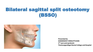
  • 1. Bilateral sagittal split osteotomy (BSSO) Presented by KANIMOZHIY SENGUTTUVAN 2nd year post graduate Thaimoogambigai Dental College and Hospital
  • 2. • Bilateral sagittal split osteotomy (BSSO) of the mandible is one of the most frequently performed surgical procedures. • This is a very popular, most versatile procedure performed on the mandibular ramus and body. • First described by Obwegeser and Trauner and later modified by Dal Pont, Hunsuck and Epker. INTRODUCTION
  • 3. • The osteotomy splits the ramus and the posterior body of the mandible sagittally, which allows either setback or advancement. • This is a highly cosmetic procedure, as it is done intraorally. • For mandibular advancement, there is no need for bone grafts. • Thus donor site morbidity and second operative site for the bone graft is totally avoided. • BSSO gives excellent results. Only drawback is the technique demands high level of operative skill and experience to minimize the surgical complications.
  • 4. • In 1957, Obwegeser and Trauner first described the sagittal split osteotomy of the ramus region, only by placing buccal and lingual cortical horizontal cuts. • The procedure has undergone numerous modifications over the years. HUGO OBWEGESER & TRAUNER 1957 HISTORY
  • 5. • In 1961, Dal Pont changed the lower horizontal cut to a vertical cut on the buccal cortex between the first and the second molars, thereby obtaining broader contact surfaces and requiring minimal muscular displacement with improved access. DALPONT (1961)
  • 6. • In 1968, Hunsuck further modified the technique, advocating a shorter, horizontal medial cut, just posterior to the lingula, to minimize soft tissue dissection. • His anterior vertical cut was similar to Dal Pont’s. HUNSUCK (1968)
  • 7. • In 1977, Epker suggested several modifications. • These include minimal stripping of the masseter muscle and limited medial dissection. • These modifications helped to reduce postoperative swelling, oedema, haemorrhage • Epker et al described completing the osteotomy through the inferior border of the mandible.
  • 8. INDICATIONS 1.Mandibular deficiency -with normal or short face, -with long face- increase maxillary vertical dimension -excessive chin height -for correction of sleep apnea Limitation- Additional surgery for most dentofacial deformity 2.Mandibular prognathism Limitation -Large setbacks of more than 7 -8 mm, IVRO/ inverted L osteotomy should be considered 3.Mandibular asymmetry - Hemi mandibular hypertrophy -Hemi mandibular elongation 4. Open bite 5. Cross bite
  • 9. CONTRAINDICATIONS • Severe decreased posterior mandibular body height • Extremely thin medial –lateral width of ramus • Severe ramus hypoplasia and • Severe asymmetries
  • 10. Surgical procedure Incision & Dissection • The incision begins at the anterior aspect of the ramus at a midpoint between upper and lower molars. • The incision is about 2 cm long. • It runs down the lateral crest of the external oblique ridge and ends in the facial vestibule at approximately the first molar region. • After sharp dissection of the mucosa, the buccinator muscle over the ramus is dissected off at its medial aspect so as to keep as much soft tissue laterally as possible. • Then, the cut is completed through periosteum to bone.
  • 11. Osteotomy cut • The procedure starts with three corticotomies. • The first cut is made through the lingual cortex just above the mandibular foramen parallel to the occlusion. • The corticotomy is extended from the anterior border of the ramus to just behind the entrance of the inferior alveolar canal (lingula).
  • 12. • The second corticotomy is made through the buccal cortex in a vertical direction at the level of the first or second molar.
  • 13. • We place the vertical cut at the horizontal ramus between the first and second molars and extend it downward to the inferior border, curving around it. • Cortical bone below the second molar is thick; this thickness protects the neurovascular bundle. • Nevertheless, the neurovascular bundle lies just below the cortical bone; thus, the osteotomy is required to go only through cortical bone.
  • 14. • The third corticotomy connects the first two osteotomy lines along the anterior border of the ascending ramus.
  • 15. The sagittal split • The final split is completed with a thin osteotome, splitting the entire ascending ramus from the anterior to the posterior border of the ramus.
  • 16. Mobilization • A special bone spreader can be used to mobilize the segments
  • 17. • After the bilateral split is completed the large tooth bearing segment can be moved three dimensionally.
  • 18. • Once the osteotomy has been completed on both sides, a prefabricated interocclusal acrylic wafer splint is placed. • We achieve maxillo-mandibular fixation (MMF) with three wire ligatures. • Some centers do not use an occlusal wafer splint. • In this case, the teeth must be placed in good three-point contact for a stable MMF to avoid any shift when screws or plate and screws are set. Positioning of the tooth bearing segment
  • 19. • Care must be taken to maintain the normal fossa-condyle relation (see upper insert) and to avoid condylar displacement (see lower insert). • Usually this is achieved by manual positioning of the condyle bearing segment superiorly into the glenoid fossa Positioning of condyle bearing segment
  • 20. A. Custom splint that lies along the lateral surface of the maxillary teeth is placed before osteotomy. The splint has a guide wire attached, extending along the lateral surface of the ramus. A saw is used to score a groove in the bone along this guide wire. B. After the osteotomy, the distal segment is wired into the final splint (MMF). The condylar positioning splint is reapplied and alignment of the scored mark in the ramus with the guide wire is examined. In this illustration, the ramus has rotated counterclockwise. C. The ramus can be rotated around the condyle until the scored groove aligns with the guide wire. Fixation between the proximal and distal segments can then be performed. Condylar positioning technique described in 1976 by Leonard.’
  • 21. Condylar positioning device connecting the maxillary dentition to the ramus with a stiff wire. A.Placement of device before osteotomy. The device is removed, the osteotomy performed, and the distal segment wired into the final splint (MMF). B. The positioning device is replaced and fixation between the proximal and distal segments can be performed. Such devices prevent rotation of the proximal segment but do not provide three-dimensional control.
  • 22. Condylar positioning technique that produce three-dimensional control of the proximal segment. A.A bone plate is adapted and secured between the proximal segment and zygomaticomaxillary buttress. The screws in the buttress are removed, the osteotomy performed, and the distal segment wired into the final splint (MMF). B. The screws are then replaced in the buttress, repositioning the ramus into its preosteotomy position.
  • 23. Fixation techniques • With wire at upper and lower border • Lag screws/Bicortical screws • Mini plates • Bioresorbable plates and screws
  • 24. • In the past, osteosynthesis was achieved with upper border wire, lower border wire, or circum-ramus body wire. • Today, this inveterate method is no longer justifiable for use on a routine basis because MMF is mandatory for weeks following surgery with this type of fixation. UPPER AND LOWER BORDER OR CIRCUM-RAMUS WIRING
  • 25. • Lag screws are indicated only when passive bone contact with no tension or torque to the condyle is sufficient. • Two or three 2.0-mm position screws are used for stabilization. • These are placed transorally at the superior border of the osteotomy site. Lag Screws/ position screws
  • 26. • It is important to place the first screw where bone contact is sufficient and passive with no tension, to avoid displacement of the condyle. • The proximal segment is positioned slightly into the fossa and is held with a ramus pusher.
  • 27. • For placement of a 2.0-mm screw, a 1.5-mm drill hole is positioned through the proximal and distal segment. • The two segments must be kept aligned and are held together as the first hole is drilled with a 1.5-mm wire-passing bur. • Then, a 2-mm self-tapping screw engages both segments of cortical bone.
  • 28. Osteosythesis With Miniplates • Fixation can be accomplished by bridging the osteotomy with a bone plate. • Usually, a four-hole bone plate with two screws on each side of the cut is sufficient for stable fixation. • Self retaining monocortical screws provide adequate stability. • The plates we use are 2.3-mm rigid standard titanium plates (Martin).
  • 29. • More flexible plates such as low-profile plates with 1.8- mm diameter do not provide enough strength for function. • Therefore, if we use non-rigid plates for fixation, we add a second plate or position screws to give additional stability. • Usually, two screws on each side of the cut is sufficient. • In unusual splits (bad splits), it is sometimes necessary to use longer plates to stabilize the osteotomy. • After the segments have been stabilized
  • 30. • Before the osteotomy site is closed, it is wise to proof the occlusion. • Therefore, the MMF is released, the splint is removed, and functions such as extent of mouth opening and left and right lateral and protrusive functions are checked and compared with original data.
  • 31. • The need for screws or plates and screws is temporary until the sagittal split has healed. • Some centers remove metallic hardware on a routine base; others do so only if symptoms are present. • The wounds are then irrigated meticulously with saline. • For wound closure, we use a 4.0 non-resorbable suture in a horizontal mattress suture technique; this suture is removed 10 to 12 days after completion of surgery.
  • 32. Osteosynthesis With Resorbable Fixation Implants • The use of biologically inert resorbable implants eliminates the need for a second operation to remove fixation material. • Polyglycolic acid, polylactic acid, and polyester polyparadioxanon are organic macromolecular compounds that are degradable and absorbable by the body.
  • 33. • These compounds possess physical properties such as rigidity and tensile and flex strength that are necessary for their use as internal fixation devices in orthognathic surgery. • As experiments have shown, the degradation process in itself does not imply immediate absorption of an implant.
  • 34. • About 70% of the material in the implant (depending on the material) remains in situ for about 3 months before the degradation process starts. • The rate of degradation is dependant on several factors: molecular weight, crystalline, and thermal history and geometry of the implant.
  • 36. Injury to the Inferior Alveolar Nerve (IAN) • Postoperative neurosensory deficit generally is considered to be caused by mechanical damage of the IAN (open injuries). • Even in cases in which the IAN is not visibly traumatized during surgery, postoperative neurosensory deficit can occur (closed injury). • This may result from edema, hematoma, or direct damage to cancellous bone fragments after stabilization or incorrect placement of screws.
  • 37. • Nerve injuries are described clinically as “open” or “closed.” • Open injuries are those observed by the surgeon at the time they occur. • Immediate primary repair by suturing of the nerve guarantees the best result. • Closed injuries are not visualized at the time of their occurrence (e.g., laceration of nerves by osteosynthesis screws), and they may or may not be suspected. • Closed injuries are followed by repeat examination every 4 weeks. • If unacceptable loss of sensation or painful or unpleasant sensation occurs, microsurgical repair is the only way to regain sensation of the nerve
  • 38. • The IAN is occasionally lacerated during osteotomy at the site of its entrance into the mandible. • This can be prevented by protecting this site during osteotomy with a freer. • Lesions of the IAN in the anterior vertical part of the osteotomy are most often due to the close relationship of the mandibular canal to the lateral mandibular cortex in the second molar. • The risk for neurosensory disturbance is significantly greater if the distance between the mandibular canal and the buccal cortex of the mandible is 2 mm or less.
  • 39. • Tear of the IAN can be the consequence of forced manipulation during the splitting process. • To avoid this, only rotating instruments and saws may be used to open the mandibular cortex. • Therefore, the osteotome may penetrate only very superficially into the mandible during splitting.
  • 40. • The IAN is variable with respect to its anatomic location in the mandible. • However, some mandibular deformities seem to be associated with certain abnormal locations of the IAN. • The presence of an unerupted third molar during surgery has been proposed as a factor that increases the risk of neural deficit. • In addition, roots of third molars are frequently located close to the mandibular canal and tear the nerve upon mobilization.
  • 41. • Paresthesia is an expression of increased manipulation that is performed if the neurovascular bundle has to be dissected from the proximal segment or released from the bone canal in the proximal segment.
  • 42. Injury to the Lingual Nerve • Occasionally, a temporary disturbance of the lingual nerve occurs. • Two well-known reasons have been put forth for lingual nerve disturbance: bicortical screw placement and pressure of a hematoma at the lingual side of the mandible. • Bicortical screws that are placed near the superior border of the mandible in the region of the third molar must not extend beyond the inner cortex of the mandible because they may damage the lingual nerve in this region.
  • 43. • Hematoma following severe bleeding at the inner side of the mandible exerts pressure on the lingual nerve. • This can lead to temporary paresthesia of the tongue that is characterized by sensible defects of the affected side of the tongue
  • 44. Displacement of the Condyle • Failure to seat the proximal segments properly into the fossa during surgery may result in condylar displacement. • This is the main reason for TMJ symptoms after BSSO • Rotation of the proximal segment • Condylar sag • Condylar torque
  • 45. Condylar Sag • Condylar sag can be defined as an immediate or late caudal movement of the condyle in the glenoid fossa after surgical establishment of a pre-planned occlusion and rigid fixation of the bone fragments, leading to a change in the occlusion.
  • 46.
  • 47. CENTRAL CONDYLAR SAG • In central condylar sag, the condyle is positioned inferiorly in the glenoid fossa and makes no contact with any part of the fossa • After removal of the IMF and in the absence of intracapsular edema or hemarthrosis (causing hydraulic pressure), the condyle will move superiorly after removal of maxillo-mandibular fixation, leading to a malocclusion
  • 48.
  • 49. PERIPHERAL CONDYLAR SAG - I • Two types of peripheral condylar sag may occur. • In Type 1, the condyle is positioned inferiorly, with some fossa contact (lateral, medial, posterior, or anterior) with the maxillo-mandibular fixation in position (teeth in occlusion) and rigid fixation placed. • This type of condylar malpositioning provides physical support to the occlusion. • Postoperative resorption or change in condylar shape will lead to late relapse.
  • 50. PERIPHERAL CONDYLAR SAG - II • In Type II, the condyle is positioned correctly in the fossa with the maxillo- mandibular fixation in position (teeth in occlusion); however, with the placement of rigid fixation, a torqueing force is applied to the condyle and ramus of the mandible. • The tension on the ramus is released when the maxillo-mandibular fixation is removed, and the condyle will move either laterally or medially and slide inferiorly in the fossa.
  • 51.
  • 52. • The incidence of condylar resorption has been reported at between 1% and 31%. Resorption of Condyle
  • 53. • Predisposing factors for condyle resorption include the following: • High mandibular plane angle, condyle with posterior inclination, and a low posterior-to-anterior face/height ratio are predisposing skeletal factors for condylar resorption. • Contributing are surgical factors such as counter-clockwise rotation of the proximal segment, preoperative TMJ dysfunction, and vascular necrosis of the condyle that occurs as a consequence of traumatic stress during surgery. • Proper positioning of the proximal segment into the fossa after the mandible is split is of utmost importance for avoiding postoperative TMJ problems such as condylar resorption.
  • 54. UNFAVORABLE BONY SPLIT • Poor split of the distal segment most often affects the lingual cortical bone posterior to the third molar. • This may result in sequestration of the fragment, delayed union, malunion of the osteotomy site, or even infection. • Stabilization of the unfavourable fracture prevents postsurgical instability. • Unanticipated splits of the proximal segment may occur before or after splitting of the mandible
  • 55. • Often, the reason for a poor split is improper wedging with osteotome, especially in cases where bone is used as a fulcrum during splitting. • Therefore, careful inspection of the inferior border and the outer cortex is mandatory. • Predisposing factors include • difficult anatomy, • poor osteotomy design, and • the presence of impacted third molars. • Anatomic features such as width and thickness of the ascending ramus and the relationship between positions in the canal have to be studied previously. • Lack of cancellous bone between the two cortical bone layers makes the split more difficult
  • 56. • Correct cuts through cortical bone with the use of a reciprocating saw or a bur are essential for avoiding a bad split. • This is especially true for the cut at the inner side of the ascending ramus and the inferior border prior to application of the chisel to the osteotomy. • An increased risk of unanticipated fracture during BSSO has been associated with the presence of impacted third molars, which have been postulated to compromise the anatomic structure of the mandible, causing adverse proximal and distal segment splits. • Stabilization is more challenging than in non-fractured splits, and more osteosynthesis material is required. • Plates, bicortical screws, or a combination of these can be used to stabilize fractured bony segments.
  • 59. (a)The buccal cortex fractures separately from the lower border, while the fracture runs superiorly toward the coronoid notch. The splitting maneuver is stopped and the lower border captured to form part of the proximal segment. (b) The buccal cortex fracture continues superiorly and includes the coronoid process. The buccal osteotomy is redefined and the lower border captured. Rigid fixation is achieved using a bone plate and by securing the fractured cortex by means of bicortical screws,
  • 60. NERVE CANAL IS STILL ATTACHED TO THE PROXIMAL BONE SEGMENT
  • 62. • Reason for bleeding in this phase is inadvertent laceration of the masseter muscle during incision of the vestibular mucosa. • This can be avoided by meticulous cutting of the mucosa all the way through to the periosteum. HEMORRHAGE • Uncontrolled hemorrhage is a rare condition in mandibular surgery. • It occurs in most cases as the consequence of clumsy use of rotating instruments or chisels, which reflects obvious inexperience of the surgeon in performing the surgical procedure. • Bleeding from the pterygoideus medialis muscle may occur during osteotomy at the lingual surface of the ascending mandibular ramus, if the periosteum is not completely released from the bone.
  • 63. • Bleedings from the facial artery may occur during removal of soft tissue from the mandibular border; this must be treated with a suture. • Bleeding from the inferior alveolar artery can occur when bone fragments after osteotomy are separated via forced use of a bibeveled osteotome. • This complication can be avoided by a cautious split performed under good vision. • This can be achieved by placement of the suction tip into the bottom of the split. In addition, careful preparation and release of vessels (and nerves) from the proximal fragment is necessary
  • 64. INFECTION • Wound dehiscence and/or infection is now a rare complication and is most often related to poor oral hygiene. • Tobacco smoking seems to be an additional risk factor. • Contributing factors include long duration of the surgical procedure, the patient’s age, the presence of foreign bodies, sequestering after bone fracture, and hematoma. • The mandatory use of prophylactic antibiotics during and after surgery has substantially reduced the risk of infection.
  • 65. • Relapse is a multifactorial problem that cannot be attributed to a single cause. • Proper diagnoses, adequate treatment planning, correct and skillful surgical technique are essential contributions of the surgeon. • Soft tissue and muscle tension may influence postoperative stability. • Factors that have an effect on relapse include type of fixation, magnitude and type of mandibular movement, position of proximal segment, and disk displacement. Relapse
  • 66. Stability depends on : • Adequate presurgical orthodontics. • Long-term maxillomandibular fixation (MMF). • Non-rigid fixation that allow muscular adaptation. • Minimal muscle alteration, Good bony contact, and control of the proximal segment
  • 67. Factors : • Magnitude of mandibular advancement or setback, • Stretch of surrounding soft tissue, • Positioning of mandibular condyles • Method of fixation • Growth of mandible • skeletal behaviour among hyper/ hypo divergent skeletal patterns
  • 68. • Obligate relapse after mandibular advancements >7mm • Mandibular setback >12 mm - skeletal relapse • Closure of anterior open bite with only mandibular osteotomies How to reduce/avoid : • Counter-clockwise rotation of the mandible be avoided • Mandibular advancement limited to < 7mm • Bimaxillary surgery
  • 69. REFERENCES • FONSECA -Oral and Maxillofacial Surgery, Second edition Volume 3, Orthognathic surgery Esthetic surgery. • Johan P. Reyneke, Essential of orthognathic surgery • Textbook of Oral and Maxillofacial Surgery, Third Edition- Neelima Anil Malik. • Intraoperative diagnosis of condylar sag after bilateral sagittal split ramus osteotomy, Johan. P. Reyneke, C. Ferretti , British Journal of Oral and Maxillofacial Surgery (2002) 40, 285–292 • Condylar Positioning Devices For Orthognathic Surgery, EDWARD ELLIS III, DDS, MS, J Oral Maxillofacial Surgery 52 536.552(1994).