Download as PDF, PPTX


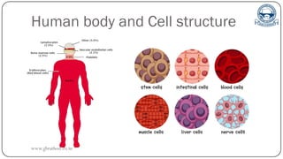
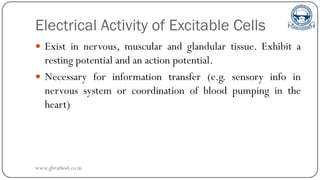
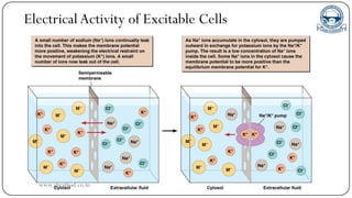

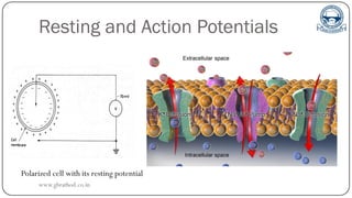


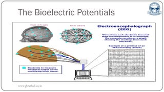
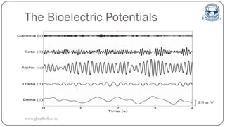
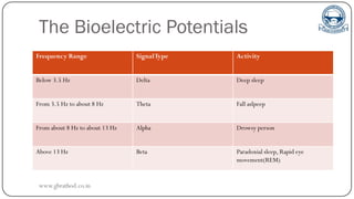
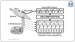


The document outlines the structure and electrical activity of cells in the human body, emphasizing the role of bioelectric potentials in nerve, muscle, and glandular tissues. It explains the concepts of resting and action potentials, their significance in cellular communication, and how these potentials are measured through various bioelectrical signals like ECG, EEG, and EMG. Additionally, it discusses the propagation of action potentials and their measurement rates, highlighting the all-or-nothing law and refractory periods.


